Related Research Articles

Orthopedic surgery or orthopedics is the branch of surgery concerned with conditions involving the musculoskeletal system. Orthopedic surgeons use both surgical and nonsurgical means to treat musculoskeletal trauma, spine diseases, sports injuries, degenerative diseases, infections, tumors, and congenital disorders.
An osteotomy is a surgical operation whereby a bone is cut to shorten or lengthen it or to change its alignment. It is sometimes performed to correct a hallux valgus, or to straighten a bone that has healed crookedly following a fracture. It is also used to correct a coxa vara, genu valgum, and genu varum. The operation is done under a general anaesthetic.

Bisphosphonates are a class of drugs that prevent the loss of bone density, used to treat osteoporosis and similar diseases. They are the most commonly prescribed drugs used to treat osteoporosis. They are called bisphosphonates because they have two phosphonate groups. They are thus also called diphosphonates.

A bone fracture is a medical condition in which there is a partial or complete break in the continuity of any bone in the body. In more severe cases, the bone may be broken into several fragments, known as a comminuted fracture. A bone fracture may be the result of high force impact or stress, or a minimal trauma injury as a result of certain medical conditions that weaken the bones, such as osteoporosis, osteopenia, bone cancer, or osteogenesis imperfecta, where the fracture is then properly termed a pathologic fracture.

Hip replacement is a surgical procedure in which the hip joint is replaced by a prosthetic implant, that is, a hip prosthesis. Hip replacement surgery can be performed as a total replacement or a hemi/semi(half) replacement. Such joint replacement orthopaedic surgery is generally conducted to relieve arthritis pain or in some hip fractures. A total hip replacement consists of replacing both the acetabulum and the femoral head while hemiarthroplasty generally only replaces the femoral head. Hip replacement is one of the most common orthopaedic operations, though patient satisfaction varies widely between different techniques and implants. Approximately 58% of total hip replacements are estimated to last 25 years. The average cost of a total hip replacement in 2012 was $40,364 in the United States, and about $7,700 to $12,000 in most European countries.
Perioperative mortality has been defined as any death, regardless of cause, occurring within 30 days after surgery in or out of the hospital. Globally, 4.2 million people are estimated to die within 30 days of surgery each year. An important consideration in the decision to perform any surgical procedure is to weigh the benefits against the risks. Anesthesiologists and surgeons employ various methods in assessing whether a patient is in optimal condition from a medical standpoint prior to undertaking surgery, and various statistical tools are available. ASA score is the most well known of these.
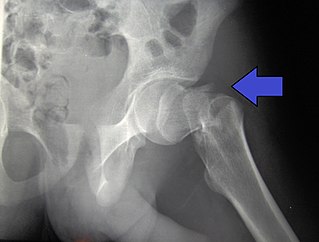
A hip fracture is a break that occurs in the upper part of the femur, at the femoral neck or (rarely) the femoral head. Symptoms may include pain around the hip, particularly with movement, and shortening of the leg. Usually the person cannot walk.
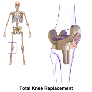
Knee replacement, also known as knee arthroplasty, is a surgical procedure to replace the weight-bearing surfaces of the knee joint to relieve pain and disability, most commonly offered when joint pain is not diminished by conservative sources. It may also be performed for other knee diseases, such as rheumatoid arthritis. In patients with severe deformity from advanced rheumatoid arthritis, trauma, or long-standing osteoarthritis, the surgery may be more complicated and carry higher risk. Osteoporosis does not typically cause knee pain, deformity, or inflammation, and is not a reason to perform knee replacement.

Traction is a set of mechanisms for straightening broken bones or relieving pressure on the spine and skeletal system. There are two types of traction: skin traction and skeletal traction. They are used in orthopedic medicine.

Osteochondritis dissecans is a joint disorder primarily of the subchondral bone in which cracks form in the articular cartilage and the underlying subchondral bone. OCD usually causes pain during and after sports. In later stages of the disorder there will be swelling of the affected joint which catches and locks during movement. Physical examination in the early stages does only show pain as symptom, in later stages there could be an effusion, tenderness, and a crackling sound with joint movement.

Ibandronic acid is a bisphosphonate medication used in the prevention and treatment of osteoporosis and metastasis-associated skeletal fractures in people with cancer. It may also be used to treat hypercalcemia. It is typically formulated as its sodium salt ibandronate sodium.

The AO Foundation is a nonprofit organization dedicated to improving the care of patients with musculoskeletal injuries or pathologies and their sequelae through research, development, and education of surgeons and operating room personnel. The AO Foundation is credited with revolutionizing operative fracture treatment and pioneering the development of bone implants and instruments.
An open fracture, also called a compound fracture, is a type of bone fracture that has an open wound in the skin near the fractured bone. The skin wound is usually caused by the bone breaking through the surface of the skin. An open fracture can be life threatening or limb-threatening due to the risk of a deep infection and/or bleeding. Open fractures are often caused by high energy trauma such as road traffic accidents and are associated with a high degree of damage to the bone and nearby soft tissue. Other potential complications include nerve damage or impaired bone healing, including malunion or nonunion. The severity of open fractures can vary. For diagnosing and classifying open fractures, Gustilo-Anderson open fracture classification is the most commonly used method. This classification system can also be used to guide treatment, and to predict clinical outcomes. Advanced trauma life support is the first line of action in dealing with open fractures and to rule out other life-threatening condition in cases of trauma. The person is also administered antibiotics for at least 24 hours to reduce the risk of an infection.
The Gustilo open fracture classification system is the most commonly used classification system for open fractures. It was created by Ramón Gustilo and Anderson, and then further expanded by Gustilo, Mendoza, and Williams.

A femoral fracture is a bone fracture that involves the femur. They are typically sustained in high-impact trauma, such as car crashes, due to the large amount of force needed to break the bone. Fractures of the diaphysis, or middle of the femur, are managed differently from those at the head, neck, and trochanter; those are conventionally called hip fractures. Thus, mentions of femoral fracture in medicine usually refer implicitly to femoral fractures at the shaft or distally.

A tibial plateau fracture is a break of the upper part of the tibia (shinbone) that involves the knee joint. This could involve the medial, lateral, central, or bicondylar. Symptoms include pain, swelling, and a decreased ability to move the knee. People are generally unable to walk. Complication may include injury to the artery or nerve, arthritis, and compartment syndrome.

Ultrasonography of suspected or previously confirmed chronic venous insufficiency of leg veins is a risk-free, non-invasive procedure. It gives information about the anatomy, physiology and pathology of mainly superficial veins. As with heart ultrasound (echocardiography) studies, venous ultrasonography requires an understanding of hemodynamics in order to give useful examination reports. In chronic venous insufficiency, sonographic examination is of most benefit; in confirming varicose disease, making an assessment of the hemodynamics, and charting the progression of the disease and its response to treatment. It has become the reference standard for examining the condition and hemodynamics of the lower limb veins. Particular veins of the deep venous system (DVS), and the superficial venous system (SVS) are looked at. The great saphenous vein (GSV), and the small saphenous vein (SSV) are superficial veins which drain into respectively, the common femoral vein and the popliteal vein. These veins are deep veins. Perforator veins drain superficial veins into the deep veins. Three anatomic compartments are described, (N1) containing the deep veins, (N2) containing the perforator veins, and (N3) containing the superficial veins, known as the saphenous compartment. This compartmentalisation makes it easier for the examiner to systematize and map. The GSV can be located in the saphenous compartment where together with the Giacomini vein and the accessory saphenous vein (ASV) an image resembling an eye, known as the 'eye sign' can be seen. The ASV which is often responsible for varicose veins, can be located at the 'alignment sign', where it is seen to align with the femoral vessels.

Pain in the hip is the experience of pain in the muscles or joints in the hip/ pelvic region, a condition commonly arising from any of a number of factors. Sometimes it is closely associated with lower back pain.
Transcatheter pulmonary valve replacement (TPVR), also known as percutaneous pulmonary valve implantation (PPVI), is the replacement of the pulmonary valve via catheterization through a vein. It is a significantly less invasive procedure in comparison to open heart surgery and is commonly used to treat conditions such as pulmonary atresia.
Bone malrotation refers to the situation that results when a bone heals out of rotational alignment from another bone, or part of bone. It often occurs as the result of a surgical complication after a fracture where intramedullary nailing (IMN) occurs, especially in the femur and tibial bones, but can also occur genetically at birth. The severity of this complication is often neglected due to its complexity to detect and treat, yet if left untreated, bone malrotation can significantly impact regular bodily functioning, and even lead to severe arthritis. Detection throughout history has become more advanced and accurate, ranging from clinical assessment to ultrasounds to CT scans. Treatment can include an osteotomy, a major surgical procedure where bones are cut and realigned correctly, or compensatory methods, where individuals learn to externally or internally rotate their limb to compensate for the rotation. Further research is currently being examined in this area to reduce occurrences of malrotation, including detailed computer navigation to improve visual accuracy during surgery.
References
- ↑ Seinsheimer, F. "Subtrochanteric fractures of the femur." The Journal of Bone and Joint Surgery 60.3 (1978): 300-306.
- ↑ Gehrchen, P. Martin, et al. "Seinsheimer's classification of subtrochanteric fractures: Poor reproducibility of 4 observers' evaluation of 50 cases." Acta Orthopaedica 68.6 (1997): 524-526.
- ↑ Guyver, P. M., et al. "Is there any purpose in classifying subtrochanteric fractures? The reproducibility of four classification systems." European Journal of Orthopaedic Surgery and Traumatology 24.4 (2014): 513-518.