Related Research Articles
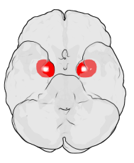
The amygdala is a paired nuclear complex present in the cerebral hemispheres of vertebrates. It is considered part of the limbic system. In primates, it is located medially within the temporal lobes. It consists of many nuclei, each made up of further subnuclei. The subdivision most commonly made is into the basolateral, central, cortical, and medial nuclei together with the intercalated cell clusters. The amygdala has a primary role in the processing of memory, decision-making, and emotional responses. The amygdala was first identified and named by Karl Friedrich Burdach in 1822.

The limbic system, also known as the paleomammalian cortex, is a set of brain structures located on both sides of the thalamus, immediately beneath the medial temporal lobe of the cerebrum primarily in the forebrain.
Hypersexuality is a medical condition that causes unwanted or excessive sexual arousal, causing people to engage in or think about sexual activity to a point of distress or impairment. It is controversial whether it should be included as a clinical diagnosis used by mental healthcare professionals. Nymphomania, satyromania and sex maniac were terms previously used for the condition in women and men, respectively.

The temporal lobe is one of the four major lobes of the cerebral cortex in the brain of mammals. The temporal lobe is located beneath the lateral fissure on both cerebral hemispheres of the mammalian brain.

Brodmann area 38, also BA38 or temporopolar area 38 (H), is part of the temporal cortex in the human brain. BA 38 is at the anterior end of the temporal lobe, known as the temporal pole.
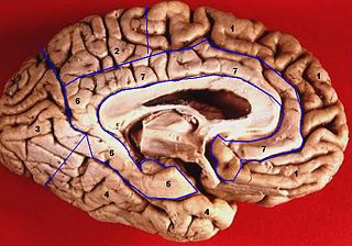
The limbic lobe is an arc-shaped cortical region of the limbic system, on the medial surface of each cerebral hemisphere of the mammalian brain, consisting of parts of the frontal, parietal and temporal lobes. The term is ambiguous, with some authors including the paraterminal gyrus, the subcallosal area, the cingulate gyrus, the parahippocampal gyrus, the dentate gyrus, the hippocampus and the subiculum; while the Terminologia Anatomica includes the cingulate sulcus, the cingulate gyrus, the isthmus of cingulate gyrus, the fasciolar gyrus, the parahippocampal gyrus, the parahippocampal sulcus, the dentate gyrus, the fimbrodentate sulcus, the fimbria of hippocampus, the collateral sulcus, and the rhinal sulcus, and omits the hippocampus.
Witzelsucht is a set of rare neurological symptoms characterized by a tendency to make puns, or tell inappropriate jokes or pointless stories in socially inappropriate situations. It makes one unable to read sarcasm.
Auditory verbal agnosia (AVA), also known as pure word deafness, is the inability to comprehend speech. Individuals with this disorder lose the ability to understand language, repeat words, and write from dictation. Some patients with AVA describe hearing spoken language as meaningless noise, often as though the person speaking was doing so in a foreign language. However, spontaneous speaking, reading, and writing are preserved. The maintenance of the ability to process non-speech auditory information, including music, also remains relatively more intact than spoken language comprehension. Individuals who exhibit pure word deafness are also still able to recognize non-verbal sounds. The ability to interpret language via lip reading, hand gestures, and context clues is preserved as well. Sometimes, this agnosia is preceded by cortical deafness; however, this is not always the case. Researchers have documented that in most patients exhibiting auditory verbal agnosia, the discrimination of consonants is more difficult than that of vowels, but as with most neurological disorders, there is variation among patients.

The hippocampal formation is a compound structure in the medial temporal lobe of the brain. It forms a c-shaped bulge on the floor of the temporal horn of the lateral ventricle. There is no consensus concerning which brain regions are encompassed by the term, with some authors defining it as the dentate gyrus, the hippocampus proper and the subiculum; and others including also the presubiculum, parasubiculum, and entorhinal cortex. The hippocampal formation is thought to play a role in memory, spatial navigation and control of attention. The neural layout and pathways within the hippocampal formation are very similar in all mammals.

Frontal lobe disorder, also frontal lobe syndrome, is an impairment of the frontal lobe of the brain due to disease or frontal lobe injury. The frontal lobe plays a key role in executive functions such as motivation, planning, social behaviour, and speech production. Frontal lobe syndrome can be caused by a range of conditions including head trauma, tumours, neurodegenerative diseases, neurodevelopmental disorders, neurosurgery and cerebrovascular disease. Frontal lobe impairment can be detected by recognition of typical signs and symptoms, use of simple screening tests, and specialist neurological testing.
Klüver–Bucy syndrome is a syndrome resulting from lesions of the medial temporal lobe, particularly Brodmann area 38, causing compulsive eating, hypersexuality, a compulsive need to insert inappropriate objects in the mouth (hyperorality), visual agnosia, and docility. Klüver–Bucy syndrome is more commonly found in rhesus monkeys, where the condition was first documented, than in humans. The underlying pathology of the syndrome is still controversial, with Muller theory and a theory by Norman Geschwind offering different explanations for the condition. Treatment for Klüver–Bucy syndrome is usually with mood stabilizers, anti-psychotics, and anti-depressants.

Cortical deafness is a rare form of sensorineural hearing loss caused by damage to the primary auditory cortex. Cortical deafness is an auditory disorder where the patient is unable to hear sounds but has no apparent damage to the structures of the ear. It has been argued to be as the combination of auditory verbal agnosia and auditory agnosia. Patients with cortical deafness cannot hear any sounds, that is, they are not aware of sounds including non-speech, voices, and speech sounds. Although patients appear and feel completely deaf, they can still exhibit some reflex responses such as turning their head towards a loud sound.
Paul Bucy was an American neurosurgeon and neuropathologist who was a native of Hubbard, Iowa. He is known both for his part in describing the Klüver–Bucy syndrome, his academic life as a teacher in the neurosciences, and for his founding in 1972 and editing Surgical Neurology – An International Journal of Neurosurgery and Neuroscience from 1972 to 1987.

Urbach–Wiethe disease is a very rare recessive genetic disorder, with approximately 400 reported cases since its discovery. It was first officially reported in 1929 by Erich Urbach and Camillo Wiethe, although cases may be recognized dating back as early as 1908.
Charcot–Wilbrand syndrome (CWS) is dream loss following focal brain damage specifically characterised by visual agnosia and loss of ability to mentally recall or "revisualize" images. The name of this condition dates back to the case study work of Jean-Martin Charcot and Hermann Wilbrand, and was first described by Otto Potzl as "mind blindness with disturbance of optic imagination". MacDonald Critchley, former president of the World Federation of Neurology, more recently summarized CWS as "a patient loses the power to conjure up visual images or memories, and furthermore, ceases to dream during his sleeping hours". This condition is quite rare and affects only a handful of brain damage patients. Further study could help illuminate the neurological pathway for dream formation.
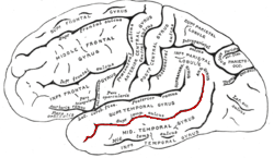
In the human brain, the superior temporal sulcus (STS) is the sulcus separating the superior temporal gyrus from the middle temporal gyrus in the temporal lobe of the brain. A sulcus is a deep groove that curves into the largest part of the brain, the cerebrum, and a gyrus is a ridge that curves outward of the cerebrum.
Phonagnosia is a type of agnosia, or loss of knowledge, that involves a disturbance in the recognition of familiar voices and the impairment of voice discrimination abilities in which the affected individual does not suffer from comprehension deficits. Phonagnosia is an auditory agnosia, an acquired auditory processing disorder resulting from brain damage. Other auditory agnosias include cortical deafness and auditory verbal agnosia also known as pure word deafness.
S.M., sometimes referred to as SM-046, is an American woman with a peculiar type of brain damage that physiologically reduces her ability to feel fear. First described by scientists in 1994, she has had exclusive and complete bilateral amygdala destruction since late childhood as a consequence of Urbach–Wiethe disease. Dubbed by the media as the "woman with no fear", S.M. has been studied extensively in scientific research; she has helped researchers elucidate the function of the amygdala.
Nonverbal autism, also called nonspeaking autism, is a subset of autism spectrum disorder (ASD) where the person does not learn how to speak. One study has shown that 64% of autistic children who are nonverbal at age 5 are still nonverbal 10 years later.
Amygdalotomy, also known as amygdalectomy, is a form of psychosurgery which involves the surgical removal or destruction of the amygdala, or parts of the amygdala. It is usually a last-resort treatment for severe aggressive behavioral disorders and similar behaviors including hyperexcitability, violent outbursts, and self-mutilation. The practice of medical amygdalotomy typically involves the administration of general anesthesia and is achieved through the application of cranial stereotactic surgery to target regions of the amygdala for surgical destruction. While some studies have found stereotactic amygdalotomy in humans to be an effective treatment for severe cases of intractable aggressive behavior that has not responded to standard treatment methods, other studies remain inconclusive. In most cases of amygdalotomy in humans, there is no substantial evidence of impairment in overall cognitive function, including intelligence and working memory, however, deficits in specific areas of memory have been noted pertaining to the recognition and emotional interpretation of facial stimuli. This is because there are specialized cells in the amygdala which attend to facial stimuli.
References
- ↑ "Medical Education for Undergraduate MD Students ." Agnosia. N.p., 14 Apr 2011. Web. 28 Nov 2011.
- ↑ Schmitz, Bettina, and Michael Trimble. The Neuropsychiatry of Epilepsy. 1st. London: Cambridge University Press, 2002: 110-111.
- 1 2 Baron-Cohen, S.; Ring, H.A.; Bullmore, E.T.; Wheelwright, S.; Ashwin, C.; Williams, S.C.R. (2000). "The amygdala theory of autism". Neuroscience & Biobehavioral Reviews. 24 (3): 355–364. doi:10.1016/s0149-7634(00)00011-7. PMID 10781695. S2CID 7455984.
- 1 2 3 Salloway, Stephen, Paul Mallory, and Jeffrey L. Cummings. The neuropsychiatry of limbic and subcortical disorders. 1997.
- 1 2 Amaral, D. G.; Corbett, B. A. (2003). "The Amygdala, Autism and Anxiety". Autism: Neural Basis and Treatment Possibilities. Novartis Foundation Symposia. Vol. 251. pp. 177–87, discussion 187-97, 281–97. doi:10.1002/0470869380.ch11. ISBN 978-0-470-85099-2. PMID 14521193.
{{cite book}}:|journal=ignored (help) - ↑ Schmitz, Bettina, and Michael Trimble. The Neuropsychiatry of Epilepsy. 1st. London: Cambridge University Press, 2002: 114-115.
- ↑ Stone, Valerie E.; Baron-Cohen, Simon; Knight, Robert T. (1998). "Frontal Lobe Contributions to Theory of Mind" (PDF). Journal of Cognitive Neuroscience. 10 (5): 640–656. doi:10.1162/089892998562942. PMID 9802997. S2CID 207724498.