Spemann-Mangold organizer

Last updated

The Spemann-Mangold organizer is a group of cells that are responsible for the induction of the neural tissues during development in amphibian embryos. First described in 1924 by Hans Spemann and Hilde Mangold, the introduction of the organizer provided evidence that the fate of cells can be influenced by factors from other cell populations. [1] This discovery significantly impacted the world of developmental biology and fundamentally changed the understanding of early development.

Contents

Discovery

The Spemann-Mangold organizer was first described in 1924 by Hans Spemann and Hilde Mangold. Prior to its discovery, it had been hypothesized by multiple groups that there exists a portion of the developing embryo that serves as an "organization center". In 1918 and 1921, Hans Spemann showed that transplanting presumptive epidermis into the area of presumptive neural tissue would change the fate of the transplanted cells to that of their new destination, and likewise when he transplanted presumptive neural tissue to where the presumptive epidermis was forming. Spemann also showed that by transplanting a piece from the upper blastopore lip into an area of presumptive epidermis, a secondary embryonic primordium formed, including a secondary neural tube, notochord and somites. Additionally, splitting the embryo in half and rotating the animal pole in respect to the vegetal pole resulted in determination spreading from the lower vegetal pole, where the upper blastopore lip was located, to the upper animal half. He also fused together two identical halves from different embryos and observed formation of the neural plate. This work provided the initial evidence to support the notion that there existed some “organization center” that was determined prior to the other embryonic tissue and influenced the determination of surrounding cells. [1]

To test this Spemann's hypothesis, one of his PhD students, Hilde Mangold, performed experiments between 1921 and 1922 using embryos from Triturus cristatus and Triturus taeniatus that were undergoing gastrulation. The experiment performed resembled the one done in 1918, however instead of a homoplastic transplantation she used embryos from two species of newt that are closely related. One of the benefits of using the cristatus and taeniatus embryos was that the cristatus embryo cells lacked pigment so the fate of the transplant could be easily tracked when placed among the pigmented taeniatus cells. A piece from the upper blastopore lip was removed from the cristatus embryo and transplanted into a ventral region of presumptive epidermis in the taeniatus embryo, away from the developing host blastopore. Following this transplant, she observed the formation of a secondary embryonic primordium, consistent with their previous work. This secondary embryo had the normal features of the primary embryo, including structures such as the neural plate and notochord, although they lagged slightly in development. Sectioning of the embryo showed that cells from the transplant were incorporated into the mesoderm, the neural plate, and constituted almost the entire notochord of the secondary embryo. It was further shown that the neural plate was almost entirely composed of cells from the host taeniatus embryo. These experiments concluded that a piece of the upper blastopore lip can be transplanted into the indifferent tissue of another embryo and induce the host tissue into the formation of a secondary embryo, therefore implicating the transplanted tissue as an "organization center". [1]

The discovery of the Spemann-Mangold Organizer is considered one of the most influential findings in the field of developmental biology and resulted in Hans Spemann being awarded the Nobel Prize in 1935 for his work (Mangold tragically died before the Nobel prize was awarded, thus was not eligible). The mechanisms of how this organizer operates has been the subject of decades of follow up research.

Mechanism

The Spemann-Mangold organizer refers to the population of cells in the Xenopus laevis embryo that establishes the dorso-ventral and antero-posterior axes. [2] While an organizer exists in other species, the term Spemann-Mangold organizer is specifically reserved for the amphibian embryo. The Spemann-Mangold organizer is located in the dorsal blastopore lip, where gastrulation movements originate. Initial organizer cells migrate and localize anteriorly. The organizer cells are subdivided into head, trunk, and tail organizers. These distinct cell populations have different inducers and set up unique growth factor gradients as they migrate. Secondary cell-cell interactions further establish the axes as gastrulation and neurulation continues. [3]

The Spemann-Mangold organizer is particularly important in mesoderm induction. In the three signal model, the dorsalizing signal from the organizer is mediated by bone morphogenic protein (BMP) gradients to give rise to cells of mesodermal fate. The other two signals arise from the vegetal pole and induce the extreme ventral and dorsal mesoderm in the overlying marginal zone. [4]

In order for the Spemann-Mangold organizer to form, maternal factors, such as mVegT must be present in the vegetal cap. [5] Wnt pathway signaling is the other major maternal cue in the formation of the organizer and is required autonomously for expression of organizer genes. [2] Siamois (Sia) and Twin (Xtwn) are expressed at the onset of zygotic gene expression in the blastula and become activated by Wnt signaling in the blastula Chordin- and Noggin-expressing (BCNE) center. [6] [5] Sia and Xtwn can function as homo- or heterodimers to bind a conserved P3 site within the proximal element (PE) of the goosecoid (Gsc) promoter. [6] Wnt signaling also acts with mVegT to upregulate Xnr5, secreted from the Nieuwkoop center, in the interior dorso-vegetal region, which will then induce additional transcription factors such as Xnr1, Xnr2, Gsc, chordin (chd). The final cue is mediated by Nodal/activin signaling, inducing transcription factors, that in combination with Sia, will induce the cerberus (cer) gene. [5]

The organizer has both transcription and secreted factors. Transcription factors include goosecoid, Lim1, and Xnot, which are all homeodomain proteins. Goosecoid was the first organizer gene discovered, providing “the first visualization of Spemann-Mangold organizer cells and of their dynamic changes during gastrulation”. [7] While it was the first to be studied, it is not the first gene to be activated. Following transcriptional activation by Sia and Xtwn, Gsc is expressed in a subset of cells encompassing 60° of arc on the dorsal marginal zone. [8] Expression of Gsc activates the expression of secreted signaling molecules. [7] Ventral injection of Gsc leads to a phenotype as seen in Spemann and Mangold's original experiment: a twinned axis. [8]

Secreted factors from the organizer form gradients in the embryo to differentiate the tissues.

FactorMechanism
Chordin BMP antagonist
Noggin BMP antagonist
Follistatin Activin and BMP antagonist
Frzb1 Wnt antagonist
Secreted frizzled-related protein-2 (sFrp2)Wnt antagonist
crescentWnt antagonist
dickkopf-1 Wnt antagonist
cerberus Nodal, Wnt, and BMP antagonist
anti-dorsalizing morphogenic protein (Admp)Growth factor

International recognition

After the discovery of the Sepmann-Mangold Organizer, many labs rushed to be the first to discover the inducing factors responsible for this organization. [9] This created a large international impact with labs in Japan, Russia, and Germany changing the way they viewed and studied developmental organization. [9] [10] [11] However, due to the slow progress in the field, many labs move research interests away from the organizer, but not before the impact of the discovery was made. [9] 60 years after the discovery of the organizer, many Nobel Prizes were given to developmental biologists for work that was influenced by the Organizer. [10]

Japan

Until the mid 19th century, Japan was a closed society that did not participate in advances in modern biology until later in that century. At that time, many students who went abroad to study in American and European labs, came back with new ideas about approaches to developmental sciences. When the returning students would try to incorporate their new ideas into the Japanese experimental embryology, they were rejected by the members of Japanese Biological Society. After the publication of the Spemann-Mangold organizer, many more students went to study abroad in European Labs, to learn much more about this organizer and returned to use that knowledge to aid in huge advantages in embryonic biology at the time. The discovery of the organizer influenced many embryonic induction projects in Japan. For example, T. Yamada created the double potential theory for the induction process in embryos. Another discovery after the organizer discovery was the modified Vogt fate map using newt and Xenopus blastula by researcher Osamu Nakamura. The new concept of transdifferentiation was proposed by T.S. Okada and G. Eguchi. These discoveries and many more in Japan were inspired by the publication of the organizer by Spemann and Mangold. [9]

Russia

The publication of the Sepmann-Mangold organizer also has a huge influence on the Russian developmental research. At first the Spemann's organizer was not accepted in Russia. The Russian scientists did not agree with the idea of embryonic inducers (morphogens) because the Russian researchers focused on developmental patterns in evolution. It was not until another researcher, A. Gurwitch, published his theory of embryonic fields that Russian scientists began to accept other theories of development, including the Sepmann-Mangold organizer, as it agreed with many of the concepts of Gurwitch's theory. With this new influence, labs in Moscow and Leningrad began to focus on the genetic control of individual development instead of evolutionary development. Russia began to analyze morphogenetic tissue interactions in a similar manner as Spemann by using the eye-lens system. From this research, Russia was able to add to the field with their research on lens and neural induction, and the discovery of the lens induction influenced the beginning of developmental mechanic labs to open in Russia. [11]

Germany

In Germany, the period immediately following the Spemann-Mangold publication was known as a period with little progress, as many questions that the new organizer produced were left unsolved. The holistic view of the Spemann-Mangold organizer needed supplemental research since many methods were not available at the time of that publication. Spemann initiated the movement of developmental and molecular biology and influenced many projects in Germany based on his findings. Spemann's work with the minced organizer tissue indicated the presence of morphogens which then lead to the double gradient hypothesis of Toivonen and Saxén. This led to the discovery that the tissues studies contained factors that caused inducing activity. Because of the Spemann-Mangold organizer discovery and suggestion of morphogens, labs in Germany were able to further learn about the mechanisms behind development with new methods to further the knowledge in the field. [10]

Related Research Articles

<span class="mw-page-title-main">Blastulation</span> Sphere of cells formed during early embryonic development in animals

Blastulation is the stage in early animal embryonic development that produces the blastula. In mammalian development the blastula develops into the blastocyst with a differentiated inner cell mass and an outer trophectoderm. The blastula is a hollow sphere of cells known as blastomeres surrounding an inner fluid-filled cavity called the blastocoel. Embryonic development begins with a sperm fertilizing an egg cell to become a zygote, which undergoes many cleavages to develop into a ball of cells called a morula. Only when the blastocoel is formed does the early embryo become a blastula. The blastula precedes the formation of the gastrula in which the germ layers of the embryo form.

<span class="mw-page-title-main">Gastrulation</span> Stage in embryonic development in which germ layers form

Gastrulation is the stage in the early embryonic development of most animals, during which the blastula, or in mammals the blastocyst is reorganized into a multilayered structure known as the gastrula. Before gastrulation, the embryo is a continuous epithelial sheet of cells; by the end of gastrulation, the embryo has begun differentiation to establish distinct cell lineages, set up the basic axes of the body, and internalized one or more cell types including the prospective gut.

<span class="mw-page-title-main">Hans Spemann</span>

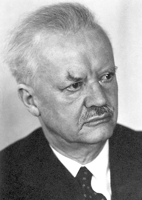
Hans Spemann was a German embryologist who was awarded a Nobel Prize in Physiology or Medicine in 1935 for his student Hilde Mangold's discovery of the effect now known as embryonic induction, an influence, exercised by various parts of the embryo, that directs the development of groups of cells into particular tissues and organs. Spemann added his name as an author to Hilde Mangold's dissertation and won a Nobel Prize for her work.

<span class="mw-page-title-main">Neurulation</span> Embryological process forming the neural tube

Neurulation refers to the folding process in vertebrate embryos, which includes the transformation of the neural plate into the neural tube. The embryo at this stage is termed the neurula.

Organogenesis is the phase of embryonic development that starts at the end of gastrulation and continues until birth. During organogenesis, the three germ layers formed from gastrulation form the internal organs of the organism.

The primitive node is the organizer for gastrulation in most amniote embryos. In birds it is known as Hensen's node, and in amphibians it is known as the Spemann-Mangold organizer. It is induced by the Nieuwkoop center in amphibians, or by the posterior marginal zone in amniotes including birds.

<span class="mw-page-title-main">Primitive streak</span> Structure in early amniote embryogenesis

The primitive streak is a structure that forms in the early embryo in amniotes. In amphibians the equivalent structure is the blastopore. During early embryonic development, the embryonic disc becomes oval shaped, and then pear-shaped with the broad end towards the anterior, and the narrower region projected to the posterior. The primitive streak forms a longitudinal midline structure in the narrower posterior (caudal) region of the developing embryo on its dorsal side. At first formation the primitive streak extends for half the length of the embryo. In the human embryo this appears by stage 6, about 17 days.

<span class="mw-page-title-main">Epiblast</span> Embryonic inner cell mass tissue that forms the embryo itself, through the three germ layers

In amniote embryonic development, the epiblast is one of two distinct cell layers arising from the inner cell mass in the mammalian blastocyst, or from the blastula in reptiles and birds, the other layer is the hypoblast. It derives the embryo proper through its differentiation into the three primary germ layers, ectoderm, mesoderm and endoderm, during gastrulation. The amnionic ectoderm and extraembryonic mesoderm also originate from the epiblast.

Chordin is a protein with a prominent role in dorsal–ventral patterning during early embryonic development. In humans it is encoded for by the CHRD gene.

In the field of developmental biology, regional differentiation is the process by which different areas are identified in the development of the early embryo. The process by which the cells become specified differs between organisms.

Convergent extension (CE), sometimes called convergence and extension (C&E), is the process by which the tissue of an embryo is restructured to converge (narrow) along one axis and extend (elongate) along a perpendicular axis by cellular movement.

<span class="mw-page-title-main">Fish development</span>

The development of fishes is unique in some specific aspects compared to the development of other animals.

<span class="mw-page-title-main">Homeobox protein goosecoid</span> Protein-coding gene in the species Homo sapiens

Homeobox protein goosecoid(GSC) is a homeobox protein that is encoded in humans by the GSC gene. Like other homeobox proteins, goosecoid functions as a transcription factor involved in morphogenesis. In Xenopus, GSC is thought to play a crucial role in the phenomenon of the Spemann-Mangold organizer. Through lineage tracing and timelapse microscopy, the effects of GSC on neighboring cell fates could be observed. In an experiment that injected cells with GSC and observed the effects of uninjected cells, GSC recruited neighboring uninjected cells in the dorsal blastopore lip of the Xenopus gastrula to form a twinned dorsal axis, suggesting that the goosecoid protein plays a role in the regulation and migration of cells during gastrulation.

<span class="mw-page-title-main">Hilde Mangold</span> German biologist

Hilde Mangold was a German embryologist who was best known for her 1923 dissertation which was the foundation for her mentor, Hans Spemann's, 1935 Nobel Prize in Physiology or Medicine for the discovery of the embryonic organizer, "one of the very few doctoral theses in biology that have directly resulted in the awarding of a Nobel Prize". The general effect she demonstrated is known as embryonic induction, that is, the capacity of some cells to direct the developmental trajectory of other cells. Induction remains a fundamental concept and area of ongoing research in the field.

<span class="mw-page-title-main">Koller's sickle</span>

In avian gastrulation, Koller's sickle is a local thickening of cells at the posterior edge of the upper layer of the area pellucida called the epiblast. Koller's sickle is crucial for avian development, due to its critical role in inducing the differentiation of various avian body parts. Koller's sickle induces primitive streak and Hensen's node, which are major components of avian gastrulation. Avian gastrulation is a process by which developing cells in an avian embryo move relative to one another in order to form the three germ layers.

<span class="mw-page-title-main">Johannes Holtfreter</span> American embryologist (1901–1992)

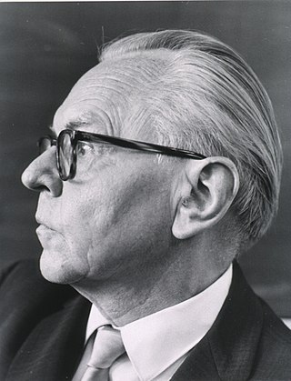
Johannes Holtfreter was a German-American developmental biologist whose primary focus was the “organizer,” a part of the embryo essential for the development of the proper body plan.

<span class="mw-page-title-main">Edward M. De Robertis</span> American embryologist

Edward Michael De Robertis is an American embryologist and Professor at the University of California, Los Angeles. His work has contributed to the finding of conserved molecular processes of embryonic inductions that result in tissue differentiations during animal development. He was elected to the National Academy of Sciences in 2013, worked for the Howard Hughes Medical Institute for 26 years, and holds a Distinguished Professor at the University of California, Los Angeles. In 2009 Pope Benedict XVI appointed De Robertis to a lifetime position in the Pontifical Academy of Sciences, and in 2022 Pope Francis appointed him Councillor of the Academy for four years.

<span class="mw-page-title-main">Embryonic differentiation waves</span>

A mechanochemical based model for primary neural induction was first proposed in 1985 by Brodland and Gordon. They proposed that there is a mechanically sensitive bistable organelle made of microtubules and microfilaments in the apical ends of cells within cell sheets that are about to differentiate and these cells are under mechanical tension. The microtubules and microfilaments are in mechanical opposition in a proposed embryonic organelle they called the cell state splitter. Depending on where the cell is within a sheet, the tension will be resolved by either the apical end contracting or the apical end expanding. The resolution will begin at one point and spread over the rest of the tissue limited by other mechanical forces at boundaries. An actual physical wave of contraction has been found which traverses the presumptive neural epithelium of the developing salamander, the axolotl. The contraction wave's trajectory was more complex than predicted in the original model however it did originate from the precise location of the Spemann organizer and traversed only the presumptive neural epithelium. Electron microscopy showed intermediate filaments are also present in the cell state splitter. Additional waves of both contraction and expansion were also discovered by time lapse photography of axolotl gastrulation. Among them was a wave of expansion that occurs in ectoderm only in the presumptive epithelium. When the trajectories of the waves were superimposed on the fate map of the axolotl it was shown that there is a unique combination of expansion and contraction waves that correlates with the tissue types determined during gastrulation and that this set of wave trajectories could explain the shape of the fate map.

<span class="mw-page-title-main">Dorsal lip</span>

The dorsal lip of the blastopore is a structure that forms during early embryonic development and is important for its role in organizing the germ layers. The dorsal lip is formed during early gastrulation as folding of tissue along the involuting marginal zone of the blastocoel forms an opening known as the blastopore. It is particularly important for its role in neural induction through the default model, where signaling from the dorsal lip protects a region of the epiblast from becoming epidermis, thus allowing it to develop to its default neural tissue.

A developmental signaling center is defined as a group of cells that release various morphogens which can determine the fates, or destined cell types, of adjacent cells. This process in turn determines what tissues the adjacent cells will form. Throughout the years, various development signaling centers have been discovered.

References

  1. 1 2 3 Spemann, Hans (2001). "Induction of embryonic primordia by implantation of organizers from a different species". International Journal of Developmental Biology. 45 (1): 13–38. PMID   11291841.
  2. 1 2 Vonica, Alin (Dec 1, 2007). "The Xenopus Nieuwkoop Center and Spemann–Mangold Organizer Share Molecular Components and a Requirement for Maternal Wnt Activity". Developmental Biology. 312 (1): 90–102. doi:10.1016/j.ydbio.2007.09.039. PMC   2170525 . PMID   17964564.
  3. Niehrs, Christof (June 1, 2004). "Regionally Specific Induction by the Spemann–Mangold Organizer". Nature Reviews Genetics. 5 (6): 425–434. doi:10.1038/nrg1347. PMID   15153995. S2CID   13176134.
  4. Pownall, Mary Elizabeth (2010). FGF Signalling in Vertebrate Development (1 ed.). Morgan & Claypool Life Sciences. ISBN   978-1615040636.
  5. 1 2 3 Sudou, N (May 2012). "Dynamic in Vivo Binding of Transcription Factors to Cis-Regulatory Modules of Cer and Gsc in the Stepwise Formation of the Spemann-Mangold Organizer". Development. 139 (9): 1651–1661. doi:10.1242/dev.068395. PMC   4074222 . PMID   22492356.
  6. 1 2 Bae, Sangwoo (April 15, 2011). "Siamois and Twin Are Redundant and Essential in Formation of the Spemann Organizer". Developmental Biology. 352 (2): 367–381. doi:10.1016/j.ydbio.2011.01.034. PMC   3065516 . PMID   21295564.
  7. 1 2 De Robertis, Edward M (April 2006). "Spemann's Organizer and Self-Regulation in Amphibian Embryos". Nature Reviews Molecular Cell Biology. 7 (4): 296–302. doi:10.1038/nrm1855. PMC   2464568 . PMID   16482093.
  8. 1 2 Cho, Ken W.y. (December 20, 1991). "Molecular Nature of Spemann's Organizer: the Role of the Xenopus Homeobox Gene Goosecoid". Cell. 67 (6): 1111–1120. doi:10.1016/0092-8674(91)90288-a. PMC   3102583 . PMID   1684739.
  9. 1 2 3 4 Asashima, Makoto (2001). "Spemann's influence on Japanese developmental biology". International Journal of Developmental Biology. 45 (1): 57–65. PMID   11291871.
  10. 1 2 3 Grunz, Horst (2001). "Developmental biology of amphibians after Hans Spemann in Germany". International Journal of Developmental Biology. 45 (1): 39–50. PMID   11291869.
  11. 1 2 Mikhailov, Alexander (2001). "Consequences of the Spemann-Mangold organizer concept for embryological research in Russia: Personal impressions". International Journal of Developmental Biology. 45 (1): 83–96. PMID   11291874.