Structured Illumination Light Sheet Microscopy

Last updated

Structured illumination light sheet microscopy (SI-LSM) is an optical imaging technique used for achieving volumetric imaging with high temporal and spatial resolution in all three dimensions. It combines the ability of light sheet microscopy to maintain spatial resolution throughout relatively thick samples with the higher axial and spatial resolution characteristic of structured illumination microscopy. SI-LSM can achieve lateral resolution below 100 nm [1] in biological samples hundreds of micrometers thick. [2]

Contents

SI-LSM is most often used for fluorescent imaging of living biological samples, such as cell cultures. It is particularly useful for longitudinal studies, where high-rate imaging must be performed over long periods of time without damaging the sample. [3] [4] The two methods most used for fluorescent imaging of 3D samples – confocal microscopy and widefield microscopy – both have significant drawbacks for this type of application. In widefield microscopy, both in-focus light from the plane of interest as well as out-of-focus light from the rest of the sample is acquired together, creating the “missing cone problem” which makes high resolution imaging difficult. [5] Although confocal microscopy largely solves this problem by using a pinhole to block unfocused light, this technique also inevitably blocks useful signal, which is particularly detrimental in fluorescent imaging when the signal is already very weak. [6] In addition, both widefield and confocal microscopy illuminate the entirety of the sample throughout imaging, which leads to problems with photobleaching and phototoxicity in some samples. [3] [4] While light-field microscopy alone can address most of these issues, its achieved resolution is still fundamentally limited by the diffraction of light and it is unable to achieve super-resolution. [3] [5]

SI-LSM works by using a patterned rather than uniform light sheet to illuminate a single plane of a volume being imaged. In this way, it maintains the many benefits of light-sheet microscopy while achieving the high resolution of structured illumination microscopy.

Background and Theory

The theory behind SI-LSM is best understood by considering the separate development of structured illumination and light sheet microscopy.

Structured Illumination Microscopy

Moire Effect Moire pattern.svg
Moiré Effect

Structured illumination microscopy (SIM) is a method of super-resolution microscopy which is performed by acquiring multiple images of the same sample under different patterns of illumination, then computationally combining these images to achieve a single reconstruction with up to 2x improvement over the diffraction limited lateral resolution. The theory was first proposed and implemented in a 1995 paper by John M. Guerra [7] in which a silicon grating with 50 nm lines and spaces was resolved with 650 nm wavelength (in air) illumination structured by a transparent replica proximal to said grating. The name “structured illumination microscopy” was coined in 2000 by M.G.L. Gustafsson. [6] SIM takes advantage of the “Moiré Effect”, which occurs when two patterns are multiplicatively superimposed. [6] The superimposition causes “Moiré Fringes” to appear, which are coarser than either original pattern but still contain information about the high frequency patterns which would otherwise not be visible. [8]

The theory behind SIM is best understood in the Fourier or frequency domain. In general, imaging systems can only resolve frequencies below the diffraction limit. Thus, in the Fourier domain, all recorded frequencies from the imaged sample would reside within a circle of a fixed radius. Any frequencies outside this limit cannot be resolved. However, the frequency spectrum can be shifted by imaging the sample with patterned illumination. Most often, the pattern is a 1D sinusoidal gradient, such as the pattern used to create the Moiré fringes in the above image. Because the Fourier transform of a sinusoid is a shifted delta function, the transform of this pattern will consist of three delta functions: one at the zero frequency and two corresponding to the positive and negative frequency components of the sinusoid (see below image). When the target is illuminated using this pattern, the target and illumination pattern are multiplicatively superimposed, which means the Fourier transform of the resulting image is the convolution of the individual transforms of the target and the illumination pattern. Convolving any function with a delta function has the effect of shifting the center of the original function to the location of the delta function. Thus, in this situation, the frequency spectrum of the target is shifted and frequencies that were previously too high to resolve now lie within the circle of resolvable frequencies. The result is that for a single image acquisition with SIM, the frequency components from three separate regions in the Fourier domain (corresponding to the center and the positive and negative shifts) are all captured together. Finally, because rotation in the spatial domain results in the same rotation in the Fourier domain, high frequencies over the full 360° can be captured by rotating the illumination pattern. Figure b) in the image below shows which frequency components would be captured by acquiring 4 separate images and rotating the illumination pattern by 45° in between each acquisition.

Once all images have been captured, a single final image can be computationally reconstructed. [2] Using this technique, resolution can be improved up to 2x over the diffraction limit. [9] This 2x limit is imposed because the illumination pattern itself is still diffraction limited. [2]

SIM in the Fourier domain. a) Acquisition of single SIM image. The Fourier transform of the imaged sample is convolved with the Fourier transform of a 1D sinusoidal gradient. b) All frequencies obtained after rotating illumination pattern 4 times. SIM theory.jpg
SIM in the Fourier domain. a) Acquisition of single SIM image. The Fourier transform of the imaged sample is convolved with the Fourier transform of a 1D sinusoidal gradient. b) All frequencies obtained after rotating illumination pattern 4 times.

The concepts behind 2D SIM can be expanded to 3D volumetric imaging. [5] By using three mutually coherent beams of excitation light, interference patterns with multiple frequency components can be created in the imaged sample. This ultimately makes it possible to perform 3D reconstructions with up to 2x improved resolution along all three axes. [5] However, due to the strong scattering coefficient of biological tissues, this theoretical resolution can only be achieved in samples thinner than about 10 um. [3] Beyond that, the scattering leads to an excess of background signal which makes accurate reconstruction impossible.

Light Sheet Microscopy

Typical light sheet microscopy setup. Structure de MFL.png
Typical light sheet microscopy setup.

Light sheet microscopy (LSM) was developed to allow for fine optical sectioning of thick biological samples without the need for physical sectioning or clearing, which are both time consuming and detrimental to in-vivo imaging. [10] While most fluorescent imaging techniques use aligned illumination and detection axes, LSM utilizes orthogonal axes. A focused light sheet is used to illuminate the sample from the side, while the fluorescent signal is detected from above. [10] This both eliminates the “cone problem” of widefield microscopy by eliminating out-of-focus contributions from planes not being actively imaged and reduces the impact of photobleaching since the entire sample is not illuminated throughout imaging. In addition, because the sample is illuminated from the side, the focus of the illumination light is not depth-dependent, making volumetric imaging of biological samples far more feasible. A major ongoing challenge in LSM is in shaping the light sheet. In general, there is a tradeoff between the thickness of the light sheet at the optical axis (which largely determines axial resolution) and the field-of-view over which the light sheet maintains adequate thickness. [11] This problem can be partially addressed by the added resolution from SI-LSM.

Techniques

SI-LSM can be divided into two main categories. Optical Sectioning SI-LSM is the most common approach and improves axial resolution by further reducing the impact of un-focused background signal. Super-resolution SI-LSM uses the illumination and reconstruction techniques of 2D SIM to achieve super-resolution in 3D samples.

Optical Sectioning SI-LSM

Optical sectioning SI-LSM (OS-SI-LSM) was first described in a 1997 paper by M.A. Neil et al. [12] Rather than achieving super-resolution, this technique uses the ideas behind structured illumination to improve axial resolution by removing background haze from layers other than where the illuminating light sheet is most focused. [12] [13] While there are several approaches for achieving this, the most common approach is known as “three-phase” SIM, which will be described here.

It is shown in the Neil paper [12] that the signal acquired by imaging a target with a grid illumination pattern can be represented by the following equation:

Here, is the background signal, while and are signals from the region of the target illuminated by the cosine and sine components of the grid. It is also shown that an in-focus image of the plane of interest could be reconstructed using the equation:

This can be achieved by acquiring three separate images under the grid illumination conditions, rotating the grid by 60° between each acquisition. The desired 2D image can then be reconstructed using the equation:

This creates a 2D image containing only information from most focused region of the grid illumination pattern. If this pattern is created using a light sheet, the sheet can then be scanned in the axial direction to generate a full 3D reconstruction of a sample. [13] The primary drawback of using this approach for reducing background signal is that it ultimately relies on subtracting out the shared background signal between two images. Some in-focus signal will inevitably be subtracted alongside the background haze. [3] This will result in an overall reduction of signal, which can be detrimental in low-signal fluorescent imaging. Nevertheless, this technique is the most common use of SI-LSM and has shown improved axial resolution over LSM alone. [13]

Super-resolution SI-LSM

Super-resolution SI-LSM (SR-SI-LSM) uses the techniques from 2D or 3D SIM while using a light sheet as the illumination source to achieve the spatial resolution of SIM alongside the depth independent imaging and low photobleaching of LSM. In the most common application, a light sheet is used to create a 1D sinusoidal pattern at a single plane of the 3D target sample. [1] The pattern is then rotated multiple times at this single plane to acquire enough images for a high resolution 2D reconstruction. The light is scanned in the axial resolution and the process is repeated until there are enough 2D images for a full 3D reconstruction. In general, this approach demonstrates not only improved resolution but also improved SNR over OS-SI-LSM, because no information is discarded in the reconstruction. [3] In addition, although the theoretical resolution for SR-SI-LSM is slightly lower than 3D SIM, in depths >10 um this technique shows improved performance over 3D SIM due to the depth-independent focusing of illumination light characteristic of LSM. [3]

Implementation

A major challenge in SI-LSM is engineering systems which are physically capable of generating structured patterns in light sheets. The three main approaches for accomplishing this are using interfering light sheets, digital LSM, and spatial light modulators.

With interfering light sheets, two coherent counterpropagating sheets are sent into the sample. [1] The interference pattern between these sheets creates the desired illumination pattern, which can be rotated and scanned using rotating mirrors to deflect the sheets. Additional flexibility can be added by using digital light-sheet microscopy to generate the illumination patterns. In digital LSM, the light sheet is created by rapidly scanning a laser beam through the sample. [3] [2] [14] This allows for fine control over the specific illumination pattern by modulating the intensity of the laser as it scans. This technique has been used to create systems capable of multiple types of light sheet microscopy in addition to SI-LSM. [2] [3] Finally, spatial light modulators can be used to electronically control the light patterns, which has the advantage of allowing for very fine control of and fast switching between patterns. [15] [16]

In addition, much of the recent work around SI-LSM focuses on combining the approach with other techniques for deep imaging in biological tissues. For instance, a 2021 paper demonstrated the use of SI-LSM with NIR-II illumination to improve resolution of transcranial mouse brain imaging by ~1.7x with a penetration depth of ~750 um and almost 16x improvement in the signal to background ratio. [14] Other promising directions include combining SIM with other techniques for shaping the light sheets in LSM, [11] combining SI-LSM with two-photon excitation, [14] or using non-linear fluorescence to further push the resolution limits. [9]

Related Research Articles

<span class="mw-page-title-main">Microscopy</span> Viewing of objects which are too small to be seen with the naked eye

Microscopy is the technical field of using microscopes to view objects and areas of objects that cannot be seen with the naked eye. There are three well-known branches of microscopy: optical, electron, and scanning probe microscopy, along with the emerging field of X-ray microscopy.

<span class="mw-page-title-main">Optical microscope</span> Microscope that uses visible light

The optical microscope, also referred to as a light microscope, is a type of microscope that commonly uses visible light and a system of lenses to generate magnified images of small objects. Optical microscopes are the oldest design of microscope and were possibly invented in their present compound form in the 17th century. Basic optical microscopes can be very simple, although many complex designs aim to improve resolution and sample contrast.

<span class="mw-page-title-main">Diffraction-limited system</span> Optical system with resolution performance at the instruments theoretical limit

In optics, any optical instrument or system – a microscope, telescope, or camera – has a principal limit to its resolution due to the physics of diffraction. An optical instrument is said to be diffraction-limited if it has reached this limit of resolution performance. Other factors may affect an optical system's performance, such as lens imperfections or aberrations, but these are caused by errors in the manufacture or calculation of a lens, whereas the diffraction limit is the maximum resolution possible for a theoretically perfect, or ideal, optical system.

<span class="mw-page-title-main">Immunofluorescence</span> Technique used for light microscopy

Immunofluorescence(IF) is a light microscopy-based technique that allows detection and localization of a wide variety of target biomolecules within a cell or tissue at a quantitative level. The technique utilizes the binding specificity of antibodies and antigens. The specific region an antibody recognizes on an antigen is called an epitope. Several antibodies can recognize the same epitope but differ in their binding affinity. The antibody with the higher affinity for a specific epitope will surpass antibodies with a lower affinity for the same epitope.

<span class="mw-page-title-main">Fluorescence microscope</span> Optical microscope that uses fluorescence and phosphorescence

A fluorescence microscope is an optical microscope that uses fluorescence instead of, or in addition to, scattering, reflection, and attenuation or absorption, to study the properties of organic or inorganic substances. "Fluorescence microscope" refers to any microscope that uses fluorescence to generate an image, whether it is a simple set up like an epifluorescence microscope or a more complicated design such as a confocal microscope, which uses optical sectioning to get better resolution of the fluorescence image.

<span class="mw-page-title-main">Confocal microscopy</span> Optical imaging technique

Confocal microscopy, most frequently confocal laser scanning microscopy (CLSM) or laser scanning confocal microscopy (LSCM), is an optical imaging technique for increasing optical resolution and contrast of a micrograph by means of using a spatial pinhole to block out-of-focus light in image formation. Capturing multiple two-dimensional images at different depths in a sample enables the reconstruction of three-dimensional structures within an object. This technique is used extensively in the scientific and industrial communities and typical applications are in life sciences, semiconductor inspection and materials science.

Super-resolution imaging (SR) is a class of techniques that enhance (increase) the resolution of an imaging system. In optical SR the diffraction limit of systems is transcended, while in geometrical SR the resolution of digital imaging sensors is enhanced.

<span class="mw-page-title-main">Near-field scanning optical microscope</span>

Near-field scanning optical microscopy (NSOM) or scanning near-field optical microscopy (SNOM) is a microscopy technique for nanostructure investigation that breaks the far field resolution limit by exploiting the properties of evanescent waves. In SNOM, the excitation laser light is focused through an aperture with a diameter smaller than the excitation wavelength, resulting in an evanescent field on the far side of the aperture. When the sample is scanned at a small distance below the aperture, the optical resolution of transmitted or reflected light is limited only by the diameter of the aperture. In particular, lateral resolution of 6 nm and vertical resolution of 2–5 nm have been demonstrated.

RESOLFT, an acronym for REversible Saturable OpticaLFluorescence Transitions, denotes a group of optical fluorescence microscopy techniques with very high resolution. Using standard far field visible light optics a resolution far below the diffraction limit down to molecular scales can be obtained.

<span class="mw-page-title-main">Ptychography</span>

Ptychography is a computational method of microscopic imaging. It generates images by processing many coherent interference patterns that have been scattered from an object of interest. Its defining characteristic is translational invariance, which means that the interference patterns are generated by one constant function moving laterally by a known amount with respect to another constant function. The interference patterns occur some distance away from these two components, so that the scattered waves spread out and "fold" into one another as shown in the figure.

<span class="mw-page-title-main">Vertico spatially modulated illumination</span>

Vertico spatially modulated illumination (Vertico-SMI) is the fastest light microscope for the 3D analysis of complete cells in the nanometer range. It is based on two technologies developed in 1996, SMI and SPDM. The effective optical resolution of this optical nanoscope has reached the vicinity of 5 nm in 2D and 40 nm in 3D, greatly surpassing the λ/2 resolution limit applying to standard microscopy using transmission or reflection of natural light according to the Abbe resolution limit That limit had been determined by Ernst Abbe in 1873 and governs the achievable resolution limit of microscopes using conventional techniques.

<span class="mw-page-title-main">Optical sectioning</span> Imaging of focal planes within a thick sample

Optical sectioning is the process by which a suitably designed microscope can produce clear images of focal planes deep within a thick sample. This is used to reduce the need for thin sectioning using instruments such as the microtome. Many different techniques for optical sectioning are used and several microscopy techniques are specifically designed to improve the quality of optical sectioning.

Super-resolution microscopy is a series of techniques in optical microscopy that allow such images to have resolutions higher than those imposed by the diffraction limit, which is due to the diffraction of light. Super-resolution imaging techniques rely on the near-field or on the far-field. Among techniques that rely on the latter are those that improve the resolution only modestly beyond the diffraction-limit, such as confocal microscopy with closed pinhole or aided by computational methods such as deconvolution or detector-based pixel reassignment, the 4Pi microscope, and structured-illumination microscopy technologies such as SIM and SMI.

Photo-activated localization microscopy and stochastic optical reconstruction microscopy (STORM) are widefield fluorescence microscopy imaging methods that allow obtaining images with a resolution beyond the diffraction limit. The methods were proposed in 2006 in the wake of a general emergence of optical super-resolution microscopy methods, and were featured as Methods of the Year for 2008 by the Nature Methods journal. The development of PALM as a targeted biophysical imaging method was largely prompted by the discovery of new species and the engineering of mutants of fluorescent proteins displaying a controllable photochromism, such as photo-activatible GFP. However, the concomitant development of STORM, sharing the same fundamental principle, originally made use of paired cyanine dyes. One molecule of the pair, when excited near its absorption maximum, serves to reactivate the other molecule to the fluorescent state.

<span class="mw-page-title-main">Light sheet fluorescence microscopy</span> Fluorescence microscopy technique

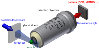
Light sheet fluorescence microscopy (LSFM) is a fluorescence microscopy technique with an intermediate-to-high optical resolution, but good optical sectioning capabilities and high speed. In contrast to epifluorescence microscopy only a thin slice of the sample is illuminated perpendicularly to the direction of observation. For illumination, a laser light-sheet is used, i.e. a laser beam which is focused only in one direction. A second method uses a circular beam scanned in one direction to create the lightsheet. As only the actually observed section is illuminated, this method reduces the photodamage and stress induced on a living sample. Also the good optical sectioning capability reduces the background signal and thus creates images with higher contrast, comparable to confocal microscopy. Because light sheet fluorescence microscopy scans samples by using a plane of light instead of a point, it can acquire images at speeds 100 to 1,000 times faster than those offered by point-scanning methods.

<span class="mw-page-title-main">Live-cell imaging</span> Study of living cells using time-lapse microscopy

Live-cell imaging is the study of living cells using time-lapse microscopy. It is used by scientists to obtain a better understanding of biological function through the study of cellular dynamics. Live-cell imaging was pioneered in the first decade of the 21st century. One of the first time-lapse microcinematographic films of cells ever made was made by Julius Ries, showing the fertilization and development of the sea urchin egg. Since then, several microscopy methods have been developed to study living cells in greater detail with less effort. A newer type of imaging using quantum dots have been used, as they are shown to be more stable. The development of holotomographic microscopy has disregarded phototoxicity and other staining-derived disadvantages by implementing digital staining based on cells’ refractive index.

Lattice light-sheet microscopy is a modified version of light sheet fluorescence microscopy that increases image acquisition speed while decreasing damage to cells caused by phototoxicity. This is achieved by using a structured light sheet to excite fluorescence in successive planes of a specimen, generating a time series of 3D images which can provide information about dynamic biological processes.

Wide-field multiphoton microscopy refers to an optical non-linear imaging technique tailored for ultrafast imaging in which a large area of the object is illuminated and imaged without the need for scanning. High intensities are required to induce non-linear optical processes such as two-photon fluorescence or second harmonic generation. In scanning multiphoton microscopes the high intensities are achieved by tightly focusing the light, and the image is obtained by beam scanning. In wide-field multiphoton microscopy the high intensities are best achieved using an optically amplified pulsed laser source to attain a large field of view (~100 µm). The image in this case is obtained as a single frame with a CCD without the need of scanning, making the technique particularly useful to visualize dynamic processes simultaneously across the object of interest. With wide-field multiphoton microscopy the frame rate can be increased up to a 1000-fold compared to multiphoton scanning microscopy. Wide-field multiphoton microscopes are not yet commercially available, but working prototypes exist in several optics laboratories.

Super-resolution dipole orientation mapping (SDOM) is a form of fluorescence polarization microscopy (FPM) that achieved super resolution through polarization demodulation. It was first described by Karl Zhanghao and others in 2016. Fluorescence polarization (FP) is related to the dipole orientation of chromophores, making fluorescence polarization microscopy possible to reveal structures and functions of tagged cellular organelles and biological macromolecules. In addition to fluorescence intensity, wavelength, and lifetime, the fourth dimension of fluorescence—polarization—can also provide intensity modulation without the restriction to specific fluorophores; its investigation in super-resolution microscopy is still in its infancy.

Super-resolution photoacoustic imaging is a set of techniques used to enhance spatial resolution in photoacoustic imaging. Specifically, these techniques primarily break the optical diffraction limit of the photoacoustic imaging system. It can be achieved in a variety of mechanisms, such as blind structured illumination, multi-speckle illumination, or photo-imprint photoacoustic microscopy in Figure 1.

References

  1. 1 2 3 Chang, Bo-Jui; Perez Meza, Victor Didier; Stelzer, Ernst H. K. (9 May 2017). "csiLSFM combines light-sheet fluorescence microscopy and coherent structured illumination for a lateral resolution below 100 nm". Proceedings of the National Academy of Sciences. 114 (19): 4869–4874. doi: 10.1073/pnas.1609278114 . PMC   5441750 . PMID   28438995.
  2. 1 2 3 4 5 Keller, Philipp J; Schmidt, Annette D; Santella, Anthony; Khairy, Khaled; Bao, Zhirong; Wittbrodt, Joachim; Stelzer, Ernst H K (August 2010). "Fast, high-contrast imaging of animal development with scanned light sheet–based structured-illumination microscopy". Nature Methods. 7 (8): 637–642. doi:10.1038/nmeth.1476. PMC   4418465 . PMID   20601950.
  3. 1 2 3 4 5 6 7 8 9 Gao, Liang; Shao, Lin; Higgins, Christopher D.; Poulton, John S.; Peifer, Mark; Davidson, Michael W.; Wu, Xufeng; Goldstein, Bob; Betzig, Eric (December 2012). "Noninvasive Imaging beyond the Diffraction Limit of 3D Dynamics in Thickly Fluorescent Specimens". Cell. 151 (6): 1370–1385. doi:10.1016/j.cell.2012.10.008. PMC   3615549 . PMID   23217717.
  4. 1 2 Chen, Bi-Chang; Legant, Wesley R.; Wang, Kai; Shao, Lin; Milkie, Daniel E.; Davidson, Michael W.; Janetopoulos, Chris; Wu, Xufeng S.; Hammer, John A.; Liu, Zhe; English, Brian P.; Mimori-Kiyosue, Yuko; Romero, Daniel P.; Ritter, Alex T.; Lippincott-Schwartz, Jennifer; Fritz-Laylin, Lillian; Mullins, R. Dyche; Mitchell, Diana M.; Bembenek, Joshua N.; Reymann, Anne-Cecile; Böhme, Ralph; Grill, Stephan W.; Wang, Jennifer T.; Seydoux, Geraldine; Tulu, U. Serdar; Kiehart, Daniel P.; Betzig, Eric (24 October 2014). "Lattice light-sheet microscopy: Imaging molecules to embryos at high spatiotemporal resolution". Science. 346 (6208): 1257998. doi:10.1126/science.1257998. PMC   4336192 . PMID   25342811.
  5. 1 2 3 4 Frohn, J. T.; Knapp, H. F.; Stemmer, A. (1 June 2001). "Three-dimensional resolution enhancement in fluorescence microscopy by harmonic excitation". Optics Letters. 26 (11): 828–830. doi:10.1364/ol.26.000828. PMID   18040464.
  6. 1 2 3 Gustafsson, M. G. L. (May 2000). "Surpassing the lateral resolution limit by a factor of two using structured illumination microscopy". Journal of Microscopy. 198 (2): 82–87. doi: 10.1046/j.1365-2818.2000.00710.x . PMID   10810003. S2CID   9257781.
  7. Guerra, John M. (26 June 1995). "Super‐resolution through illumination by diffraction‐born evanescent waves". Applied Physics Letters. 66 (26): 3555–3557. doi:10.1063/1.113814.
  8. Rego, E. Hesper; Shao, Lin (2015). "Practical Structured Illumination Microscopy". Advanced Fluorescence Microscopy. Methods in Molecular Biology. Vol. 1251. pp. 175–192. doi:10.1007/978-1-4939-2080-8_10. ISBN   978-1-4939-2079-2. PMID   25391800.
  9. 1 2 Gustafsson, Mats G. L. (13 September 2005). "Nonlinear structured-illumination microscopy: Wide-field fluorescence imaging with theoretically unlimited resolution". Proceedings of the National Academy of Sciences. 102 (37): 13081–13086. doi: 10.1073/pnas.0406877102 . PMC   1201569 . PMID   16141335.
  10. 1 2 Huisken, Jan; Stainier, Didier Y. R. (15 June 2009). "Selective plane illumination microscopy techniques in developmental biology". Development. 136 (12): 1963–1975. doi:10.1242/dev.022426. PMC   2685720 . PMID   19465594.
  11. 1 2 Planchon, Thomas A; Gao, Liang; Milkie, Daniel E; Davidson, Michael W; Galbraith, James A; Galbraith, Catherine G; Betzig, Eric (May 2011). "Rapid three-dimensional isotropic imaging of living cells using Bessel beam plane illumination". Nature Methods. 8 (5): 417–423. doi:10.1038/nmeth.1586. PMC   3626440 . PMID   21378978.
  12. 1 2 3 Neil, M. A. A.; Juškaitis, R.; Wilson, T. (15 December 1997). "Method of obtaining optical sectioning by using structured light in a conventional microscope". Optics Letters. 22 (24): 1905–1907. doi:10.1364/ol.22.001905. PMID   18188403.
  13. 1 2 3 Mertz, Jerome; Kim, Jinhyun (2010). "Scanning light-sheet microscopy in the whole mouse brain with HiLo background rejection". Journal of Biomedical Optics. 15 (1): 016027. doi:10.1117/1.3324890. PMC   2917465 . PMID   20210471.
  14. 1 2 3 Wang, Feifei; Ma, Zhuoran; Zhong, Yeteng; Salazar, Felix; Xu, Chun; Ren, Fuqiang; Qu, Liangqiong; Wu, Anna M.; Dai, Hongjie (9 February 2021). "In vivo NIR-II structured-illumination light-sheet microscopy". Proceedings of the National Academy of Sciences. 118 (6): e2023888118. doi:10.1073/pnas.2023888118. PMC   8017937 . PMID   33526701.
  15. Landry, Joseph R. (9 April 2019). "Tunable structured illumination light sheet microscopy for background rejection and imaging depth in minimally processed tissues". Journal of Biomedical Optics. 24 (4): 1–6. doi:10.1117/1.Jbo.24.4.046501. PMC   6454294 . PMID   30968649.
  16. Garbellotto, Chiara; Taylor, Jonathan M. (1 November 2018). "Multi-purpose SLM-light-sheet microscope". Biomedical Optics Express. 9 (11): 5419–5436. doi:10.1364/BOE.9.005419. PMC   6238942 . PMID   30460137.