Related Research Articles

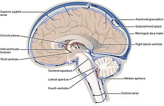
Cerebrospinal fluid (CSF) is a clear, colorless body fluid found within the tissue that surrounds the brain and spinal cord of all vertebrates.

The blood–brain barrier (BBB) is a highly selective semipermeable border of endothelial cells that regulates the transfer of solutes and chemicals between the circulatory system and the central nervous system, thus protecting the brain from harmful or unwanted substances in the blood. The blood–brain barrier is formed by endothelial cells of the capillary wall, astrocyte end-feet ensheathing the capillary, and pericytes embedded in the capillary basement membrane. This system allows the passage of some small molecules by passive diffusion, as well as the selective and active transport of various nutrients, ions, organic anions, and macromolecules such as glucose and amino acids that are crucial to neural function.

Viral meningitis, also known as aseptic meningitis, is a type of meningitis due to a viral infection. It results in inflammation of the meninges. Symptoms commonly include headache, fever, sensitivity to light and neck stiffness.

In neuroanatomy, the ventricular system is a set of four interconnected cavities known as cerebral ventricles in the brain. Within each ventricle is a region of choroid plexus which produces the circulating cerebrospinal fluid (CSF). The ventricular system is continuous with the central canal of the spinal cord from the fourth ventricle, allowing for the flow of CSF to circulate.

Lumbar puncture (LP), also known as a spinal tap, is a medical procedure in which a needle is inserted into the spinal canal, most commonly to collect cerebrospinal fluid (CSF) for diagnostic testing. The main reason for a lumbar puncture is to help diagnose diseases of the central nervous system, including the brain and spine. Examples of these conditions include meningitis and subarachnoid hemorrhage. It may also be used therapeutically in some conditions. Increased intracranial pressure is a contraindication, due to risk of brain matter being compressed and pushed toward the spine. Sometimes, lumbar puncture cannot be performed safely. It is regarded as a safe procedure, but post-dural-puncture headache is a common side effect if a small atraumatic needle is not used.

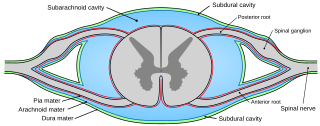
In anatomy, the meninges are the three membranes that envelop the brain and spinal cord. In mammals, the meninges are the dura mater, the arachnoid mater, and the pia mater. Cerebrospinal fluid is located in the subarachnoid space between the arachnoid mater and the pia mater. The primary function of the meninges is to protect the central nervous system.

Pia mater, often referred to as simply the pia, is the delicate innermost layer of the meninges, the membranes surrounding the brain and spinal cord. Pia mater is medieval Latin meaning "tender mother". The other two meningeal membranes are the dura mater and the arachnoid mater. Both the pia and arachnoid mater are derivatives of the neural crest while the dura is derived from embryonic mesoderm. The pia mater is a thin fibrous tissue that is permeable to water and small solutes. The pia mater allows blood vessels to pass through and nourish the brain. The perivascular space between blood vessels and pia mater is proposed to be part of a pseudolymphatic system for the brain. When the pia mater becomes irritated and inflamed the result is meningitis.

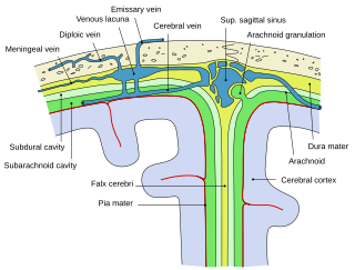
Arachnoid granulations are small protrusions of the arachnoid mater into the outer membrane of the dura mater. They protrude into the dural venous sinuses of the brain, and allow cerebrospinal fluid (CSF) to exit the subarachnoid space and enter the blood stream.

The choroid plexus, or plica choroidea, is a plexus of cells that arises from the tela choroidea in each of the ventricles of the brain. Regions of the choroid plexus produce and secrete most of the cerebrospinal fluid (CSF) of the central nervous system. The choroid plexus consists of modified ependymal cells surrounding a core of capillaries and loose connective tissue. Multiple cilia on the ependymal cells move to circulate the cerebrospinal fluid.

In neuroanatomy, dura mater is a thick membrane made of dense irregular connective tissue that surrounds the brain and spinal cord. It is the outermost of the three layers of membrane called the meninges that protect the central nervous system. The other two meningeal layers are the arachnoid mater and the pia mater. It envelops the arachnoid mater, which is responsible for keeping in the cerebrospinal fluid. It is derived primarily from the neural crest cell population, with postnatal contributions of the paraxial mesoderm.

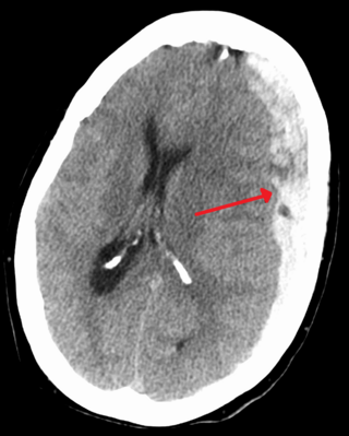
A subdural hematoma (SDH) is a type of bleeding in which a collection of blood—usually but not always associated with a traumatic brain injury—gathers between the inner layer of the dura mater and the arachnoid mater of the meninges surrounding the brain. It usually results from tears in bridging veins that cross the subdural space.

The arachnoid mater is one of the three meninges, the protective membranes that cover the brain and spinal cord. It is so named because of its resemblance to a spider web. The arachnoid mater is a derivative of the neural crest mesoectoderm in the embryo.

The cranial cavity, also known as intracranial space, is the space within the skull that accommodates the brain. The skull minus the mandible is called the cranium. The cavity is formed by eight cranial bones known as the neurocranium that in humans includes the skull cap and forms the protective case around the brain. The remainder of the skull is called the facial skeleton. Meninges are protective membranes that surround the brain to minimize damage to the brain in the case of head trauma. Meningitis is the inflammation of meninges caused by bacterial or viral infections.

The glia limitans, or the glial limiting membrane, is a thin barrier of astrocyte foot processes associated with the parenchymal basal lamina surrounding the brain and spinal cord. It is the outermost layer of neural tissue, and among its responsibilities is the prevention of the over-migration of neurons and neuroglia, the supporting cells of the nervous system, into the meninges. The glia limitans also plays an important role in regulating the movement of small molecules and cells into the brain tissue by working in concert with other components of the central nervous system (CNS) such as the blood–brain barrier (BBB).

Leptomeningeal cancer is a rare complication of cancer in which the disease spreads from the original tumor site to the meninges surrounding the brain and spinal cord. This leads to an inflammatory response, hence the alternative names neoplastic meningitis (NM), malignant meningitis, or carcinomatous meningitis. The term leptomeningeal describes the thin meninges, the arachnoid and the pia mater, between which the cerebrospinal fluid is located. The disorder was originally reported by Eberth in 1870. It is also known as leptomeningeal carcinomatosis, leptomeningeal disease (LMD), leptomeningeal metastasis, meningeal metastasis and meningeal carcinomatosis.

Tarlov cysts, are type II innervated meningeal cysts, cerebrospinal-fluid-filled (CSF) sacs most frequently located in the spinal canal of the sacral region of the spinal cord (S1–S5) and much less often in the cervical, thoracic or lumbar spine. They can be distinguished from other meningeal cysts by their nerve-fiber-filled walls. Tarlov cysts are defined as cysts formed within the nerve-root sheath at the dorsal root ganglion. The etiology of these cysts is not well understood; some current theories explaining this phenomenon have not yet been tested or challenged but include increased pressure in CSF, filling of congenital cysts with one-way valves, inflammation in response to trauma and disease. They are named for American neurosurgeon Isadore Tarlov, who described them in 1938.

The arachnoid trabeculae are delicate strands of connective tissue that loosely connect the two innermost layers of the meninges – the arachnoid mater and the pia mater. They are found within the subarachnoid space where cerebrospinal fluid is also found. Embryologically, the trabeculae are the remnants of the common precursor that forms both the arachnoid and pial layers of the meninges.

The glymphatic system is a system for waste clearance in the central nervous system (CNS) of vertebrates. According to this model, cerebrospinal fluid (CSF) flows into the paravascular space around cerebral arteries, combining with interstitial fluid (ISF) and parenchymal solutes, and exiting down venous paravascular spaces. The pathway consists of a para-arterial influx route for CSF to enter the brain parenchyma, coupled to a clearance mechanism for the removal of interstitial fluid (ISF) and extracellular solutes from the interstitial compartments of the brain and spinal cord. Exchange of solutes between CSF and ISF is driven primarily by arterial pulsation and regulated during sleep by the expansion and contraction of brain extracellular space. Clearance of soluble proteins, waste products, and excess extracellular fluid is accomplished through convective bulk flow of ISF, facilitated by astrocytic aquaporin 4 (AQP4) water channels.
Maiken Nedergaard is a Danish neuroscientist most well known for discovering the glymphatic system. She is a jointly appointed professor in the Departments of Neuroscience and Neurology at the University of Rochester Medical Center. She holds a part-time appointment in the Department of Neurosurgery within the University of Rochester Center for Translational Neuromedicine, where she is the principal investigator of the Division of Glial Disease and Therapeutics laboratory. She is also Professor of Glial Cell Biology at the University of Copenhagen, Center for Translational Neuromedicine.

The meningeal lymphatic vessels are a network of conventional lymphatic vessels located parallel to the dural venous sinuses and middle meningeal arteries of the mammalian central nervous system (CNS). As a part of the lymphatic system, the meningeal lymphatics are responsible for draining immune cells, small molecules, and excess fluid from the CNS into the deep cervical lymph nodes. Cerebrospinal fluid, and interstitial fluid are exchanged, and drained by the meningeal lymphatic vessels.
References
- 1 2 3 4 5 6 7 8 9 Møllgård, Kjeld; Beinlich, Felix R. M.; Kusk, Peter; et al. (2023). "A mesothelium divides the subarachnoid space into functional compartments". Science . 379 (6627): 84–88. Bibcode:2023Sci...379...84M. doi:10.1126/science.adc8810. PMID 36603070. S2CID 255440992.
- ↑ Mestre, Humberto; Tithof, Jeffrey; Du, Ting; Song, Wei; Peng, Weiguo; Sweeney, Amanda M.; Olveda, Genaro; Thomas, John H.; Nedergaard, Maiken; Kelley, Douglas H. (2018-11-19). "Flow of cerebrospinal fluid is driven by arterial pulsations and is reduced in hypertension". Nature Communications. 9 (1): 4878. doi:10.1038/s41467-018-07318-3. ISSN 2041-1723. PMC 6242982 .
- ↑ Mestre, Humberto; Mori, Yuki; Nedergaard, Maiken (July 2020). "The Brain's Glymphatic System: Current Controversies". Trends in Neurosciences. 43 (7): 458–466. doi:10.1016/j.tins.2020.04.003. PMC 7331945 .
- ↑ Plá, Virginia; Bitsika, Styliani; Giannetto, Michael J; Ladron-de-Guevara, Antonio; Gahn-Martinez, Daniel; Mori, Yuki; Nedergaard, Maiken; Møllgård, Kjeld (2023-12-14). "Structural characterization of SLYM—a 4th meningeal membrane". Fluids and Barriers of the CNS. 20 (1). doi: 10.1186/s12987-023-00500-w . ISSN 2045-8118. PMC 10722698 . PMID 38098084.
- ↑ Pietilä, Riikka; Del Gaudio, Francesca; He, Liqun; Vázquez-Liébanas, Elisa; Vanlandewijck, Michael; Muhl, Lars; Mocci, Giuseppe; Bjørnholm, Katrine D.; Lindblad, Caroline; Fletcher-Sandersjöö, Alexander; Svensson, Mikael; Thelin, Eric P.; Liu, Jianping; van Voorden, A. Jantine; Torres, Monica (December 2023). "Molecular anatomy of adult mouse leptomeninges". Neuron. 111 (23): 3745–3764.e7. doi:10.1016/j.neuron.2023.09.002.
- ↑ Mapunda, Josephine A.; Pareja, Javier; Vladymyrov, Mykhailo; Bouillet, Elisa; Hélie, Pauline; Pleskač, Petr; Barcos, Sara; Andrae, Johanna; Vestweber, Dietmar; McDonald, Donald M.; Betsholtz, Christer; Deutsch, Urban; Proulx, Steven T.; Engelhardt, Britta (2023-09-20). "VE-cadherin in arachnoid and pia mater cells serves as a suitable landmark for in vivo imaging of CNS immune surveillance and inflammation". Nature Communications. 14 (1). doi:10.1038/s41467-023-41580-4. ISSN 2041-1723. PMC 10511632 . PMID 37730744.