Sublingua

Last updated

The sublingua is found on the underside of the primary tongue in tarsiers, lemuriform primates, and some other mammals. Sublingua - Jones 1918.jpg
The sublingua is found on the underside of the primary tongue in tarsiers, lemuriform primates, and some other mammals.

The sublingua ("under-tongue") is a muscular secondary tongue found below the primary tongue in tarsiers and living strepsirrhine primates, which includes lemurs and lorisoids (collectively called "lemuriforms"). [lower-alpha 1] Although it is most fully developed in these primates, similar structures can be found in some other mammals, such as marsupials, treeshrews, and colugos. This "second tongue" lacks taste buds, and in lemuriforms, it is thought to be used to remove hair and other debris from the toothcomb, a specialized dental structure used to comb the fur during oral grooming.

Contents

A rigid structure called the plica mediana or lytta runs from the front to the back, down the center of the sublingua to give it support. The plica mediana is usually made of cartilage and attaches the sublingua to the underside of the tongue. In lemuriforms, the sublingua mostly consists of two plicae fimbriatae (singular: plica fimbriata), which run along the sides of the plica mediana and end in comb-like serrated edges that are hardened with keratin. The plicae fimbriatae move freely over a limited range. The plica sublingualis, which is found in all primates, but is particularly small in lemuriforms, attaches the tongue and sublingua to the floor of the mouth. Tarsiers have a large but highly generalized sublingua, but their closest living relatives, monkeys and apes, lack one.

The sublingua is thought to have evolved from specialized folds of tissue below the tongue, which can be seen in some marsupials and other mammals. Simians do not have a sublingua, but the fimbria linguae found on the underside of ape tongues may be a vestigial version of the sublingua. Because of widely variable appearance of sublingual tissue in primates, the term "sublingua" is often confused with the frenal lamella, lingual frenulum, and other sublingual tissues.

Anatomical structure

The sublingua, or "under-tongue", is a secondary tongue located below the primary tongue in tarsiers, lemuriform primates, and some other mammals. [4] This structure does not have taste buds or salivary glands. [5] In lemuriforms, the sublingua is relatively large and its front edge is usually lined with keratinized serrations (sometimes called "denticles"). [6] [7]

The serrated edges of a strepsirrhine sublingua are part of the plicae fimbriatae, while the plica sublingualis, which attaches the sublingua and tongue to the floor of the mouth, is small. Lemur catta tongue and sublingua - Jones 1918.jpg
The serrated edges of a strepsirrhine sublingua are part of the plicae fimbriatae, while the plica sublingualis, which attaches the sublingua and tongue to the floor of the mouth, is small.

Down the middle of the sublingua is a thick strengthening rod called the plica mediana or lytta, which connects the sublingua to the underside of the tongue, [7] [8] [9] and is part of the lingual septum (septum of the tongue). [8] The thickness and size of the plica mediana can vary between species, [10] and except in treeshrews, it is cartilaginous and provides support for the sublingua. [5]

Only the serrated and often keratinized tip of the sublingua is free to move small distances along the underside of the tongue, while the majority of its length adheres to the underside of the tongue. [7] [11] These free-moving folds or filaments are called the plica fimbriata and attach to the base of the sublingua [10] and are supported at the midline by the plica mediana. The plica fimbriata is highly developed and specialized in lemurs, and makes up the majority of the sublingua. [11]

The fold that connects the back of the sublingua and tongue to the rear floor of the mouth is called the plica sublingualis. [10] In lemurs, this is an underdeveloped structure consisting of only a tiny outgrowth on the floor of the mouth. [12] The point where the plica sublingualis attaches to the rear floor of the mouth marks the location of the submandibular salivary glands. [6] [13]

Differences between species

In the aye-aye, the sublingua is not shaped like a brush like it is in most lemurs. Instead, there is a thickened area along the plica mediana or lytta which has a hook-shaped structure on the end. [6] [7] Within cheirogaleids, the sublingua lacks cartilage, and the sublingua of the gray mouse lemur has a distinct plica mediana and ends in two lobe-like projections that lack keratinized serrations, but have three keratinized ridges which make the sublingua rigid. [14]

In tarsiers, the sublingua does not have serrations along its tip and is much simpler and generalized in structure, making it clearly distinguishable from that of the lemuriform primates. [15] The tarsier has a distinct plica mediana and its plicae fimbriatae are large and stick to the entire underside of the tongue. The plica sublingualis is also prominent. [16]

Marsupials such as opossums and the common brushtail possum have also developed noticeable sublingua with a plica mediana and a less specialized, but conspicuous, plica fimbriata. [12]

Function

Sublingua of a slow loris 001.jpg
Lemur catta toothcomb.jpg
The sublingua (above) is a secondary tongue below the primary tongue and is used to remove hair and debris from the toothcomb (below) of lemurs and other lemuriform primates.

Originally, the sublingua in lemurs was thought to be a vestigial organ inherited from their mammalian ancestors. [17] In lemuriform primates, the sublingua is used to remove hair and debris from the highly specialized toothcomb, [6] [17] an arrangement of four or six long, forward-facing teeth in the lower jaw used in oral grooming. [18] The toothcomb of lemuriforms consists of both incisors and canine teeth (which reinforce the incisors), and together, these finely spaced teeth act like teeth on a comb. [19] Although the cleaning function has been suspected for nearly a century, there has been no clear confirmation of this. [20] However, a study from 1941 presented evidence that the toothcomb accumulated a mat of hair during oral grooming, and the author did observe that lemurs extend and retract their tongue rapidly, possibly to use the sublingua to clean the toothcomb. [21]

In the aye-aye, which has replaced the toothcomb by evolving continually growing, rodent-like incisors, the hook-shaped tip of the sublingua fits precisely within the gap between the two lower incisors and keeps the area clean. [6] [7] [22] Tarsiers lack a toothcomb, which may explain why their sublingua lacks the serrations typically found on the sublingua of lemuriforms. [15] Although colugos also have a toothcomb, consisting of serrated edges on the tips of their incisors instead of finely spaced, elongated teeth, they do not have a sublingua. Instead, their toothcomb is cleaned by the tongue, which has serrated edges at the front that match the serrations on the incisors. [17]

Evolution and development

The sublingua in lemuriform primates and tarsiers may have evolved from the specialized folds of tissue below that tongue, as seen in some marsupials, such as sugar gliders, as well as some embryonic eutherian mammals, such as whales and dogs. [9] [23] It is also found in some adult eutherian mammals, such as treeshrews, colugos, and rodents. [4] [9] The sublingua of treeshrews, close relatives of primates, is less developed than in lemuriforms and tarsiers, [5] but suggests a phylogenetic relationship. [14]

The sublingua in lemuriform primates is fully developed and particularly unique. [7] Tarsiers, which are most closely related to monkeys and apes (collectively called simians), also have a well-developed but non-specialized sublingua. Simians, however, do not have a sublingua, although some, such as titis have a highly specialized frenal lamella (plica sublingualis). [24] All primates have a plica sublingualis, [5] and the fimbria linguae (plica fimbriata) found under the tongue of apes may be a vestigial version, although that is still disputed. [7] [9] [14] The structure and appearance of the sublingua, frenal lamella, lingual frenulum, and other sublingual tissue vary greatly between primates, and as a result, their terminology is often confused. [25]

In the species that have cartilage in the sublingua or lytta, that cartilage is not derived from the hyoid bone or hyoid arch (the bone and cartilage that supports the tongue). [5] [14] Instead, the cartilage of the sublingua is a separately developed structure specifically adapted to support the sublingua. [14]

Notes

  1. Although the monophyletic relationship between lemurs and lorisoids is widely accepted, their clade name is not. The term "lemuriform" is used here because it derives from one popular taxonomy that clumps the clade of toothcombed primates into one infraorder and the extinct, non-toothcombed adapiforms into another, both within the suborder Strepsirrhini. [1] [2] However, another popular alternative taxonomy places the lorisoids in their own infraorder, Lorisiformes. [3]

Related Research Articles

Strepsirrhini Suborder of primates which includes lemurs, galagos, pottos and lorises

Strepsirrhini or Strepsirhini is a suborder of primates that includes the lemuriform primates, which consist of the lemurs of Madagascar, galagos ("bushbabies") and pottos from Africa, and the lorises from India and southeast Asia. Collectively they are referred to as strepsirrhines. Also belonging to the suborder are the extinct adapiform primates which thrived during the Eocene in Europe, North America, and Asia, but disappeared from most of the Northern Hemisphere as the climate cooled. Adapiforms are sometimes referred to as being "lemur-like", although the diversity of both lemurs and adapiforms does not support this comparison.

Prosimian Obsolete primate taxon

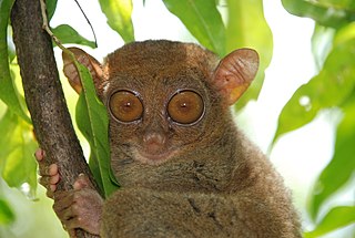
Prosimians are a group of primates that includes all living and extinct strepsirrhines, as well as the haplorhine tarsiers and their extinct relatives, the omomyiforms, i.e. all primates excluding the simians. They are considered to have characteristics that are more "primitive" than those of simians.

Lemur Clade of primates endemic to the island of Madagascar

Lemurs are mammals of the order Primates, divided into 8 families and consisting of 15 genera and around 100 existing species. They are native only to the island of Madagascar. Most existing lemurs are small, have a pointed snout, large eyes, and a long tail. They chiefly live in trees (arboreal), and are active at night (nocturnal).

Ring-tailed lemur A large lemur from Madagascar

The ring-tailed lemur is a large strepsirrhine primate and the most recognized lemur due to its long, black and white ringed tail. It belongs to Lemuridae, one of five lemur families, and is the only member of the Lemur genus. Like all lemurs it is endemic to the island of Madagascar. Known locally in Malagasy as maky or hira, it inhabits gallery forests to spiny scrub in the southern regions of the island. It is omnivorous and the most terrestrial of extant lemurs. The animal is diurnal, being active exclusively in daylight hours.

Lemuriformes Infraorder of primates

Lemuriformes is an infraorder of primate that falls under the suborder Strepsirrhini. It includes the lemurs of Madagascar, as well as the galagos and lorisids of Africa and Asia, although a popular alternative taxonomy places the lorisoids in their own infraorder, Lorisiformes.

Slow loris Genus of primates from Southeast Asia

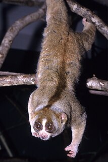
Slow lorises are a group of several species of nocturnal strepsirrhine primates that make up the genus Nycticebus. Found in Southeast Asia and bordering areas, they range from Bangladesh and Northeast India in the west to the Sulu Archipelago in the Philippines in the east, and from Yunnan province in China in the north to the island of Java in the south.

Rhinarium

The rhinarium is the furless skin surface surrounding the external openings of the nostrils in many mammals. Commonly it is referred to as the tip of the snout, and breeders of cats and dogs sometimes use the term nose leather. Informally, it may be called a "truffle", "wet snout" or "wet nose," because its surface is moist in some species: for example, healthy dogs and cats.

Masoala fork-marked lemur Species of lemur

The Masoala fork-marked lemur, also known as the eastern fork-marked lemur or Masoala fork-crowned lemur, is a species of lemur found in the coastal forests of northeastern Madagascar. It is a small nocturnal animal with large eyes, greyish fur and a long tail.

Fork-marked lemur Genus of Madagascan primates

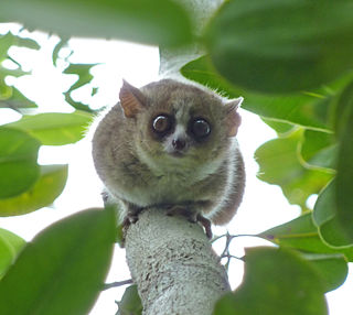
Fork-marked lemurs or fork-crowned lemurs are strepsirrhine primates; the four species comprise the genus Phaner. Like all lemurs, they are native to Madagascar, where they are found only in the west, north, and east sides of the island. They are named for the two black stripes which run up from the eyes, converge on the top of the head, and run down the back as a single black stripe. They were originally placed in the genus Lemur in 1839, later moved between the genera Cheirogaleus and Microcebus, and given their own genus in 1870 by John Edward Gray. Only one species was recognized, until three subspecies described in 1991 were promoted to species status in 2001. New species may yet be identified, particularly in northeast Madagascar.

Toothcomb Dental structure found in some mammals, comprising a group of front teeth arranged in a manner that facilitates grooming

A toothcomb is a dental structure found in some mammals, comprising a group of front teeth arranged in a manner that facilitates grooming, similar to a hair comb. The toothcomb occurs in lemuriform primates, treeshrews, colugos, hyraxes, and some African antelopes. The structures evolved independently in different types of mammals through convergent evolution and varies both in dental composition and structure. In most mammals the comb is formed by a group of teeth with fine spaces between them. The toothcombs in most mammals include incisors only, while in lemuriform primates they include incisors and canine teeth that tilt forward at the front of the lower jaw, followed by a canine-shaped first premolar. The toothcombs of colugos and hyraxes take a different form with the individual incisors being serrated, providing multiple tines per tooth.

<i>Babakotia</i> Extinct genus of lemurs

Babakotia is an extinct genus of medium-sized lemur, or strepsirrhine primate, from Madagascar that contains a single species, Babakotia radofilai. Together with Palaeopropithecus, Archaeoindris, and Mesopropithecus, it forms the family Palaeopropithecidae, commonly known as the sloth lemurs. The name Babakotia comes from the Malagasy name for the indri, babakoto, to which it and all other sloth lemurs are closely related. Due to its mix of morphological traits that show intermediate stages between the slow-moving smaller sloth lemurs and the suspensory large sloth lemurs, it has helped determine the relationship between both groups and the closely related and extinct monkey lemurs.

<i>Mesopropithecus</i> Extinct genus of small to medium-sized lemur from Madagascar

Mesopropithecus is an extinct genus of small to medium-sized lemur, or strepsirrhine primate, from Madagascar that includes three species, M. dolichobrachion, M. globiceps, and M. pithecoides. Together with Palaeopropithecus, Archaeoindris, and Babakotia, it is part of the sloth lemur family (Palaeopropithecidae). Once thought to be an indriid because its skull is similar to that of living sifakas, a recently discovered postcranial skeleton shows Mesopropithecus had longer forelimbs than hindlimbs—a distinctive trait shared by sloth lemurs but not by indriids. However, as it had the shortest forelimbs of all sloth lemurs, it is thought that Mesopropithecus was more quadrupedal and did not use suspension as much as the other sloth lemurs.

Evolution of lemurs History of primate evolution on Madagascar

Lemurs, primates belonging to the suborder Strepsirrhini which branched off from other primates less than 63 million years ago, evolved on the island of Madagascar, for at least 40 million years. They share some traits with the most basal primates, and thus are often confused as being ancestral to modern monkeys, apes, and humans. Instead, they merely resemble ancestral primates.

Taxonomy of lemurs The science of describing species and defining the evolutionary relationships between taxa of lemurs

Lemurs were first classified in 1758 by Carl Linnaeus, and the taxonomy remains controversial today, with approximately 70 to 100 species and subspecies recognized, depending on how the term "species" is defined. Having undergone their own independent evolution on Madagascar, lemurs have diversified to fill many ecological niches normally filled by other types of mammals. They include the smallest primates in the world, and once included some of the largest. Since the arrival of humans approximately 2,000 years ago, lemurs have become restricted to 10% of the island, or approximately 60,000 square kilometers (23,000 sq mi), and many face extinction. Concerns over lemur conservation have affected lemur taxonomy, since distinct species receive increased conservation attention compared to subspecies.

Azibiidae is an extinct family of fossil primate from the late early or early middle Eocene from the Glib Zegdou Formation in the Gour Lazib area of Algeria. They are thought to be related to the living toothcombed primates, the lemurs and lorisoids, although paleoanthropologists such as Marc Godinot have argued that they may be early simians. It includes the genera Azibius and Algeripithecus, the latter of which was originally considered the oldest known simian, not a strepsirrhine.

Djebelemur is an extinct genus of early strepsirrhine primate from the late early or early middle Eocene period from the Chambi locality in Tunisia. Although they probably lacked a toothcomb, a specialized dental structure found in living lemuriforms, they are thought to be a related stem group. The one recognized species, Djebelemur martinezi, was very small, approximately 100 g (3.5 oz).

Plesiopithecus is an extinct genus of early strepsirrhine primate from the late Eocene.

<i>Nycticebus kayan</i> Species of primate

The Kayan River slow loris is a strepsirrhine primate and a species of slow loris that is native to the northern and central highland region of the island of Borneo. The species was originally thought to be a part of the Bornean slow loris (N. menagensis) population until 2013, when a study of museum specimens and photographs identified distinct facial markings, which helped to differentiate it. It is distinguished by the high contrast of its black and white facial features, as well as the shape and width of the stripes of its facial markings.

The Bangka slow loris is a strepsirrhine primate and a species of slow loris that is native to southwestern Borneo and the island of Bangka. Originally considered a subspecies or synonym of the Bornean slow loris (N. menagensis), it was promoted to full species status in 2013 when a study of museum specimens and photographs identified distinct facial markings, which helped to differentiate it as a separate species. It is distinguished by the crimson red fur on its back, light-colored facial features, as well as the shape and width of the stripes of its facial markings.

<i>Nycticebus borneanus</i> Species of primate

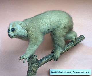
Nycticebus borneanus, the Bornean slow loris, is a strepsirrhine primate and a species of slow loris that is native to central south Borneo in Indonesia. Formerly considered a subspecies or synonym of N. menagensis, it was promoted to full species status in 2013 when a study of museum specimens and photographs identified distinct facial markings, which helped to differentiate it as a separate species. It is distinguished by its dark, contrasting facial features, as well as the shape and width of the stripes of its facial markings.

References

  1. Szalay & Delson 1980, p. 149.
  2. Cartmill 2010, p. 15.
  3. Hartwig 2011, pp. 20–21.
  4. 1 2 Ankel-Simons 2007, p. 421.
  5. 1 2 3 4 5 Rommel 1981, p. 153.
  6. 1 2 3 4 5 Osman Hill 1953, p. 73.
  7. 1 2 3 4 5 6 7 Ankel-Simons 2007, p. 422.
  8. 1 2 Wood Jones 1918, p. 349.
  9. 1 2 3 4 Hershkovitz 1977, p. 110.
  10. 1 2 3 Wood Jones 1918, pp. 349–350.
  11. 1 2 Wood Jones 1918, pp. 350–351.
  12. 1 2 Wood Jones 1918, p. 351.
  13. Wood Jones 1918, p. 350.
  14. 1 2 3 4 5 Hofer 1989, p. 25.
  15. 1 2 Ankel-Simons 2007, pp. 422–423.
  16. Wood Jones 1918, pp. 351–353.
  17. 1 2 3 Wood Jones 1918, p. 347.
  18. Tattersall 2006, pp. 7–8.
  19. Wood Jones 1918, p. 346.
  20. Ankel-Simons 2007, p. 423.
  21. Roberts 1941, p. 237.
  22. Sonntag 1921, p. 757 (?).
  23. Wood Jones 1918, pp. 348–349.
  24. Hofer 1977, p. 297.
  25. Hershkovitz 1977, p. 110–111.

Literature cited

  • Ankel-Simons, F. (2007). Primate Anatomy (3rd ed.). Academic Press. ISBN   978-0-12-372576-9.
  • Cartmill, M. (2010). "Primate Classification and Diversity". In Platt, M.; Ghazanfar, A (eds.). Primate Neuroethology. Oxford University Press. pp. 10–30. ISBN   978-0-19-532659-8.
  • Hartwig, W. (2011). "Chapter 3: Primate evolution". In Campbell, C. J.; Fuentes, A.; MacKinnon, K. C.; Bearder, S. K.; Stumpf, R. M (eds.). Primates in Perspective (2nd ed.). Oxford University Press. pp. 19–31. ISBN   978-0-19-539043-8.
  • Hershkovitz, P. (1977). Living New World monkeys (Platyrrhini): with an introduction to Primates. 1. University Of Chicago Press. ISBN   978-0-226-32788-4.
  • Hofer, H.O. (1989). "Lightmicroscopical investigations of the sublingua of Microcebus murinus (Cheirogaleidae, Lemuriformes) with remarks on the phylogenetic relations of the tree shrews (Scandentia) to primates". Zeitschrift für Morphologie und Anthropologie. 78 (1): 25–42. PMID   2603472.
  • Hofer, H.O. (1977). "On the sublingual structures of Tarsius (Prosimiae, Tarsiiformes) and some platyrrhine monkeys (Platyrrhina, Simiae, Primates) with casual remarks on the histology of the tongue". Folia Primatologica. 27 (4): 297–314. doi:10.1159/000155793. PMID   407139.
  • Wood Jones, F. (1918). "The sublingua and the plica fimbriata". Journal of Anatomy. 52 (Pt 4): 345–353. PMC   1262842 . PMID   17103846.
  • Osman Hill, W.C. (1953). Primates Comparative Anatomy and Taxonomy I—Strepsirhini. Edinburgh Univ Pubs Science & Maths, No 3. Edinburgh University Press. OCLC   500576914.
  • Roberts, D. (1941). "The dental comb of lemurs". Journal of Anatomy. 75 (Pt 2): 236–238. PMC   1252661 . PMID   17104854.
  • Rommel, C. (1981). "Sublingual structures in primates. Part 1: Prosimiae, Platyrrhini and Cercopithecinae". Gegenbaurs morphologisches Jahrbuch (in German). 127 (2): 153–175. PMID   6788642.
  • Sonntag, C.F. (1921). "39. The comparative anatomy of the tongues of the Mammalia. - VI. Summary and classification of the tongues of the primates". Proceedings of the Zoological Society of London. 91 (4): 757–767. doi:10.1111/j.1096-3642.1921.tb03290.x.
  • Szalay, F.S.; Delson, E. (1980). Evolutionary History of the Primates. Academic Press. ISBN   978-0126801507. OCLC   893740473.
  • Tattersall, I. (2006). "Chapter 1: Origin of the Malagasy strepsirhine primates". In Gould, L.; Sauther, M.L (eds.). Lemurs: Ecology and Adaptation. Springer. ISBN   0-387-34585-X.