The humerus is a long bone in the arm that runs from the shoulder to the elbow. It connects the scapula and the two bones of the lower arm, the radius and ulna, and consists of three sections. The humeral upper extremity consists of a rounded head, a narrow neck, and two short processes. The body is cylindrical in its upper portion, and more prismatic below. The lower extremity consists of 2 epicondyles, 2 processes, and 3 fossae. As well as its true anatomical neck, the constriction below the greater and lesser tubercles of the humerus is referred to as its surgical neck due to its tendency to fracture, thus often becoming the focus of surgeons.

At the base of the skull, the foramen ovale is one of the larger of the several holes that transmit nerves through the skull. The foramen ovale is situated in the posterior part of the sphenoid bone, posterolateral to the foramen rotundum.

The foramen lacerum is a triangular hole in the base of skull, located between the sphenoid, the apex of the petrous temporal and the basilar part of the occipital.
The foramen spinosum is one of two foramina located in the base of the human skull, on the sphenoid bone. It is situated just anterior to the spine of the sphenoid bone, and just lateral to the foramen ovale. The middle meningeal artery, middle meningeal vein, and the meningeal branch of the mandibular nerve pass through the foramen.

In human cranial neuroanatomy, the supratrochlear nerve is a branch of the frontal nerve, which itself comes from the ophthalmic division of the trigeminal cranial nerve. It is smaller than the nearby supraorbital nerve. It passes above the pulley of the superior oblique muscle, and gives off a descending filament that joins the infratrochlear branch of the nasociliary nerve.
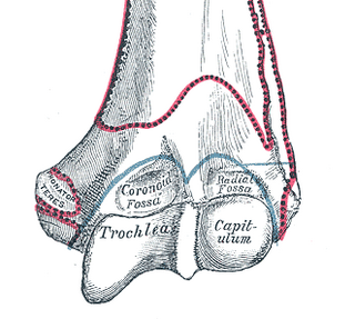
In the human arm, the humeral trochlea is the medial portion of the articular surface of the elbow joint which articulates with the trochlear notch on the ulna in the forearm.

At either posterior angle of the hard palate is the greater palatine foramen, for the transmission of the descending palatine vessels and greater palatine nerve; and running anteriorly (forward) and medially from it is a groove, for the same vessels and nerve.
In the base of the skull, in the great wings of the sphenoid bone, medial to the foramen ovale, a small aperture, the sphenoidal emissary foramen, may occasionally be seen opposite the root of the pterygoid process. When present, it opens below near the scaphoid fossa. Vesalius was the first to describe and illustrate this foramen, and it thus sometimes bears the name of foramen Vesalii. Other names include foramen venosum and canaliculus sphenoidalis.

The mastoid foramen is a hole in the posterior border of the temporal bone. It transmits a Mastoid emissary vein to the sigmoid sinus and a small branch of the occipital artery, the posterior meningeal artery to the dura mater.
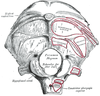
The condylar canal is a canal in the condyloid fossa of the lateral parts of occipital bone behind the occipital condyle. Resection of the rectus capitis posterior major and minor muscles reveals the bony recess leading to the condylar canal, which is situated posterior and lateral to the occipital condyle. It is immediately superior to the extradural vertebral artery, which makes a loop above the posterior C1 ring to enter the foramen magnum. The anteriomedial wall of the condylar canal thickens to join the foramen magnum rim and connect to the occipital condyle.

The lower extremity of the humerus is flattened from before backward, and curved slightly forward; it ends below in a broad, articular surface, which is divided into two parts by a slight ridge.
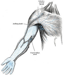
One or two supratrochlear lymph nodes are placed above the medial epicondyle of the humerus, medial to the basilic vein.

The anatomical neck of the humerus is obliquely directed, forming an obtuse angle with the body of the humerus. It represents the fused epiphyseal plate.
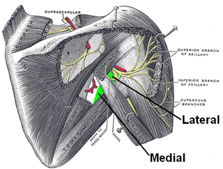
The quadrangular space is one of the three spaces in the axillary space. The other two spaces are: triangular space and triangular interval.

The following outline is provided as an overview of and topical guide to human anatomy:

In human anatomy, the omental foramen, is the passage of communication, or foramen, between the greater sac, and the lesser sac.
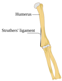
Struthers' ligament is a feature of human anatomy consisting of a band of connective tissue at the medial aspect of the distal humerus. It courses from the supracondylar process of the humerus to the medial humeral epicondyle. It is not a constant ligament, and can be acquired or congenital. The structure was highlighted by John Struthers, who discussed the feature's evolutionary significance with Charles Darwin. Struthers originally reported that the ligament usually arose at a position 3.2 to 6.4 cm from the medial condyle, being 1.2 to 1.9 cm in length, and nearer to the anterior than the medial border of the humerus.
The entepicondylar foramen is an opening in the distal (far) end of the humerus present in some mammals. It is often present in primitive placentals, such as the enigmatic Madagascan Plesiorycteropus. In most Neotominae and all Tylomyinae among cricetid rodents, it is located above the medial epicondyle of the humerus, but it is absent in all Sigmodontinae and Arvicolinae and this trait has been suggested as a synapomorphy for the former subfamily.
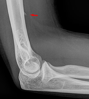
The supracondylar process of the humerus is a bony projection on the anteromedial aspect of the upper arm bone (humerus), about 5 cm above the medial epicondyle. It is directed downward, forward and medially pointing to the medial epicondyle. It is an anatomical variation which occurs in about one percent of all people. A fibrous band, Struthers ligament, may connect this process to the medial epicondyle.

Many anatomical terms descriptive of bone are defined in anatomical terminology, and are often derived from Greek and Latin.