Related Research Articles

Cooper's ligaments are connective tissue in the breast that help maintain structural integrity. They are named for Astley Cooper, who first described them in 1840. Their anatomy can be revealed using Transmission diffraction tomography.

The axilla is the area on the human body directly under the shoulder joint. It includes the axillary space, an anatomical space within the shoulder girdle between the arm and the thoracic cage, bounded superiorly by the imaginary plane between the superior borders of the first rib, clavicle and scapula, medially by the serratus anterior muscle and thoracolumbar fascia, anteriorly by the pectoral muscles and posteriorly by the subscapularis, teres major and latissimus dorsi muscle.

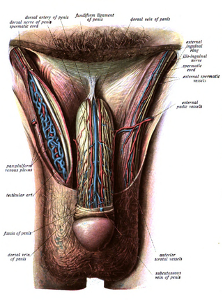
The fundiform ligament of the penis is a structure of the external genitalia whose descriptions differ according to the source consulted.

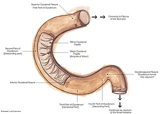
The suspensory muscle of duodenum is a thin muscle connecting the junction between the duodenum and jejunum, as well as the duodenojejunal flexure to connective tissue surrounding the superior mesenteric and coeliac arteries. The suspensory muscle most often connects to both the third and fourth parts of the duodenum, as well as the duodenojejunal flexure, although the attachment is quite variable.
A suspensory ligament is a ligament that supports a body part, especially an organ.

The suspensory ligament of the ovary, also infundibulopelvic ligament, is a fold of peritoneum that extends out from the ovary to the wall of the pelvis.

The clavipectoral fascia is a strong fascia situated under cover of the clavicular portion of the pectoralis major.
The pectoral fascia is very thin over the upper part of the pectoralis major, but thicker in the interval between it and the latissimus dorsi, where it closes in the axillary space and forms the axillary fascia. Axillary fascia, together with the skin, forms the base of the axilla.

The dorsal artery of the penis is a bilaterally paired terminal branch of the internal pudendal artery which passes upon the dorsum of the penis to the base of the glans penis, where it unites with its contralateral partner and supply the glans and foreskin.

The pectoral fascia is a thin lamina, covering the surface of the pectoralis major, and sending numerous prolongations between its fasciculi: it is attached, in the middle line, to the front of the sternum; above, to the clavicle; laterally and below it is continuous with the fascia of the shoulder, axilla, and thorax.

In human male anatomy, the dorsal veins of the penis are blood vessels that drain the shaft, the skin and the glans of the human penis. They are typically located in the midline on the dorsal aspect of the penis and they comprise the superficial dorsal veinof the penis, that lies in the subcutaneous tissue of the shaft, and the deep dorsal veinof the penis, that lies beneath the deep fascia.

Buck's fascia is a layer of deep fascia covering the three erectile bodies of the penis.

Pierre Nicolas Gerdy was a French physician who was a native of Loches-sur-Ource.

Václav Treitz was a Czech pathologist.

The following outline is provided as an overview of and topical guide to human anatomy:

In human male anatomy, the radix or root of the penis is the internal and most proximal portion of the human penis that lies in the perineum. Unlike the pendulous body of the penis, which is suspended from the pubic symphysis, the root is attached to the pubic arch of the pelvis and is not visible externally. It is triradiate in form, consisting of three masses of erectile tissue; the two diverging crura, one on either side, and the median bulb of the penis or urethral bulb. Approximately one third to one half of the penis is embedded in the pelvis and can be felt through the scrotum and in the perineum.

The fascia of perineum is the fascia which covers the muscles of the superficial perineal pouch. The muscles surrounded by the deep perineal fascia are the bulbospongiosus, ischiocavernosus, and superficial transverse perineal. The fascia is attached laterally to the ischiopubic rami and fused anteriorly with the suspensory ligament of the penis or clitoris. It is continuous anteriorly with the deep investing fascia of the abdominal wall muscles, and in males, it is continuous with Buck's fascia.
Degenerative suspensory ligament desmitis, commonly called DSLD, also known as equine systemic proteoglycan accumulation (ESPA), is a systemic disease of the connective tissue of the horse and other equines. It is a disorder akin to Ehlers–Danlos syndrome being researched in multiple horse breeds. Originally thought to be a condition of overwork and old age, the disease is now recognized as hereditary and has been seen in horses of all ages, including foals. The latest research (2010) has led to the proposed renaming of the disease from DSLD to ESPA because of the systemic and hereditary components now being found.

The suspensory ligament of the clitoris is a fibrous band at the deep fascial level that extends from the pubic symphysis to the deep fascia of the clitoris, anchoring the clitoris to the pubic symphysis. By virtue of this connection, the pubic symphysis supports the clitoris.
The suspensory ligament of the thyroid gland, or Berry's ligament, is a suspensory ligament that passes from the thyroid gland to the trachea.
References
- ↑ The American Heritage Medical Dictionary (Revised edition of 2nd ed.). Houghton Mifflin Company. 2007. ISBN 9780618824359. OCLC 71223299 . Retrieved 2 October 2012.