Thymoproteasome

Last updated

Thymoproteasome is a special kind of proteasome, which is present in vertebrates. [1] In the body it is located in thymus, exclusively in cortical thymic epithelial cells (cTECs). But in thymus we can also find another type of specific proteasome, immunoproteasome, which is present in thymocytes, dendritic cells and medular thymic epithelial cells. [2] It was first described in 2007 during a search for non-intronic sequence proximal to PSMB5 locus in mouse genome. The PSMB5 locus encodes the standard β5 proteasome subunit, while this sequence encodes a variant subunit β5t (PSMB11) specific to thymoproteasome. [3] The importance of this protein complex is its involvement in positive selection of T cells.

Contents

Generally proteasomes are protein complexes in cells, which degrade proteins marked by ubiquitin systems. Proteasomes are present in all eukaryotes. It has been shown that there exist modifications, which have different catalytic subunits. The first type of proteasome which was described is immunoproteasome, which is present in all jawed vertebrates. [1] Its function is degrading proteins for presentation on the surface of plasmatic membrane bound to MHC I complex. Immunoproteasome is activated when cells detect pro-inflammatory stimuli such as interferon gamma or other pro-inflammatory cytokines. The next type is thymoproteasome, which is involved in degradation of proteins, whose fragments are then presented to T cells in thymus.

Structure

Proteasomes consist of 20S catalytic core, which itself is created from two outer rings of alpha subunits and two inner rings of beta subunits. The catalytic activity is possessed only by β1, β2 and β5 subunits. In the basic type of proteasome these subunits have caspase, trypsin-like and chymotrypsin-like activities respectively. [4] To form the complete proteasome two 19S regulatory complexes are attached to both sides of the 20S core. This whole complex, designed as 26S, is then fully functional.

In thymoproteasome the catalytic subunits are replaced by their variants β1i, β2i and β5t. The first two subunits are also present in immunoproteasome, while the last one is specific to thymoproteasome. [1] The β5t subunit active proteolytic center contains mostly hydrophilic amino acid residues. In contrast both the β5 and β5i subunits contain mostly hydrophobic residues. [3] This change leads to their different proteolytic activities and thus to specific protein fragment production. β5t subunit also creates different S1 pockets, which determine the C-terminus of the processed peptide. This leads to decrease in chymotrypsin-like activity without affecting the trypsin-like and caspase-like activities. [5] Thymoproteasome produces different sequences and quantities of peptides due to the β5t subunit. [6]

Duplication of the original proteasome gene is the most likely mechanism of the development of specific subunits (β5t/i). Those duplications may have played a role in the development of adaptive immunity. [7]

Function

The function of thymoproteasome is the cleavage of proteins to peptides for display in the MHC I complex. These peptides serve in thymus during the positive selection of CD8+ T lymphocytes. [8] The thymoproteasome is capable of providing unique self, MHC-associated peptides, or the self-peptides that vary from conventional self-peptides expressed in other parts of the body. [9]

β5t deficient mice were used to study the function of thymoproteasome. Such mice developed thymuses as large as healthy mice, but their cTECs no longer contained thymoproteasomes. In these mice it was replaced by immunoproteasome, which did not replicate its function sufficiently. This then decreased their CD8+ T lymphocyte count by about 20%, which shows the important role of thymoproteasome in T cell development. [3]

In a knockout model of nude mice with deficiency of Foxn-1 gene it is obvious that Foxn1 transcriptional factor is involved in specific expression of β5t in thymus. [7] Its promoter (with sequence 5′ -ACGC-3′) is highly conserved to Foxn1 transcriptional factor. Mutation in this promoter leads to decrease in β5t expression and CD8+ T cells production. [10]

β5t subunit in diagnostic of thymic epithelilal tumors

Thymic epithelial tumors form a group of six categories (A, AB, B1, B2, B3 and C), which are based on its histology and morphology. Diagnostic of those tumors is problematic because of their rarity and variablitity in the cytological and structural patterns. Above-mentioned Foxn1 was used as a marker, but there is a problem of it appearing in other tumors like lungs carcinome. Recent research into β5t subunit has shown, that it can be used as a diagnostic marker of some categories of thymic epithelial tumors. β5t subunit is expressed the most in the B type of tumors, less so in AB and not at all expressed in the A type. [11]

Related Research Articles

<span class="mw-page-title-main">Proteasome</span> Protein complexes which degrade unnecessary or damaged proteins by proteolysis

Proteasomes are protein complexes which degrade unneeded or damaged proteins by proteolysis, a chemical reaction that breaks peptide bonds. Enzymes that help such reactions are called proteases.

<span class="mw-page-title-main">Thymus</span> Endocrine gland

The thymus is a specialized primary lymphoid organ of the immune system. Within the thymus, thymus cell lymphocytes or T cells mature. T cells are critical to the adaptive immune system, where the body adapts to specific foreign invaders. The thymus is located in the upper front part of the chest, in the anterior superior mediastinum, behind the sternum, and in front of the heart. It is made up of two lobes, each consisting of a central medulla and an outer cortex, surrounded by a capsule.

<span class="mw-page-title-main">T cell</span> White blood cells of the immune system

T cells are one of the important types of white blood cells of the immune system and play a central role in the adaptive immune response. T cells can be distinguished from other lymphocytes by the presence of a T-cell receptor (TCR) on their cell surface.

Cross-presentation is the ability of certain professional antigen-presenting cells (mostly dendritic cells) to take up, process and present extracellular antigens with MHC class I molecules to CD8 T cells (cytotoxic T cells). Cross-priming, the result of this process, describes the stimulation of naive cytotoxic CD8+ T cells into activated cytotoxic CD8+ T cells. This process is necessary for immunity against most tumors and against viruses that infect dendritic cells and sabotage their presentation of virus antigens. Cross presentation is also required for the induction of cytotoxic immunity by vaccination with protein antigens, for example, tumour vaccination.

In immunology, central tolerance is the process of eliminating any developing T or B lymphocytes that are autoreactive, i.e. reactive to the body itself. Through elimination of autoreactive lymphocytes, tolerance ensures that the immune system does not attack self peptides. Lymphocyte maturation occurs in primary lymphoid organs such as the bone marrow and the thymus. In mammals, B cells mature in the bone marrow and T cells mature in the thymus.

A thymocyte is an immune cell present in the thymus, before it undergoes transformation into a T cell. Thymocytes are produced as stem cells in the bone marrow and reach the thymus via the blood.

MHC-restricted antigen recognition, or MHC restriction, refers to the fact that a T cell can interact with a self-major histocompatibility complex molecule and a foreign peptide bound to it, but will only respond to the antigen when it is bound to a particular MHC molecule.

An immunoproteasome is a type of proteasome that degrades ubiquitin-labeled proteins found in the cytoplasm in cells exposed to oxidative stress and proinflammatory stimuli. In general, proteasomes consist of a regulatory and a catalytic part. Immunoproteasomes are induced by interferon gamma and oxidative stress, which in the cell triggers the transcription of three catalytic subunits that do not occur in the classical proteasome. Another possible variation of proteasome is the thymoproteasome, which is located in the thymus and folds to present peptides to naive T cells.

<span class="mw-page-title-main">XCR1</span> Protein-coding gene in the species Homo sapiens

The "C" sub-family of chemokine receptors contains only one member: XCR1, the receptor for XCL1 and XCL2.

<span class="mw-page-title-main">Minor histocompatibility antigen</span>

Minor histocompatibility antigen are peptides presented on the cellular surface of donated organs that are known to give an immunological response in some organ transplants. They cause problems of rejection less frequently than those of the major histocompatibility complex (MHC). Minor histocompatibility antigens (MiHAs) are diverse, short segments of proteins and are referred to as peptides. These peptides are normally around 9-12 amino acids in length and are bound to both the major histocompatibility complex (MHC) class I and class II proteins. Peptide sequences can differ among individuals and these differences arise from SNPs in the coding region of genes, gene deletions, frameshift mutations, or insertions. About a third of the characterized MiHAs come from the Y chromosome. Prior to becoming a short peptide sequence, the proteins expressed by these polymorphic or diverse genes need to be digested in the proteasome into shorter peptides. These endogenous or self peptides are then transported into the endoplasmic reticulum with a peptide transporter pump called TAP where they encounter and bind to the MHC class I molecule. This contrasts with MHC class II molecules's antigens which are peptides derived from phagocytosis/endocytosis and molecular degradation of non-self entities' proteins, usually by antigen-presenting cells. MiHA antigens are either ubiquitously expressed in most tissue like skin and intestines or restrictively expressed in the immune cells.

<span class="mw-page-title-main">PSMB10</span> Protein found in humans

Proteasome subunit beta type-10 as known as 20S proteasome subunit beta-2i is a protein that in humans is encoded by the PSMB10 gene.

<span class="mw-page-title-main">PSMB8</span> Protein found in humans

Proteasome subunit beta type-8 as known as 20S proteasome subunit beta-5i is a protein that in humans is encoded by the PSMB8 gene. This protein is one of the 17 essential subunits that contributes to the complete assembly of 20S proteasome complex. In particular, proteasome subunit beta type-5, along with other beta subunits, assemble into two heptameric rings and subsequently a proteolytic chamber for substrate degradation. This protein contains "Chymotrypsin-like" activity and is capable of cleaving after large hydrophobic residues of peptide. The eukaryotic proteasome recognized degradable proteins, including damaged proteins for protein quality control purpose or key regulatory protein components for dynamic biological processes. The constitutive subunit beta1, beta2, and beta 5 can be replaced by their inducible counterparts beta1i, 2i, and 5i when cells are under the treatment of interferon-γ. The resulting proteasome complex becomes the so-called immunoproteasome. An essential function of the modified proteasome complex, the immunoproteasome, is the processing of numerous MHC class-I restricted T cell epitopes.

<span class="mw-page-title-main">PSMB9</span> Protein found in humans

Proteasome subunit beta type-9 as known as 20S proteasome subunit beta-1i is a protein that in humans is encoded by the PSMB9 gene.

<span class="mw-page-title-main">PSMB5</span> Protein found in humans

Proteasome subunit beta type-5 as known as 20S proteasome subunit beta-5 is a protein that in humans is encoded by the PSMB5 gene. This protein is one of the 17 essential subunits that contributes to the complete assembly of 20S proteasome complex. In particular, proteasome subunit beta type-5, along with other beta subunits, assemble into two heptameric rings and subsequently a proteolytic chamber for substrate degradation. This protein contains "chymotrypsin-like" activity and is capable of cleaving after large hydrophobic residues of peptide. The eukaryotic proteasome recognized degradable proteins, including damaged proteins for protein quality control purpose or key regulatory protein components for dynamic biological processes. An essential function of a modified proteasome, the immunoproteasome, is the processing of class I MHC peptides.

<span class="mw-page-title-main">FOXN1</span> Protein-coding gene in the species Homo sapiens

Forkhead box protein N1 is a protein that in humans is encoded by the FOXN1 gene.

Thymic nurse cells (TNCs) are large epithelial cells found in the cortex of the thymus and also in cortico-medullary junction. They have their own nucleus and are known to internalize thymocytes through extensions of plasma membrane. The cell surfaces of TNCs and their cytoplasmic vacuoles express MHC Class I and MHC Class II antigens. The interaction of these antigens with the developing thymocytes determines whether the thymocytes undergo positive or negative selection.

<span class="mw-page-title-main">Medullary thymic epithelial cells</span>

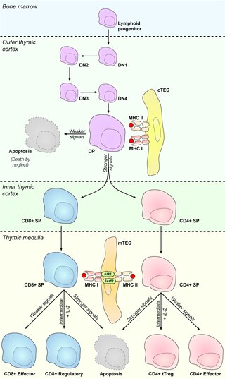
Medullary thymic epithelial cells (mTECs) represent a unique stromal cell population of the thymus which plays an essential role in the establishment of central tolerance. Therefore, mTECs rank among cells relevant for the development of functional mammal immune system.

Antigen transfer in the thymus is the transmission of self-antigens between thymic antigen-presenting cells which contributes to the establishment of T cell central tolerance.

<span class="mw-page-title-main">Cortical thymic epithelial cells</span>

Cortical thymic epithelial cells (cTECs) form unique parenchyma cell population of the thymus which critically contribute to the development of T cells.

Thymic epithelial cells (TECs) are specialized cells with high degree of anatomic, phenotypic and functional heterogeneity that are located in the outer layer (epithelium) of the thymic stroma. The thymus, as a primary lymphoid organ, mediates T cell development and maturation. The thymic microenvironment is established by TEC network filled with thymocytes in different developing stages. TECs and thymocytes are the most important components in the thymus, that are necessary for production of functionally competent T lymphocytes and self tolerance. Dysfunction of TECs causes several immunodeficiencies and autoimmune diseases.

References

  1. 1 2 3 Murata, Shigeo; Takahama, Yousuke; Tanaka, Keiji (April 2008). "Thymoproteasome: probable role in generating positively selecting peptides". Current Opinion in Immunology. 20 (2): 192–196. doi:10.1016/j.coi.2008.03.002. PMID   18403190.
  2. Ohigashi, Izumi; Takahama, Yousuke (2021-01-01), Alt, Frederick W.; Murphy, Kenneth M. (eds.), Chapter One - Thymoproteasome optimizes positive selection of CD8+ T cells without contribution of negative selection, Advances in Immunology, vol. 149, Academic Press, pp. 1–23, doi:10.1016/bs.ai.2021.03.001, PMC   8237917 , PMID   33993918
  3. 1 2 3 Murata, Shigeo; Takahama, Yousuke; Kasahara, Masanori; Tanaka, Keiji (September 2018). "The immunoproteasome and thymoproteasome: functions, evolution and human disease". Nature Immunology. 19 (9): 923–931. doi:10.1038/s41590-018-0186-z. ISSN   1529-2916. PMID   30104634. S2CID   51970543.
  4. Tomaru, Utano; Kasahara, Masanori (2013-10-01). "Thymoproteasome: Role in Thymic Selection and Clinical Significance as a Diagnostic Marker for Thymic Epithelial Tumors". Archivum Immunologiae et Therapiae Experimentalis. 61 (5): 357–365. doi:10.1007/s00005-013-0234-1. ISSN   1661-4917. PMID   23645302. S2CID   17915099.
  5. Takahama, Yousuke; Takada, Kensuke; Murata, Shigeo; Tanaka, Keiji (February 2012). "β5t-containing thymoproteasome: specific expression in thymic cortical epithelial cells and role in positive selection of CD8+ T cells". Current Opinion in Immunology. 24 (1): 92–98. doi:10.1016/j.coi.2012.01.006. PMID   22285892.
  6. Frantzeskakis, Melina; Takahama, Yousuke; Ohigashi, Izumi (2021-03-19). "The Role of Proteasomes in the Thymus". Frontiers in Immunology. 12: 646209. doi: 10.3389/fimmu.2021.646209 . ISSN   1664-3224. PMC   8017227 . PMID   33815406.
  7. 1 2 Tanaka, Keiji; Mizushima, Tsunehiro; Saeki, Yasushi (2012-04-01). "The proteasome: molecular machinery and pathophysiological roles". Biological Chemistry. 393 (4): 217–234. doi: 10.1515/hsz-2011-0285 . ISSN   1437-4315. PMID   23029643. S2CID   29581097.
  8. Kniepert, Andrea; Groettrup, Marcus (2014-01-01). "The unique functions of tissue-specific proteasomes". Trends in Biochemical Sciences. 39 (1): 17–24. doi:10.1016/j.tibs.2013.10.004. ISSN   0968-0004. PMID   24286712.
  9. Takahama, Yousuke; Ohigashi, Izumi; Murata, Shigeo; Tanaka, Keiji (2019-03-01). "Thymoproteasome and peptidic self". Immunogenetics. 71 (3): 217–221. doi:10.1007/s00251-018-1081-3. ISSN   1432-1211. PMID   30324237. S2CID   53086758.
  10. Motosugi, Ryo; Murata, Shigeo (2019). "Dynamic Regulation of Proteasome Expression". Frontiers in Molecular Biosciences. 6: 30. doi: 10.3389/fmolb.2019.00030 . ISSN   2296-889X. PMC   6504791 . PMID   31119134.
  11. Tomaru, Utano; Kasahara, Masanori (October 2013). "Thymoproteasome: Role in Thymic Selection and Clinical Significance as a Diagnostic Marker for Thymic Epithelial Tumors". Archivum Immunologiae et Therapiae Experimentalis. 61 (5): 357–365. doi:10.1007/s00005-013-0234-1. ISSN   0004-069X. PMID   23645302. S2CID   17915099.