Prostate cancer is cancer of the prostate. Prostate cancer is the second common cancerous tumor worldwide It is the fifth leading cause of cancer-related mortality among men [1]. The prostate is a gland in the male reproductive system that surrounds the urethra just below the bladder. It is located in the hypogastric region of the abdomen. To give an idea of where it is located, the bladder is superior to the prostate gland as shown in the image The rectum is posterior in perspective to the prostate gland and the ischial tuberosity of the pelvic bone is inferior. Only those who have male reproductive organs are able to get prostate cancer. Most prostate cancers are slow growing. Cancerous cells may spread to other areas of the body, particularly the bones and lymph nodes. It may initially cause no symptoms. In later stages, symptoms include pain or difficulty urinating, blood in the urine, or pain in the pelvis or back. Benign prostatic hyperplasia may produce similar symptoms. Other late symptoms include fatigue, due to low levels of red blood cells.

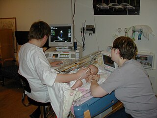
Medical ultrasound includes diagnostic techniques using ultrasound, as well as therapeutic applications of ultrasound. In diagnosis, it is used to create an image of internal body structures such as tendons, muscles, joints, blood vessels, and internal organs, to measure some characteristics or to generate an informative audible sound. It's aim is usually to find a source of disease or to exclude pathology. The usage of ultrasound to produce visual images for medicine is called medical ultrasonography or simply sonography. The practice of examining pregnant women using ultrasound is called obstetric ultrasonography, and was an early development of clinical ultrasonography.

Medical imaging is the technique and process of imaging the interior of a body for clinical analysis and medical intervention, as well as visual representation of the function of some organs or tissues (physiology). Medical imaging seeks to reveal internal structures hidden by the skin and bones, as well as to diagnose and treat disease. Medical imaging also establishes a database of normal anatomy and physiology to make it possible to identify abnormalities. Although imaging of removed organs and tissues can be performed for medical reasons, such procedures are usually considered part of pathology instead of medical imaging.

Prostate-specific antigen (PSA), also known as gamma-seminoprotein or kallikrein-3 (KLK3), P-30 antigen, is a glycoprotein enzyme encoded in humans by the KLK3 gene. PSA is a member of the kallikrein-related peptidase family and is secreted by the epithelial cells of the prostate gland.

Elastography is any of a class of medical imaging modalities that map the elastic properties and stiffness of soft tissue. The main idea is that whether the tissue is hard or soft will give diagnostic information about the presence or status of disease. For example, cancerous tumours will often be harder than the surrounding tissue, and diseased livers are stiffer than healthy ones.

A tricorder is a fictional handheld sensor that exists in the Star Trek universe. The tricorder is a multifunctional hand-held device that can perform environmental scans, data recording, and data analysis; hence the word "tricorder" to refer to the three functions of sensing, recording, and computing. In Star Trek stories the devices are issued by the fictional Starfleet organization.
Diathermy is electrically induced heat or the use of high-frequency electromagnetic currents as a form of physical therapy and in surgical procedures. The earliest observations on the reactions of high-frequency electromagnetic currents upon the human organism were made by Jacques Arsene d'Arsonval. The field was pioneered in 1907 by German physician Karl Franz Nagelschmidt, who coined the term diathermy from the Greek words dia and θέρμη therma, literally meaning "heating through".

Prostate biopsy is a procedure in which small hollow needle-core samples are removed from a man's prostate gland to be examined for the presence of prostate cancer. It is typically performed when the result from a PSA blood test is high. It may also be considered advisable after a digital rectal exam (DRE) finds possible abnormality. PSA screening is controversial as PSA may become elevated due to non-cancerous conditions such as benign prostatic hyperplasia (BPH), by infection, or by manipulation of the prostate during surgery or catheterization. Additionally many prostate cancers detected by screening develop so slowly that they would not cause problems during a man's lifetime, making the complications due to treatment unnecessary.
Prostate cancer screening is the screening process used to detect undiagnosed prostate cancer in men without signs or symptoms. When abnormal prostate tissue or cancer is found early, it may be easier to treat and cure, but it is unclear if early detection reduces mortality rates.

High-intensity focused ultrasound (HIFU) is a non-invasive therapeutic technique that uses non-ionizing ultrasonic waves to heat or ablate tissue. HIFU can be used to increase the flow of blood or lymph, or to destroy tissue, such as tumors, via thermal and mechanical mechanisms. Given the prevalence and relatively low cost of ultrasound, HIFU has been subject to much research and development. The premise of HIFU is that it is a non-invasive low-cost therapy that can at minimum outperform the current standard of care.
Ultrasound energy, simply known as ultrasound, is a type of mechanical energy called sound characterized by vibrating or moving particles within a medium. Ultrasound is distinguished by vibrations with a frequency greater than 20,000 Hz, compared to audible sounds that humans typically hear with frequencies between 20 and 20,000 Hz. Ultrasound energy requires matter or a medium with particles to vibrate to conduct or propagate its energy. The energy generally travels through most mediums in the form of a wave in which particles are deformed or displaced by the energy then reestablished after the energy passes. Types of waves include shear, surface, and longitudinal waves with the latter being one of the most common used in biological applications. The characteristics of the traveling ultrasound energy greatly depend on the medium that it is traveling through. While ultrasound waves propagate through a medium, the amplitude of the wave is continually reduced or weakened with the distance it travels. This is known as attenuation and is due to the scattering or deflecting of energy signals as the wave propagates and the conversion of some of the energy to heat energy within the medium. A medium that changes the mechanical energy from the vibrations of the ultrasound energy into thermal or heat energy is called viscoelastic. The properties of ultrasound waves traveling through the medium of biological tissues has been extensively studied in recent years and implemented into many important medical tools.
Biological imaging may refer to any imaging technique used in biology. Typical examples include:

Prostate cancer antigen 3 is a gene that expresses a non-coding RNA. PCA3 is only expressed in human prostate tissue, and the gene is highly overexpressed in prostate cancer. Because of its restricted expression profile, the PCA3 RNA is useful as a tumor marker.

John Julian Cuttance Wild was an English-born American physician who was part of the first group to use ultrasound for body imaging, most notably for diagnosing cancer. Modern ultrasonic diagnostic medical scans are descendants of the equipment Wild and his colleagues developed in the 1950s. He has been described as the "father of medical ultrasound".
The Qualcomm Tricorder XPRIZE was an inducement prize contest announced on May 10, 2011, sponsored by Qualcomm Foundation. It officially launched on January 10, 2012. The $10 million prize is awarded for creating a mobile device that can "diagnose patients better than or equal to a panel of board certified physicians". The name is taken from the tricorder device in Star Trek which can be used to instantly diagnose ailments. The focus of the competition guidelines was towards clinical symptoms, versus accurate measurement of test values.
Dong-A University Hospital (동아대학교의료원) is a major general hospital affiliated with Dong-A University in Busan, South Korea.

A medical tricorder is a handheld portable scanning device to be used by consumers to self-diagnose medical conditions within seconds and take basic vital measurements. While the device is not yet on the mass market, there are numerous reports of other scientists and inventors also working to create such a device as well as improve it. A common view is that it will be a general-purpose tool similar in functionality to a Swiss Army Knife to take health measurements such as blood pressure and temperature, and blood flow in a noninvasive way. It would diagnose a person's state of health after analyzing the data, either as a standalone device or as a connection to medical databases via an Internet connection.
Kevin M. Slawin is an American physician and the founder of Bellicum Pharmaceuticals and the Vanguard Urologic Institute at Memorial Hermann Medical Group. He was also the Director of Urology at Memorial Hermann Hospital. Slawin specializes in the diagnosis and treatment of urologic cancers and robotic surgery. He is also possesses patents related to the advancement of prostate cancer diagnosis, staging and treatment and to the cellular immunotherapy of cancer.
Caroline M. Moore is the first woman to be made a professor of urology in the United Kingdom. She works in the diagnosis and treatment of prostate cancer at University College London.
Hashim U. Ahmed is a British surgeon, medical researcher and author of publications in the field of prostate cancer diagnostics and treatment; his research has contributed to changes in the way men with suspected prostate cancer and men with prostate enlargement are diagnosed and treated. He is Professor and Chair of Urology at Imperial College Healthcare NHS Trust and Consultant Urological Surgeon at both Charing Cross Hospital and BUPA Cromwell Hospital.


