Triple-resonance nuclear magnetic resonance spectroscopy

Last updated

Triple resonance experiments are a set of multi-dimensional nuclear magnetic resonance spectroscopy (NMR) experiments that link three types of atomic nuclei, most typically consisting of 1H, 15N and 13C. These experiments are often used to assign specific resonance signals to specific atoms in an isotopically-enriched protein. The technique was first described in papers by Ad Bax, Mitsuhiko Ikura and Lewis Kay in 1990, [1] [2] and further experiments were then added to the suite of experiments. Many of these experiments have since become the standard set of experiments used for sequential assignment of NMR resonances in the determination of protein structure by NMR. They are now an integral part of solution NMR study of proteins, and they may also be used in solid-state NMR. [3] [4]

Contents

Background

There are two main methods of determining protein structure on the atomic level. The first of these is by X-ray crystallography, starting in 1958 when the crystal structure of myoglobin was determined. The second method is by NMR, which began in the 1980s when Kurt Wüthrich outlined the framework for NMR structure determination of proteins and solved the structure of small globular proteins. [5] The early method of structural determination of protein by NMR relied on proton-based homonuclear NMR spectroscopy in which the size of the protein that may be determined is limited to ~10 KDa. This limitation is due to the need to assign NMR signals from the large number of nuclei in the protein – in larger protein, the greater number of nuclei results in overcrowding of resonances, and the increasing size of the protein also broadens the signals, making resonance assignment difficult. These problems may be alleviated by using heteronuclear NMR spectroscopy which allows the proton spectrum to be edited with respect to the 15N and 13C chemical shifts, and also reduces the overlap of resonances by increasing the number of dimensions of the spectrum. In 1990, Ad Bax and coworkers developed the triple resonance technology and experiments on proteins isotopically labelled with 15N and 13C, [1] with the result that the spectra are dramatically simplified, greatly facilitating the process of resonance assignment, and increasing the size of the protein that may be determined by NMR.

These triple resonance experiments utilize the relatively large magnetic couplings between certain pairs of nuclei to establish their connectivity. Specifically, the 1JNH, 1JCH, 1JCC, and 1JCN couplings are used to establish the scalar connectivity pathway between nuclei. The magnetization transfer process takes place through multiple, efficient one-bond magnetization transfer steps, rather than a single step through the smaller and variable 3JHH couplings. The relatively large size and good uniformity of the one-bond couplings allowed the design of efficient magnetization transfer schemes that are effectively uniform across a given protein, nearly independent of conformation. [3] Triple resonance experiments involving 31P may also be use for nucleic acid studies. [6]

Suite of experiments

Sequential assignment using triple resonance experiments
HNCACB experiment.JPG
NMR sequential assignment strips.JPG
CBCACONH experiment.JPG
The top and bottom figures show the magnetization transfer pathway with the atoms that appear as peaks highlighted. The middle figure shows how the peaks appear in spectra, here presented as an overlay of strips of HNCACB and CBCA(CO)NH spectra. Peaks from CBCA(CO)NH are in intensity plot and colored yellow; they identify the resonances of the preceding Cα and Cβ. HNCACB is in contour plot and usually shows 4 peaks for each strip, two each from a residue and its preceding one. The red peaks are for Cα, and blue peaks for Cβ. Here the blue Cβ peaks of threonine and alanine are distinctive and easily identifiable, while glycine which lacks a Cβ gives only a single peak. Other residue types however may not be as easy to distinguish.

These experiments are typically named by the nuclei (H, N, and C) involved in the experiment. CO refers to the carbonyl carbon, while CA and CB refer to Cα and Cβ respectively, similarly HA and HB for Hα and Hβ (see diagram for examples of experiments). The nuclei in the name are ordered in the same sequence as in the path of magnetization transfer, those nuclei placed within parentheses are involved in the magnetization transfer pathway but are not recorded. For reason of sensitivity, these experiments generally start on a proton and end on a proton, typically via INEPT and reverse INEPT steps. Therefore, many of these experiments are what may be called "out-and-back" experiments where, although not indicated in the name, the magnetization is transferred back to the starting proton for signal acquisition.

Some of the experiments are used in tandem for the resonance assignment of protein, for example HNCACB may be used together with CBCA(CO)NH as a pair of experiments. Not all of these experiments need to be recorded for sequential assignment (it can be done with as few as two), however extra pairs of experiments are useful for independent assessment of the correctness of the assignment, and the redundancy of information may be necessary when there is ambiguity in the assignments. Other experiments are also necessary to fully assign the side chain resonances.

TROSY versions of many of these experiments exist for improvement in sensitivity. [7] Triple resonance experiments can also be used in sequence-specific backbone resonance assignment of magic angle spinning NMR spectra in solid-state NMR. [4] [8]

A large number triple-resonance NMR experiments have been created, and the experiments listed below is not meant to be exhaustive.

HNCO

The experiment provides the connectivities between the amide of a residue with the carbonyl carbon of the preceding residues. [2] It is the most sensitive of the triple resonance experiments. The sidechains carboxamides of asparagine and glutamine are also visible in this experiment. Additionally, the guanidino group of arginine, which has similar coupling constant to the carboxamide group, may also appear in this spectrum. This experiment is sometimes used together with HN(CA)CO.

HN(CA)CO

Here, the amide resonance of a residue is correlated with the carbonyl carbon of the same residue, as well as that of the preceding residue. The intra-residue resonances are usually stronger than the inter-residues one. [9]

HN(CO)CA

This experiment correlates the resonances of the amide of a residue with the Cα of the preceding residue. This experiment is often used together with HNCA. [10]

HNCA

This experiment correlates the chemical shift of amide of a residue the Cα of the same residue as well as those of the preceding residue. [2] Each strip gives two peaks, the inter and intra-residue Cα peaks. Peak from the preceding Cα may be identified from the HN(CO)CA experiment which gives only the inter-residue Cα.

CBCA(CO)NH

CBCA(CO)NH, or alternatively HN(CO)CACB, correlates the resonances of the amide of a residue with the Cα and Cβ of the preceding residue. [11] Two peaks corresponding to the Cα and Cβ are therefore visible for each residue. This experiment is normally used together with HNCACB. The sidechain carboxamide of glutamines and asparagines also appear in this spectra in this experiment. CBCA(CO)NH is sometimes more precisely called (HBHA)CBCA(CO)NH as it starts with aliphatic protons and ends on an amide proton, and is therefore not an out-and-back experiment like HN(CO)CACB.

HNCACB

HNCACB, or alternatively CBCANH, correlates the chemical shift of amide of a residue the Cα and Cβ of the same residue as well as those of the preceding residue. [12] In each strip, four peaks may be visible – 2 from the same residue and 2 from the preceding residue. Peaks from the preceding residue are usually weaker, and may be identified using CBCA(CO)NH. In this experiment, the Cα and Cβ peaks are in opposite phase, i.e. if Cα appears as a positive peak, then Cβ will be negative, making identification of Cα and Cβ straightforward. The extra information of Cβ from the CBCA(CO)NH/HNCACB set of experiments makes identification of residue type easier than HN(CO)CA/HNCA, however the HNCACB is a less sensitive experiment and may be unsuitable for some proteins.

The CBCANH experiment is less suitable for larger protein as it is more susceptible to the line-width problem than HNCACB.

CBCACO(CA)HA

This experiment provides the connectivities between the Cα and Cβ with the carbonyl carbon and Hα atoms within the same residue. [13] The sidechain carboxyl group of aspartate and glutamate may appear weakly in this spectrum.

CC(CO)NH

This experiment provides connectivities between the amide of a residue and the aliphatic carbon atoms of the preceding residue. [14]

H(CCO)NH

This experiment provides connectivities between the amide of a residue and the hydrogen atoms attached to the aliphatic carbon of the preceding residue.

HBHA(CO)NH

This experiment correlates the amide resonance to the Hα and Hβ of the preceding residue. [15]

Sequential assignment

Pairs of experiments are normally used for sequential assignment, for example, the HNCACB and CBCA(CO)NH pair, or HNCA and HNC(CO)CA. The spectra are normally analyzed as strips of peaks, and strips from the pair of experiments may be presented together side by side or as an overlay of two spectra. In the HNCACB spectra 4 peaks are usually present in each strip, the Cα and Cβ of one residue as well as those of its preceding residue. The peaks from the preceding residue can be identified from the CBCA(CO)NH experiment. Each strip of peaks can therefore be linked to the next strip of peaks from an adjoining residue, allowing the strips to be connected sequentially. The residue type can be identified from the chemical shifts of the peaks, some, such as serine, threonine, glycine and alanine, are much easier to identify than others. The resonances can then be assigned by comparing the sequence of peaks with the amino acid sequence of the protein.

Related Research Articles

The nuclear Overhauser effect (NOE) is the transfer of nuclear spin polarization from one population of spin-active nuclei to another via cross-relaxation. A phenomenological definition of the NOE in nuclear magnetic resonance spectroscopy (NMR) is the change in the integrated intensity of one NMR resonance that occurs when another is saturated by irradiation with an RF field. The change in resonance intensity of a nucleus is a consequence of the nucleus being close in space to those directly affected by the RF perturbation.

In nuclear magnetic resonance (NMR) spectroscopy, the chemical shift is the resonant frequency of a nucleus relative to a standard in a magnetic field. Often the position and number of chemical shifts are diagnostic of the structure of a molecule. Chemical shifts are also used to describe signals in other forms of spectroscopy such as photoemission spectroscopy.

Nuclear magnetic resonance spectroscopy Laboratory technique

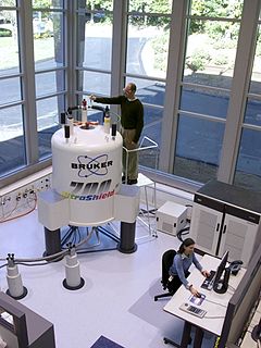
Nuclear magnetic resonance spectroscopy, most commonly known as NMR spectroscopy or magnetic resonance spectroscopy (MRS), is a spectroscopic technique to observe local magnetic fields around atomic nuclei. The sample is placed in a magnetic field and the NMR signal is produced by excitation of the nuclei sample with radio waves into nuclear magnetic resonance, which is detected with sensitive radio receivers. The intramolecular magnetic field around an atom in a molecule changes the resonance frequency, thus giving access to details of the electronic structure of a molecule and its individual functional groups. As the fields are unique or highly characteristic to individual compounds, in modern organic chemistry practice, NMR spectroscopy is the definitive method to identify monomolecular organic compounds. Similarly, biochemists use NMR to identify proteins and other complex molecules. Besides identification, NMR spectroscopy provides detailed information about the structure, dynamics, reaction state, and chemical environment of molecules. The most common types of NMR are proton and carbon-13 NMR spectroscopy, but it is applicable to any kind of sample that contains nuclei possessing spin.

Solid-state nuclear magnetic resonance

Solid-state NMR (ssNMR) spectroscopy is a special type of nuclear magnetic resonance (NMR) spectroscopy, characterized by the presence of anisotropic interactions. Compared to the more common solution NMR spectroscopy, ssNMR usually requires additional hardware for high-power radio-frequency irradiation and magic-angle spinning.

Carbon-13 (C13) nuclear magnetic resonance is the application of nuclear magnetic resonance (NMR) spectroscopy to carbon. It is analogous to proton NMR and allows the identification of carbon atoms in an organic molecule just as proton NMR identifies hydrogen atoms. As such 13C NMR is an important tool in chemical structure elucidation in organic chemistry. 13C NMR detects only the 13
C
isotope of carbon, whose natural abundance is only 1.1%, because the main carbon isotope, 12
C
, is not detectable by NMR since its nucleus has zero spin.

Proton nuclear magnetic resonance NMR via protons, hydrogen-1 nuclei

Proton nuclear magnetic resonance is the application of nuclear magnetic resonance in NMR spectroscopy with respect to hydrogen-1 nuclei within the molecules of a substance, in order to determine the structure of its molecules. In samples where natural hydrogen (H) is used, practically all the hydrogen consists of the isotope 1H.

Nuclear magnetic resonance spectroscopy of proteins is a field of structural biology in which NMR spectroscopy is used to obtain information about the structure and dynamics of proteins, and also nucleic acids, and their complexes. The field was pioneered by Richard R. Ernst and Kurt Wüthrich at the ETH, and by Ad Bax, Marius Clore, and Angela Gronenborn at the NIH, among others. Structure determination by NMR spectroscopy usually consists of several phases, each using a separate set of highly specialized techniques. The sample is prepared, measurements are made, interpretive approaches are applied, and a structure is calculated and validated.

HNCA experiment

HNCA is a 3D triple-resonance NMR experiment commonly used in the field of protein NMR. The name derives from the experiment's magnetization transfer pathway: The magnetization of the amide proton of an amino acid residue is transferred to the amide nitrogen, and then to the alpha carbons of both the starting residue and the previous residue in the protein's amino acid sequence. In contrast, the complementary HNCOCA experiment transfers magnetization only to the alpha carbon of the previous residue. The HNCA experiment is used, often in tandem with HNCOCA, to assign alpha carbon resonance signals to specific residues in the protein. This experiment requires a purified sample of protein prepared with 13C and 15N isotopic labelling, at a concentration greater than 0.1 mM, and is thus generally only applied to recombinant proteins.

HNCOCA experiment

HNCOCA is a 3D triple-resonance NMR experiment commonly used in the field of protein NMR. The name derives from the experiment's magnetization transfer pathway: The magnetization of the amide proton of an amino acid residue is transferred to the amide nitrogen, and then to the alpha carbon of the previous residue in the protein's amino acid sequence. In contrast, the complementary HNCA experiment transfers magnetization to the alpha carbons of both the starting residue and the previous residue in the sequence. The HNCOCA experiment is used, often in tandem with HNCA, to assign alpha carbon resonance signals to specific residues in the protein. This experiment requires a purified sample of protein prepared with 13C and 15N isotopic labelling, at a concentration greater than 0.1 mM, and is thus generally only applied to recombinant proteins.

The heteronuclear single quantum coherence or heteronuclear single quantum correlation experiment, normally abbreviated as HSQC, is used frequently in NMR spectroscopy of organic molecules and is of particular significance in the field of protein NMR. The experiment was first described by Geoffrey Bodenhausen and D. J. Ruben in 1980. The resulting spectrum is two-dimensional (2D) with one axis for proton (1H) and the other for a heteronucleus, which is usually 13C or 15N. The spectrum contains a peak for each unique proton attached to the heteronucleus being considered. The 2D HSQC can also be combined with other experiments in higher-dimensional NMR experiments, such as NOESY-HSQC or TOCSY-HSQC.

Two-dimensional nuclear magnetic resonance spectroscopy is a set of nuclear magnetic resonance spectroscopy (NMR) methods which give data plotted in a space defined by two frequency axes rather than one. Types of 2D NMR include correlation spectroscopy (COSY), J-spectroscopy, exchange spectroscopy (EXSY), and nuclear Overhauser effect spectroscopy (NOESY). Two-dimensional NMR spectra provide more information about a molecule than one-dimensional NMR spectra and are especially useful in determining the structure of a molecule, particularly for molecules that are too complicated to work with using one-dimensional NMR.

In nuclear chemistry and nuclear physics, Scalar or J-couplings are mediated through chemical bonds connecting two spins. It is an indirect interaction between two nuclear spins which arises from hyperfine interactions between the nuclei and local electrons. In NMR spectroscopy J-coupling contains information about relative bond distances and angles. Most importantly, J-coupling provides information on the connectivity of chemical bonds. It is responsible for the often complex splitting of resonance lines in the NMR spectra of fairly simple molecules.

Residual dipolar coupling

The residual dipolar coupling between two spins in a molecule occurs if the molecules in solution exhibit a partial alignment leading to an incomplete averaging of spatially anisotropic dipolar couplings.

Adriaan "Ad" Bax is a Dutch-American molecular biophysicist. He was born in the Netherlands and is the Chief of the Section on Biophysical NMR Spectroscopy at the National Institutes of Health. He is known for his work on the methodology of biomolecular NMR spectroscopy.

Carbohydrate NMR Spectroscopy is the application of nuclear magnetic resonance (NMR) spectroscopy to structural and conformational analysis of carbohydrates. This method allows the scientists to elucidate structure of monosaccharides, oligosaccharides, polysaccharides, glycoconjugates and other carbohydrate derivatives from synthetic and natural sources. Among structural properties that could be determined by NMR are primary structure, saccharide conformation, stoichiometry of substituents, and ratio of individual saccharides in a mixture. Modern high field NMR instruments used for carbohydrate samples, typically 500 MHz or higher, are able to run a suite of 1D, 2D, and 3D experiments to determine a structure of carbohydrate compounds.

Nuclear magnetic resonance spectroscopic technique relying on the energy difference between the quantum spin states of electrons when exposed to an external magnetic field

Nuclear magnetic resonance (NMR) is a physical phenomenon in which nuclei in a strong constant magnetic field are perturbed by a weak oscillating magnetic field and respond by producing an electromagnetic signal with a frequency characteristic of the magnetic field at the nucleus. This process occurs near resonance, when the oscillation frequency matches the intrinsic frequency of the nuclei, which depends on the strength of the static magnetic field, the chemical environment, and the magnetic properties of the isotope involved; in practical applications with static magnetic fields up to ca. 20 tesla, the frequency is similar to VHF and UHF television broadcasts (60–1000 MHz). NMR results from specific magnetic properties of certain atomic nuclei. Nuclear magnetic resonance spectroscopy is widely used to determine the structure of organic molecules in solution and study molecular physics and crystals as well as non-crystalline materials. NMR is also routinely used in advanced medical imaging techniques, such as in magnetic resonance imaging (MRI).

Nucleic acid NMR is the use of nuclear magnetic resonance spectroscopy to obtain information about the structure and dynamics of nucleic acid molecules, such as DNA or RNA. It is useful for molecules of up to 100 nucleotides, and as of 2003, nearly half of all known RNA structures had been determined by NMR spectroscopy.

Chemical shift index Laboratory technique

The chemical shift index or CSI is a widely employed technique in protein nuclear magnetic resonance spectroscopy that can be used to display and identify the location as well as the type of protein secondary structure found in proteins using only backbone chemical shift data The technique was invented by Dr. David Wishart in 1992 for analyzing 1Hα chemical shifts and then later extended by him in 1994 to incorporate 13C backbone shifts. The original CSI method makes use of the fact that 1Hα chemical shifts of amino acid residues in helices tends to be shifted upfield relative to their random coil values and downfield in beta strands. Similar kinds of upfield/downfiled trends are also detectable in backbone 13C chemical shifts.

Nuclear magnetic resonance chemical shift re-referencing is a chemical analysis method for chemical shift referencing in biomolecular nuclear magnetic resonance (NMR). It has been estimated that up to 20% of 13C and up to 35% of 15N shift assignments are improperly referenced. Given that the structural and dynamic information contained within chemical shifts is often quite subtle, it is critical that protein chemical shifts be properly referenced so that these subtle differences can be detected. Fundamentally, the problem with chemical shift referencing comes from the fact that chemical shifts are relative frequency measurements rather than absolute frequency measurements. Because of the historic problems with chemical shift referencing, chemical shifts are perhaps the most precisely measurable but the least accurately measured parameters in all of NMR spectroscopy.

Protein chemical shift re-referencing is a post-assignment process of adjusting the assigned NMR chemical shifts to match IUPAC and BMRB recommended standards in protein chemical shift referencing. In NMR chemical shifts are normally referenced to an internal standard that is dissolved in the NMR sample. These internal standards include tetramethylsilane (TMS), 4,4-dimethyl-4-silapentane-1-sulfonic acid (DSS) and trimethylsilyl propionate (TSP). For protein NMR spectroscopy the recommended standard is DSS, which is insensitive to pH variations. Furthermore, the DSS 1H signal may be used to indirectly reference 13C and 15N shifts using a simple ratio calculation [1]. Unfortunately, many biomolecular NMR spectroscopy labs use non-standard methods for determining the 1H, 13C or 15N “zero-point” chemical shift position. This lack of standardization makes it difficult to compare chemical shifts for the same protein between different laboratories. It also makes it difficult to use chemical shifts to properly identify or assign secondary structures or to improve their 3D structures via chemical shift refinement. Chemical shift re-referencing offers a means to correct these referencing errors and to standardize the reporting of protein chemical shifts across laboratories.

References

  1. 1 2 Ikura M; Kay LE; Bax A (1990). "A novel approach for sequential assignment of 1H, 13C, and 15N spectra of proteins: heteronuclear triple-resonance three-dimensional NMR spectroscopy. Application to calmodulin". Biochemistry. 29 (19): 4659–67. doi:10.1021/bi00471a022. PMID   2372549.
  2. 1 2 3 Lewis E Kay; Mitsuhiko Ikura; Rolf Tschudin, Ad Bax (1990). "Three-dimensional triple-resonance NMR spectroscopy of isotopically enriched proteins". Journal of Magnetic Resonance. 89 (3): 496–514. Bibcode:1990JMagR..89..496K. doi:10.1016/0022-2364(90)90333-5.
  3. 1 2 Ad Bax (2011). "Triple resonance three-dimensional protein NMR: before it became a black box". Journal of Magnetic Resonance. 213 (2): 442–5. Bibcode:2011JMagR.213..442B. doi:10.1016/j.jmr.2011.08.003. PMC   3235243 . PMID   21885307.
  4. 1 2 Yongchao Su; Loren Andreas & Robert G. Griffin (2015). "Magic Angle Spinning NMR of Proteins: High-Frequency Dynamic Nuclear Polarization and 1H Detection". Annual Review of Biochemistry. 84: 485–497. doi:10.1146/annurev-biochem-060614-034206. PMID   25839340.  via Annual Reviews (subscription required)
  5. Kurt Wüthrich (2001). "The way to NMR structures of proteins". Nature Structural Biology. 8 (11): 923–925. doi:10.1038/nsb1101-923. PMID   11685234. S2CID   26153265.
  6. Gabriele Varani; Fareed Aboul-ela; Frederic Allain & Charles C. Gubser (1995). "Novel three-dimensional 1H13C−31P triple resonance experiments for sequential backbone correlations in nucleic acids". Journal of Biomolecular NMR. 5 (3): 315–320. doi:10.1007/BF00211759. PMID   7540446. S2CID   31239207.
  7. Michael Salzmann; Gerhard Wider; Konstantin Pervushin; Hans Senn & Kurt Wu1thrich (1999). "TROSY-type Triple-Resonance Experiments for Sequential NMR Assignments of Large Proteins" (PDF). Journal of the American Chemical Society. 121 (4): 844–848. doi:10.1021/ja9834226.
  8. Barbet-Massin; et al. (2014). "Rapid Proton-Detected NMR Assignment for Proteins with Fast Magic Angle Spinning". Journal of the American Chemical Society. 136 (35): 12489–12497. doi:10.1021/ja507382j. PMC   4156866 . PMID   25102442.
  9. Robert T Clubb; V Thanabal; Gerhard Wagner (1992). "A constant-time three-dimensional triple-resonance pulse scheme to correlate intraresidue 1HN, 15N, and 13C′ chemical shifts in 15N/13C-labelled proteins". Journal of Magnetic Resonance. 97 (1): 213–217. Bibcode:1992JMagR..97..213C. doi:10.1016/0022-2364(92)90252-3. hdl: 2027.42/30326 .
  10. Ad Bax & Mitsuhiko Ikura (1991). "An efficient 3D NMR technique for correlating the proton and 15N backbone amide resonances with the α-carbon of the preceding residue in uniformly 15N/13C enriched proteins". Journal of Biomolecular NMR. 1 (1): 99–104. doi:10.1007/BF01874573. PMID   1668719. S2CID   20037190.
  11. Stephan Grzesiek, Ad Bax (1992). "Correlating backbone amide and side chain resonances in larger proteins by multiple relayed triple resonance NMR". Journal of the American Chemical Society. 114 (16): 6291–6293. doi:10.1021/ja00042a003.
  12. Stephan Grzesiek, Ad Bax (1992). "An efficient experiment for sequential backbone assignment of medium-sized isotopically enriched proteins". Journal of Magnetic Resonance. 99 (1): 201–207. Bibcode:1992JMagR..99..201G. doi:10.1016/0022-2364(92)90169-8.
  13. Kay, Lewis E. (1993). "Pulsed-field gradient-enhanced three-dimensional NMR experiment for correlating 13Cα/β, 13C', and 1Hα chemical shifts in uniformly carbon-13-labeled proteins dissolved in water". Journal of the American Chemical Society. 115 (5): 2055–2058. doi:10.1021/ja00058a072.
  14. S. Grzesiek; J. Anglister; A. Bax (1993). "Correlation of Backbone Amide and Aliphatic Side-Chain Resonances in 13C/15N-Enriched Proteins by Isotropic Mixing of 13C Magnetization". Journal of Magnetic Resonance, Series B. 101 (1): 114–119. Bibcode:1993JMRB..101..114G. doi:10.1006/jmrb.1993.1019.
  15. Stephan Grzesiek & Ad Bax (1993). "Amino acid type determination in the sequential assignment procedure of uniformly 13C/15N-enriched proteins". Journal of Biomolecular NMR. 3 (2): 185–204. doi:10.1007/BF00178261. PMID   8477186. S2CID   1324255.