Related Research Articles

An arteriovenous malformation (AVM) is an abnormal connection between arteries and veins, bypassing the capillary system. Usually congenital, this vascular anomaly is widely known because of its occurrence in the central nervous system, but can appear anywhere in the body. The symptoms of AVMs can range from none at all to intense pain or bleeding, and they can lead to other serious medical problems.
Hereditary hemorrhagic telangiectasia (HHT), also known as Osler–Weber–Rendu disease and Osler–Weber–Rendu syndrome, is a rare autosomal dominant genetic disorder that leads to abnormal blood vessel formation in the skin, mucous membranes, and often in organs such as the lungs, liver, and brain.

An infantile hemangioma (IH), sometimes called a strawberry mark due to appearance, is a type of benign vascular tumor or anomaly that affects babies. Other names include capillary hemangioma, strawberry hemangioma, strawberry birthmark and strawberry nevus. and formerly known as a cavernous hemangioma. They appear as a red or blue raised lesion on the skin. Typically, they begin during the first four weeks of life, growing until about five months of life, and then shrinking in size and disappearing over the next few years. Often skin changes remain after they shrink. Complications may include pain, bleeding, ulcer formation, disfigurement, or heart failure. It is the most common tumor of orbit and periorbital areas in childhood. It may occur in the skin, subcutaneous tissues and mucous membranes of oral cavities and lips as well as in extracutaneous locations including the liver and gastrointestinal tract.

A port-wine stain is a discoloration of the human skin caused by a vascular anomaly. They are so named for their coloration, which is similar in color to port wine, a fortified red wine from Portugal.

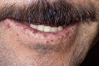
Telangiectasias, also known as spider veins, are small dilated blood vessels that can occur near the surface of the skin or mucous membranes, measuring between 0.5 and 1 millimeter in diameter. These dilated blood vessels can develop anywhere on the body, but are commonly seen on the face around the nose, cheeks and chin. Dilated blood vessels can also develop on the legs, although when they occur on the legs, they often have underlying venous reflux or "hidden varicose veins". When found on the legs, they are found specifically on the upper thigh, below the knee joint and around the ankles.

Macroglossia is the medical term for an unusually large tongue. Severe enlargement of the tongue can cause cosmetic and functional difficulties in speaking, eating, swallowing and sleeping. Macroglossia is uncommon, and usually occurs in children. There are many causes. Treatment depends upon the exact cause.

Sclerotherapy is a procedure used to treat blood vessel malformations and also malformations of the lymphatic system. A medicine is injected into the vessels, which makes them shrink. It is used for children and young adults with vascular or lymphatic malformations. In adults, sclerotherapy is often used to treat spider veins, smaller varicose veins, hemorrhoids, and hydroceles.

Lymphangiomas are malformations of the lymphatic system characterized by lesions that are thin-walled cysts; these cysts can be macroscopic, as in a cystic hygroma, or microscopic. The lymphatic system is the network of vessels responsible for returning to the venous system excess fluid from tissues as well as the lymph nodes that filter this fluid for signs of pathogens. These malformations can occur at any age and may involve any part of the body, but 90% occur in children less than 2 years of age and involve the head and neck. These malformations are either congenital or acquired. Congenital lymphangiomas are often associated with chromosomal abnormalities such as Turner syndrome, although they can also exist in isolation. Lymphangiomas are commonly diagnosed before birth using fetal ultrasonography. Acquired lymphangiomas may result from trauma, inflammation, or lymphatic obstruction.

William Bennett Bean was an internist, medical historian, teacher and collector.

Angiokeratoma is a benign cutaneous lesion of capillaries, resulting in small marks of red to blue color and characterized by hyperkeratosis. Angiokeratoma corporis diffusum refers to Fabry's disease, but this is usually considered a distinct condition.
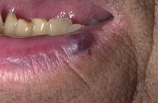
A venous lake is a generally solitary, soft, compressible, dark blue to violaceous, 0.2- to 1-cm papule commonly found on sun-exposed surfaces of the vermilion border of the lip, face and ears. Lesions generally occur among the elderly.
A vascular malformation is a blood vessel or lymph vessel abnormality. Vascular malformations are one of the classifications of vascular anomalies, the other grouping is vascular tumors. They may cause aesthetic problems as they have a growth cycle, and can continue to grow throughout life.

Caviar tongue is a condition characterized by the purplish nodular swelling of veins found on the undersurface of the tongue.
A Unilateral palmoplantar verrucous nevus is a cutaneous condition that has features of pachyonychia congenita.
Blue rubber bleb nevus syndrome is a rare disorder that consists mainly of abnormal blood vessels affecting the skin or internal organs – usually the gastrointestinal tract. The disease is characterized by the presence of fluid-filled blisters (blebs) as visible, circumscribed, chronic lesions (nevi).
A vascular anomaly is any of a range of lesions from a simple birthmark to a large tumor that may be disfiguring. They are caused by a disorder of the vascular system. A vascular anomaly is a localized defect in blood or lymph vessels. These defects are characterized by an increased number of vessels, and vessels that are both enlarged and sinuous. Some vascular anomalies are congenital, others appear within weeks to years after birth, and others are acquired by trauma or during pregnancy. Inherited vascular anomalies are also described and often present with a number of lesions that increase with age. Vascular anomalies can also be a part of a syndrome.

Parkes Weber syndrome (PWS) is a congenital disorder of the vascular system. It is an extremely rare condition, and its exact prevalence is unknown. It is named after British dermatologist Frederick Parkes Weber, who first described the syndrome in 1907.

Macrocephaly-capillary malformation (M-CM) is a multiple malformation syndrome causing abnormal body and head overgrowth and cutaneous, vascular, neurologic, and limb abnormalities. Though not every patient has all features, commonly found signs include macrocephaly, congenital macrosomia, extensive cutaneous capillary malformation, body asymmetry, polydactyly or syndactyly of the hands and feet, lax joints, doughy skin, variable developmental delay and other neurologic problems such as seizures and low muscle tone.
Hypotrichosis–lymphedema–telangiectasia syndrome is a congenital syndrome characterized by lymphedema, the presence of telegiectasias, and hypotrichosis or alopecia. Lymphedema usually develops in the lower extremities during puberty. Hair is normal at birth, but usually lost during infancy. Telangiectasias may present on the palms and soles more commonly than on the scalp, legs, and genitalia. The syndrome has been reported in association with both autosomal dominant and autosomal recessive inheritance patterns.
Victor Joel Selmanowitz was an American dermatologist. In 1970, he coined the term unilateral nevoid telangiectasia. The Victor J. Selmanowitz Memorial Lecture and chair of modern Jewish history at the Touro College Graduate School of Jewish Studies is named for him.
References
- 1 2 James, William D.; Elston, Dirk; Treat, James R.; Rosenbach, Misha A.; Neuhaus, Isaac (2020). "28. Dermal and subcutaneous tumors: Unilateral nevoid telangiectasia". Andrews' Diseases of the Skin: Clinical Dermatology (13th ed.). Edinburgh: Elsevier. pp. 604–605. ISBN 978-0-323-54753-6.
- ↑ Mulliken, John B. (2013). "13. Capillary malformations, hyperkeratotic stains, telangiectasias, and miscellaneous vascular blots". In Mulliken, John B.; Burrows, Patricia E.; Fishman, Steven J. (eds.). Mulliken and Young's Vascular Anomalies: Hemangiomas and Malformations (2nd ed.). Oxford University Press. p. 544. ISBN 978-0-19-972254-9.