Visual masking

Last updated
Visual masking involves surrounding a target image (here, the word "radio") with another image. Visual Masking.svg
Visual masking involves surrounding a target image (here, the word "radio") with another image.

Visual masking is a phenomenon of visual perception. It occurs when the visibility of one image, called a target, is reduced by the presence of another image, called a mask. [1] The target might be invisible or appear to have reduced contrast or lightness. There are three different timing arrangements for masking: forward masking, backward masking, and simultaneous masking. In forward masking, the mask precedes the target. In backward masking the mask follows the target. In simultaneous masking, the mask and target are shown together. There are two different spatial arrangements for masking: pattern masking and metacontrast. Pattern masking occurs when the target and mask locations overlap. Metacontrast masking occurs when the mask does not overlap with the target location.

Contents

Factors affecting visual masking

Target-to-mask spatial separation

Suppression can be seen in both forward and backward masking when there is pattern masking, but not when there is metacontrast. Simultaneous masking, however, will produce facilitation of target visibility during pattern masking. Facilitation also comes about when metacontrast is combined with either simultaneous or forward masking. [2] This is because it takes time for the mask to reach the target's location through lateral propagation. As the target gets further from the mask, the time required for lateral propagation increases. Thus, the masking effect will increase as the mask gets closer to the target.

Target-to-mask temporal separation

As the time difference between the target and the mask increases, the masking effect decreases. This is because the integration time of a target stimulus has an upper limit 200 ms, based on physiological experiments [3] [4] [5] and as the separation approaches this limit, the mask is able to produce less of an effect on the target, as the target has had more time to form a full neural representation in the brain. Polat, Sterkin, and Yehezkel [2] went into great detail in explaining the effect of temporal matching between target input and lateral propagation of the mask. Based on data from previous single-unit recordings, they concluded that the time window for any sort of efficient interaction with target processing is 210 to 310 ms after the target's appearance. Anything outside of this window would fail to cause any sort of masking effect. This explains why there is a masking effect when the mask is presented 50 ms after the target, but not when the inter-stimulus interval between mask and target is 150 ms. In the first case, mask response would propagate to the target location and be processed with a delay of 260 to 310 ms, whereas the ISI of 150 would result in a delay of 410 to 460 ms.

Monoptic vs. dichoptic visual masking

In dichoptic visual masking, the target is presented to one eye and the mask to the other, whereas in monoptic visual masking, both eyes are presented with the target and the mask. It was found that the masking effect was just as strong in dichoptic as it was in monoptic masking, and that it showed the same timing characteristics. [6] [7] [8]

Possible neural correlates

There are multiple theories surrounding the neural correlates of masking, but most of them agree on a few key ideas. First, backward visual masking comes about from suppression of the target's “after-discharge”, [9] where the after-discharge can be thought of as the neural response to the target's termination. Impairments in backward masking have been consistently found in those with schizophrenia [10] as well as in their unaffected siblings, [11] [12] thus suggesting that the impairments might be an endophenotype for schizophrenia. [13]

Forward masking, on the other hand, is correlated to the suppression of the target's “onset-response”, [9] which can be thought of as the neural response to the target's appearance.

Two-channel model

Originally proposed by Breitmeyer and Ganz in 1976, [14] the original version of this model stated that there were two different visual information channels- one being fast and transient, the other being slow and sustained. The theory asserts that each stimulus travels up each channel, and both channels are necessary for proper and full processing of any given stimulus. It explained backward masking by saying that the neural representation of the mask would travel up the transient channel and intercept the neural representation of the target as it travelled up the slower channel, suppressing the target's representation and decreasing its visibility. One problem with this model, as proposed by Macknik and Martinez-Conde, [9] is that it predicts masking to occur as a function of how far apart, temporally, the stimulus onset is. However, Macknik and Martinez-Conde showed that backward masking is actually more dependent on how far apart stimulus termination is.

Retino-cortical dynamics model

Breitmeyer and Ögmen modified the two-channel model in 2006, [15] renaming it to the retino-cortical dynamics (RECOD) model in the process. Their main proposed modification was that the fast and slow channels were actually feed forward and feedback channels, instead of the magnocellular and parvocellular retino-geniculocortical pathways, which is what had previously been proposed. Thus, according to this new model, backward masking is caused when feed forward input from the mask interferes with the feedback coming from the higher visual areas’ response to the target, thus reducing visibility.

Lamme's recurrent feedback hypothesis of visual awareness and masking

This model proposes that backward masking is caused by an interference with feedback from higher visual areas. [16] In this model, target duration is irrelevant because masking is supposed to occur as a function of feedback, which is generated when the target appears on screen. Lamme's group further supported their model when they described that the surgical removal of the extrastriate cortex in monkeys leads to a reduction of area V1 late responses. [17]

Lateral inhibition circuits

Proposed by Macknik and Martinez-Conde in 2008, [9] this theory proposes that masking can be explained almost entirely by feed forward lateral inhibition circuits. The idea is that the edges of the mask, if positioned in close proximity to the target, may inhibit the responses caused by the edges of the target, inhibiting perception of the target.

Coupled interactions between V1 and fusiform gyrus

Haynes, Driver, and Rees proposed this theory in 2005, [18] stating that visibility derives from the feed forward and feedback interactions between the V1 and fusiform gyrus. In their experiment, they required subjects to attend actively to the target- thus, as Macknik and Martinez-Conde point out, [9] it is possible that their results were confounded by the attentional aspect of the trials, and that the results may not accurately reflect the effects of visual masking.

Frontal lobe processing of visual masking

This was proposed by Thompson and Schall, based on experiments conducted in 1999 [19] and 2000. [20] They concluded that visual masking is processed in the frontal-eye fields, and that the neural correlate of masking lies not in the inhibition of the response to the target but in the “merging” of target and mask responses. One criticism of their experiment, however, is that their target was almost 300x dimmer than the mask, so their results may have been confounded by the different response latencies one would expect from stimuli with such differences in brightness.

Evidence from monoptic and dichoptic visual masking

Macknik & Martinez-Conde [21] recorded from neurons in the lateral geniculate nucleus (LGN) and V1 V1 while presenting monoptic and dichoptic stimuli, and found that monoptic masking occurred in all the LGN and V1 neurons that were recorded, but dichoptic masking only occurred in some of the binocular neurons in V1, which supports the hypothesis that visual masking in monoptic regions is not due to feedback from dichoptic regions. This is because, if there had been feedback from higher areas of the visual field, the early circuits would have “inherited” dichoptic masking from the feedback coming from higher levels, and so would exhibit both dichoptic and monoptic masking. Although monoptic masking is stronger in the early visual areas, monoptic and dichoptic masking are equivalent in magnitude. Thus, dichoptic masking must become stronger as it proceeds down the visual hierarchy if the preceding hypothesis is correct. In fact, dichoptic masking was shown to begin downstream of area V2. [9]

See also

Related Research Articles

<span class="mw-page-title-main">Binocular rivalry</span>

Binocular rivalry is a phenomenon of visual perception in which perception alternates between different images presented to each eye.

Neural adaptation or sensory adaptation is a gradual decrease over time in the responsiveness of the sensory system to a constant stimulus. It is usually experienced as a change in the stimulus. For example, if a hand is rested on a table, the table's surface is immediately felt against the skin. Subsequently, however, the sensation of the table surface against the skin gradually diminishes until it is virtually unnoticeable. The sensory neurons that initially respond are no longer stimulated to respond; this is an example of neural adaptation.

Microsaccades are a kind of fixational eye movement. They are small, jerk-like, involuntary eye movements, similar to miniature versions of voluntary saccades. They typically occur during prolonged visual fixation, not only in humans, but also in animals with foveal vision. Microsaccade amplitudes vary from 2 to 120 arcminutes. The first empirical evidence for their existence was provided by Robert Darwin, the father of Charles Darwin.

The concept of backward masking originated in psychoacoustics, referring to temporal masking of quiet sounds that occur moments before a louder sound.

<span class="mw-page-title-main">Filling-in</span>

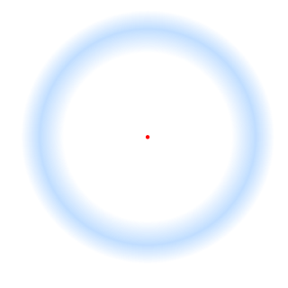
In vision, filling-in phenomena are those responsible for the completion of missing information across the physiological blind spot, and across natural and artificial scotomata. There is also evidence for similar mechanisms of completion in normal visual analysis. Classical demonstrations of perceptual filling-in involve filling in at the blind spot in monocular vision, and images stabilized on the retina either by means of special lenses, or under certain conditions of steady fixation. For example, naturally in monocular vision at the physiological blind spot, the percept is not a hole in the visual field, but the content is “filled-in” based on information from the surrounding visual field. When a textured stimulus is presented centered on but extending beyond the region of the blind spot, a continuous texture is perceived. This partially inferred percept is paradoxically considered more reliable than a percept based on external input..

Neural coding is a neuroscience field concerned with characterising the hypothetical relationship between the stimulus and the individual or ensemble neuronal responses and the relationship among the electrical activity of the neurons in the ensemble. Based on the theory that sensory and other information is represented in the brain by networks of neurons, it is thought that neurons can encode both digital and analog information.

<span class="mw-page-title-main">Chubb illusion</span> Optical illusion

The Chubb illusion is an optical illusion or error in visual perception in which the apparent contrast of an object varies substantially to most viewers depending on its relative contrast to the field on which it is displayed. These visual illusions are of particular interest to researchers because they may provide valuable insights in regard to the workings of human visual systems.

Repetition priming refers to improvements in a behavioural response when stimuli are repeatedly presented. The improvements can be measured in terms of accuracy or reaction time, and can occur when the repeated stimuli are either identical or similar to previous stimuli. These improvements have been shown to be cumulative, so as the number of repetitions increases the responses get continually faster up to a maximum of around seven repetitions. These improvements are also found when the repeated items are changed slightly in terms of orientation, size and position. The size of the effect is also modulated by the length of time the item is presented for and the length time between the first and subsequent presentations of the repeated items.

<span class="mw-page-title-main">Fixation (visual)</span> Maintaining ones gaze on a single location

Fixation or visual fixation is the maintaining of the gaze on a single location. An animal can exhibit visual fixation if it possess a fovea in the anatomy of their eye. The fovea is typically located at the center of the retina and is the point of clearest vision. The species in which fixational eye movement has been verified thus far include humans, primates, cats, rabbits, turtles, salamanders, and owls. Regular eye movement alternates between saccades and visual fixations, the notable exception being in smooth pursuit, controlled by a different neural substrate that appears to have developed for hunting prey. The term "fixation" can either be used to refer to the point in time and space of focus or the act of fixating. Fixation, in the act of fixating, is the point between any two saccades, during which the eyes are relatively stationary and virtually all visual input occurs. In the absence of retinal jitter, a laboratory condition known as retinal stabilization, perceptions tend to rapidly fade away. To maintain visibility, the nervous system carries out a procedure called fixational eye movement, which continuously stimulates neurons in the early visual areas of the brain responding to transient stimuli. There are three categories of fixational eye movement: microsaccades, ocular drifts, and ocular microtremor. At small amplitudes the boundaries between categories become unclear, particularly between drift and tremor.

Echoic memory is the sensory memory that registers specific to auditory information (sounds). Once an auditory stimulus is heard, it is stored in memory so that it can be processed and understood. Unlike most visual memory, where a person can choose how long to view the stimulus and can reassess it repeatedly, auditory stimuli are usually transient and cannot be reassessed. Since echoic memories are heard once, they are stored for slightly longer periods of time than iconic memories. Auditory stimuli are received by the ear one at a time before they can be processed and understood.

The Mauthner cells are a pair of big and easily identifiable neurons located in the rhombomere 4 of the hindbrain in fish and amphibians that are responsible for a very fast escape reflex. The cells are also notable for their unusual use of both chemical and electrical synapses.

Feature detection is a process by which the nervous system sorts or filters complex natural stimuli in order to extract behaviorally relevant cues that have a high probability of being associated with important objects or organisms in their environment, as opposed to irrelevant background or noise.

In the psychology of perception and motor control, the term response priming denotes a special form of priming. Generally, priming effects take place whenever a response to a target stimulus is influenced by a prime stimulus presented at an earlier time. The distinctive feature of response priming is that prime and target are presented in quick succession and are coupled to identical or alternative motor responses. When a speeded motor response is performed to classify the target stimulus, a prime immediately preceding the target can thus induce response conflicts when assigned to a different response as the target. These response conflicts have observable effects on motor behavior, leading to priming effects, e.g., in response times and error rates. A special property of response priming is its independence from visual awareness of the prime.

Neural decoding is a neuroscience field concerned with the hypothetical reconstruction of sensory and other stimuli from information that has already been encoded and represented in the brain by networks of neurons. Reconstruction refers to the ability of the researcher to predict what sensory stimuli the subject is receiving based purely on neuron action potentials. Therefore, the main goal of neural decoding is to characterize how the electrical activity of neurons elicit activity and responses in the brain.

Subliminal stimuli are any sensory stimuli below an individual's threshold for conscious perception, in contrast to supraliminal stimuli. A 2012 review of functional magnetic resonance imaging (fMRI) studies showed that subliminal stimuli activate specific regions of the brain despite participants' unawareness. Visual stimuli may be quickly flashed before an individual can process them, or flashed and then masked to interrupt processing. Audio stimuli may be played below audible volumes or masked by other stimuli.

<span class="mw-page-title-main">Visual processing abnormalities in schizophrenia</span>

Visual processing abnormalities in schizophrenia are commonly found, and contribute to poor social function.

Many experiments have been done to find out how the brain interprets stimuli and how animals develop fear responses. The emotion, fear, has been hard-wired into almost every individual, due to its vital role in the survival of the individual. Researchers have found that fear is established unconsciously and that the amygdala is involved with fear conditioning.

Surround suppression is where the relative firing rate of a neuron may under certain conditions decrease when a particular stimulus is enlarged. It has been observed in electrophysiology studies of the brain and has been noted in many sensory neurons, most notably in the early visual system. Surround suppression is defined as a reduction in the activity of a neuron in response to a stimulus outside its classical receptive field.

<span class="mw-page-title-main">Stephen Macknik</span> American neuroscientist and writer

Stephen Louis Macknik is an American neuroscientist and science writer. He is a Professor of Ophthalmology, Neurology, and Physiology & Pharmacology at the State University of New York, Downstate Medical Center, where he directs the Laboratory of Translational Neuroscience. He directed laboratories previously at the Barrow Neurological Institute and University College London. He is best known for his studies on illusions, consciousness, attentional misdirection in stage magic, and cerebral blood flow.

Binocular switch suppression (BSS) is a technique to suppress usually salient images from an individual's awareness, a type of experimental manipulation used in visual perception and cognitive neuroscience. In BSS, two images of differing signal strengths are repetitively switched between the left and right eye at a constant rate of 1 Hertz. During this process of switching, the image of lower contrast and signal strength is perceptually suppressed for a period of time.

References

  1. Ogmen H, Breitmeyer B (2007). "Visual masking". Scholarpedia. 2 (7): 3330. Bibcode:2007SchpJ...2.3330B. doi: 10.4249/scholarpedia.3330 .
  2. 1 2 Polat U, Sterkin A, Yehezkel O (July 2008). "Spatio-temporal low-level neural networks account for visual masking". Advances in Cognitive Psychology. 3 (1–2): 153–65. doi:10.2478/v10053-008-0021-4. PMC   2864984 . PMID   20517505.
  3. Albrecht DG (1995). "Visual cortex neurons in monkey and cat: effect of contrast on the spatial and temporal phase transfer functions". Visual Neuroscience. 12 (6): 1191–210. doi:10.1017/s0952523800006817. PMID   8962836. S2CID   14072147.
  4. Mizobe K, Polat U, Pettet MW, Kasamatsu T (2001). "Facilitation and suppression of single striate-cell activity by spatially discrete pattern stimuli presented beyond the receptive field". Visual Neuroscience. 18 (3): 377–91. doi:10.1017/s0952523801183045. PMID   11497414. S2CID   12541180.
  5. Polat U, Mizobe K, Pettet MW, Kasamatsu T, Norcia AM (February 1998). "Collinear stimuli regulate visual responses depending on cell's contrast threshold". Nature. 391 (6667): 580–4. Bibcode:1998Natur.391..580P. doi:10.1038/35372. PMID   9468134. S2CID   205024244.
  6. Crawford BH (March 1947). "Visual adaptation in relation to brief conditioning stimuli". Proceedings of the Royal Society of London. Series B, Biological Sciences. 134 (875): 283–302. Bibcode:1947RSPSB.134..283C. doi:10.1098/rspb.1947.0015. PMID   20292379.
  7. Macknik SL, Livingstone MS (June 1998). "Neuronal correlates of visibility and invisibility in the primate visual system". Nature Neuroscience. 1 (2): 144–9. doi:10.1038/393. PMID   10195130. S2CID   10520581.
  8. Macknik SL, Martinez-Conde S, Haglund MM (June 2000). "The role of spatiotemporal edges in visibility and visual masking". Proceedings of the National Academy of Sciences of the United States of America. 97 (13): 7556–60. Bibcode:2000PNAS...97.7556M. doi: 10.1073/pnas.110142097 . PMC   16584 . PMID   10852945.
  9. 1 2 3 4 5 6 Macknik SL, Martinez-Conde S (July 2008). "The role of feedback in visual masking and visual processing". Advances in Cognitive Psychology. 3 (1–2): 125–52. doi:10.2478/v10053-008-0020-5. PMC   2864985 . PMID   20517504.
  10. Green MF, Horan WP, Lee J (June 2019). "Nonsocial and social cognition in schizophrenia: current evidence and future directions". World Psychiatry. 18 (2): 146–161. doi:10.1002/wps.20624. PMC   6502429 . PMID   31059632.
  11. da Cruz JR, Shaqiri A, Roinishvili M, Favrod O, Chkonia E, Brand A, et al. (January 2020). "Neural Compensation Mechanisms of Siblings of Schizophrenia Patients as Revealed by High-Density EEG". Schizophrenia Bulletin. 46 (4): 1009–1018. doi:10.1093/schbul/sbz133. PMC   7345810 . PMID   31961928.
  12. Chkonia E, Roinishvili M, Makhatadze N, Tsverava L, Stroux A, Neumann K, et al. (December 2010). "The shine-through masking paradigm is a potential endophenotype of schizophrenia". PLOS ONE. 5 (12): e14268. Bibcode:2010PLoSO...514268C. doi: 10.1371/journal.pone.0014268 . PMC   3000331 . PMID   21151559.
  13. Green MF, Lee J, Wynn JK, Mathis KI (July 2011). "Visual masking in schizophrenia: overview and theoretical implications". Schizophrenia Bulletin. 37 (4): 700–8. doi:10.1093/schbul/sbr051. PMC   3122285 . PMID   21606322.
  14. Breitmeyer BG, Ganz L (January 1976). "Implications of sustained and transient channels for theories of visual pattern masking, saccadic suppression, and information processing". Psychological Review. 83 (1): 1–36. doi:10.1037/0033-295x.83.1.1. PMID   766038.
  15. Breitmeyer B, Öğmen H (2006). Visual Masking: Time slices through conscious and unconscious vision (2nd ed.). Oxford, UK: Oxford University Press.
  16. Lamme VA, Zipser K, Spekreijse H (October 2002). "Masking interrupts figure-ground signals in V1" (PDF). Journal of Cognitive Neuroscience. 14 (7): 1044–53. doi:10.1162/089892902320474490. PMID   12419127. S2CID   1975279.
  17. Lamme VA, Zipser K, Spekreijse H (1997). "Figure-ground signals in V1 depend on extrastriate feedback". Investigative Ophthalmology & Visual Science. 38 (4): S969.
  18. Haynes JD, Driver J, Rees G (June 2005). "Visibility reflects dynamic changes of effective connectivity between V1 and fusiform cortex". Neuron. 46 (5): 811–21. doi: 10.1016/j.neuron.2005.05.012 . PMID   15924866. S2CID   6543247.
  19. Thompson KG, Schall JD (March 1999). "The detection of visual signals by macaque frontal eye field during masking". Nature Neuroscience. 2 (3): 283–8. doi:10.1038/6398. PMID   10195223. S2CID   7085444.
  20. Thompson KG, Schall JD (2000). "Antecedents and correlates of visual detection and awareness in macaque prefrontal cortex". Vision Research. 40 (10–12): 1523–38. doi:10.1016/s0042-6989(99)00250-3. PMID   10788656. S2CID   412043.
  21. Macknik SL, Martinez-Conde S (2004). "The spatial and temporal effects of lateral inhibitory networks and their relevance to the visibility of spatiotemporal edges". Neurocomputing. 58–60: 775–782. doi:10.1016/j.neucom.2004.01.126.