Whisking in animals

Last updated

Rats using their mystacial vibrissae to perform whisking Rat noses.jpg
Rats using their mystacial vibrissae to perform whisking

Whisking is a behaviour in which the facial whiskers (vibrissae) of an animal are repetitively and rapidly swept back and forth. This behaviour occurs particularly during locomotion and exploration. The whisking movements occur in bouts of variable duration, and at rates between 3 and 25 whisks/second. Movements of the whiskers are closely co-ordinated with those of the head and body, allowing the animal to locate interesting stimuli through whisker contact, then investigate them further using both the macrovibrissae and an array of shorter, non-actuated microvibrissae on the chin and lips. Whisking has been reported in a wide range of mammals, including two species of marsupial. Whisking contributes both to exploratory movements, which function to acquire sensory inputs, and to palpation movements, which are used in the discrimination of objects and in the control of spatial navigation. [1]

Contents

Types

There are three types of whisking. Exploratory whisking occurs when animals whisk in air without contact or with only light contact. In this case, the stroke on a given whisk approaches 70°, and a total field of up to 160° may be covered as the animal slowly shifts the set point of its whisk. Such whisking is extremely regular and typically occurs with a frequency between 7 and 12 Hz and a mean of 9 Hz. Asymmetric whisking occurs when animals make contact with a large object, such as a wall, while they whisk. It also occurs if they turn their head to the side while whisking. Asymmetric whisking lasts for only one to three whisk cycles. Foveal whisking occurs when animals thrust all their vibrissae forward to palpate an object ahead of them, as occurs when they try to detect a landing on the far side of a gap. In this case the stroke is much reduced, typically to 20°. The frequency of foveal whisking is high, ranging between 15 and 25 Hz, and rats can readily switch between foveal and exploratory whisking. [2]

Facial vibrissae

Many land and marine mammals possess facial vibrissae (derived from the Latin "vibrio" meaning to vibrate). For example, rats [3] and hamsters, [4] have an arrangement of cranial (of the skull) vibrissae that includes the supraorbital (above the eyes), genal (of the cheeks), and mystacial (where a moustache would be) vibrissae, as well as mandibular (of the jaw) vibrissae under the snout. [5] Mystacial vibrissae are generally described as being further divided into two sub-groups: the large macrovibrissae that protrude to the sides, and the small microvibrissae below the nostrils that mostly point downwards. [6] The macrovibrissae are generally large, motile and used for spatial sensing, whereas microvibrissae are small, immotile and used for object identification.

Musculature and nervous system

Generally, the supraorbital, genal and macrovibrissae are reported to be motile, [4] whilst the microvibrissae are not. This is reflected in anatomical reports that have identified musculature associated with the macrovibrissae that is absent for the microvibrissae. [7]

The effector system generating whisking comprises a set of “extrinsic” muscles controlling movements of the mystacial pad and a group of “intrinsic” (follicular) muscles, producing vibrissa protraction. The extrinsic muscles in the mystacial pad move many or all of the macrovibrissae together. [7] [8] However, the individual follicles of some groups of facial vibrissae in some species are also motile. A small muscle 'sling' is attached to each macrovibrissa and can move it more-or-less independently of the others. Vibrissa retraction is thought to be a passive process produced by rebound of stretched follicular muscle. Whisking movements are amongst the fastest produced by mammals[ citation needed ]. In animals that are capable of whisking at high frequencies, the whisking musculature contains a high proportion of type 2B muscle fibers that can support faster contractions than normal skeletal muscles. [9]

Sensory innervation of the whiskers is provided by the infraorbital branch of the trigeminal maxillary nerve; motor innervation is attributable to the facial (VII) nerve. [1]

Species that whisk

Amongst those species with motile macrovibrissae, some (rats, mice, flying squirrels, gerbils, chinchillas, hamsters, shrews, porcupines, opossums) [10] perform whisking. [11] Although whisking is prominent in rodents, there are several rodent genera, such as capybara and gophers, that do not appear to whisk, and others, such as guinea pigs, that display only irregular and relatively short whisking bouts [9] Whisking behaviour has not been observed in carnivores (e.g. cats, dogs, raccoons, bears), [10] although some species, such as pinnipeds, have well-developed sinus muscles making the whiskers highly motile. [12]

Control and co-ordination

In rats, whisking movements occur in bouts of variable duration at rates between 3 and 25 whisks/second. [10] The movements have an amplitude over a range from ∼10 to 100°, at an average protraction velocity of ∼1000°/sec, and at a predominant frequency of 5–7 Hz. [1] Three types of whisk are described, single, delayed (there is an inflection point somewhere in the whisk's velocity) or double-pumped (slight retraction in the middle of the whisk followed by protraction to complete the whisk cycle). [13]

Whisking movements relative to the head are described by 3 angles of rotation (azimuth, ; elevation, ; and torsion, ) and translations of the whisker base. Azimuthal rotation moves the whiskers back and forth along the longitudinal axis. This movement co-varies with small changes in elevation. Torsion (or, roll) refers to a rotation of a whisker about its own axis. The torsional angle is correlated with the azimuth, and alters the forward facing surface of the whisker shaft that contacts the opposing surface. Because the vibrissae are curved, torsion also displaces the whisker tips relative to the head. [14] [15]

Early studies (1964) described co-ordination between vibrissae, nose, head, and sniffing movements. From these studies it was suggested that the animal's whisking behaviour is dependent on the task and that whisking during exploratory behaviors is different from whisking during discriminative behaviours. [13]

In all whisking animals in which it has so far been measured, these whisking movements are rapidly controlled in response to behavioural and environmental conditions. Movements of the whiskers are closely co-ordinated with those of the head and body. [10]

Sniffing, a high-frequency, highly rhythmic inhalation and exhalation of air through the nose, plays an important role in rodent olfaction. Whisking is thought to be co-ordinated with sniffing and normal respiratory behaviour. It has been shown that breathing and whisking movements are correlated only when the whisking rhythm is less than 5 Hz. Only 13% of whisking movements occur during high-frequency (greater than 5 Hz) respiration typically associated with sniffing, indicating that high-frequency whisking and sniffing behaviours are not correlated. [16]

Whisking and sniffing also constitute the overt expression of an animal's anticipation of a reward. [17]

Development

In rats, whiskers grow to their adult size in the first month of life, although, rats sustaining denervation of the whisker pad grow whiskers that are thinner and smaller than those of normal adults. Whisking begins around post-natal day 11 to 13, prior to the eyes opening, and achieves adult amplitudes and characteristics by the end of the third post-natal week. Prior to the onset of whisking, neonatal rats show behavioural activation in response to whisker stimulation, and tactile learning in a classical conditioning avoidance paradigm, but are not able to orient to the stimulus source. Micro-movements of the vibrissae in the first ten days of life have also been observed. [10]

Related Research Articles

Rat Several genera of rodents

Rats are various medium-sized, long-tailed rodents. Species of rats are found throughout the order Rodentia, but stereotypical rats are found in the genus Rattus. Other rat genera include Neotoma, Bandicota and Dipodomys.

Saccade Quick, simultaneous movement of both eyes between two or more phases of fixation in the same direction

A saccade is a quick, simultaneous movement of both eyes between two or more phases of fixation in the same direction. In contrast, in smooth pursuit movements, the eyes move smoothly instead of in jumps. The phenomenon can be associated with a shift in frequency of an emitted signal or a movement of a body part or device. Controlled cortically by the frontal eye fields (FEF), or subcortically by the superior colliculus, saccades serve as a mechanism for fixation, rapid eye movement, and the fast phase of optokinetic nystagmus. The word appears to have been coined in the 1880s by French ophthalmologist Émile Javal, who used a mirror on one side of a page to observe eye movement in silent reading, and found that it involves a succession of discontinuous individual movements.

Whiskers type of mammalian hair used for sensing

Whiskers or vibrissae are a type of mammalian hair that are typically characterised, anatomically, by their long length, large and well-innervated hair follicle, and by having an identifiable representation in the somatosensory cortex of the brain.

Claustrum structure in the brain, gray matter lamina located underneath the inner neocortex

The claustrum is a thin, bilateral structure that connects to cortical and subcortical regions of the brain. It is located between the insula laterally and the putamen medially, separated by the extreme and external capsules respectively. The blood supply to the claustrum is fulfilled via the middle cerebral artery. It is considered to be the most densely connected structure in the brain, allowing for integration of various cortical inputs into one experience rather than singular events. The claustrum is difficult to study given the limited number of individuals with claustral lesions and the poor resolution of neuroimaging.

Motor cortex Region of the cerebral cortex

The motor cortex is the region of the cerebral cortex involved in the planning, control, and execution of voluntary movements. Classically, the motor cortex is an area of the frontal lobe located in the posterior precentral gyrus immediately anterior to the central sulcus.

In physiology, tonotopy is the spatial arrangement of where sounds of different frequency are processed in the brain. Tones close to each other in terms of frequency are represented in topologically neighbouring regions in the brain. Tonotopic maps are a particular case of topographic organization, similar to retinotopy in the visual system.

Barrel cortex

The barrel cortex is a region of the somatosensory cortex that is identifiable in some species of rodents and species of at least two other orders and contains the barrel field. The 'barrels' of the barrel field are regions within cortical layer IV that are visibly darker when stained to reveal the presence of cytochrome c oxidase and are separated from each other by lighter areas called septa. These dark-staining regions are a major target for somatosensory inputs from the thalamus, and each barrel corresponds to a region of the body. Due to this distinctive cellular structure, organisation, and functional significance, the barrel cortex is a useful tool to understand cortical processing and has played an important role in neuroscience. The majority of what is known about corticothalamic processing comes from studying the barrel cortex, and researchers have intensively studied the barrel cortex as a model of neocortical column.

Stria terminalis Band of fibres along the thalamus

The stria terminalis is a structure in the brain consisting of a band of fibers running along the lateral margin of the ventricular surface of the thalamus. Serving as a major output pathway of the amygdala, the stria terminalis runs from its centromedial division to the ventromedial nucleus of the hypothalamus.

Head direction (HD) cells are neurons found in a number of brain regions that increase their firing rates above baseline levels only when the animal's head points in a specific direction. They have been reported in rats, monkeys, mice, chinchillas and bats, but are thought to be common to all mammals, perhaps all vertebrates and perhaps even some invertebrates, and to underlie the "sense of direction". When the animal's head is facing in the cell's "preferred firing direction" these neurons fire at a steady rate, but firing decreases back to baseline rates as the animal's head turns away from the preferred direction.

Theta waves generate the theta rhythm, a neural oscillatory pattern that can be seen on an electroencephalogram (EEG), recorded either from inside the brain or from electrodes attached to the scalp. Two types of theta rhythm have been described. The hippocampal theta rhythm is a strong oscillation that can be observed in the hippocampus and other brain structures in numerous species of mammals including rodents, rabbits, dogs, cats, bats, and marsupials. "Cortical theta rhythms" are low-frequency components of scalp EEG, usually recorded from humans. Theta rhythms can be quantified using quantitative electroencephalography (qEEG) using freely available toolboxes, such as, EEGLAB or the Neurophysiological Biomarker Toolbox (NBT).

Frontal eye fields

The frontal eye fields (FEF) are a region located in the frontal cortex, more specifically in Brodmann area 8 or BA8, of the primate brain. In humans, it can be more accurately said to lie in a region around the intersection of the middle frontal gyrus with the precentral gyrus, consisting of a frontal and parietal portion. The FEF is responsible for saccadic eye movements for the purpose of visual field perception and awareness, as well as for voluntary eye movement. The FEF communicates with extraocular muscles indirectly via the paramedian pontine reticular formation. Destruction of the FEF causes deviation of the eyes to the ipsilateral side.

Cat senses The ways in which cats experience the world. (Smell, Sight, Touch, Taste, and Hearing)

Cat senses are adaptations that allow cats to be highly efficient predators. Cats are good at detecting movement in low light, have an acute sense of hearing and smell, and their sense of touch is enhanced by long whiskers that protrude from their heads and bodies. These senses evolved to allow cats to hunt effectively at night.

Tawny-bellied cotton rat species of mammal

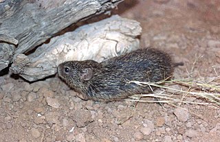
The tawny-bellied cotton rat is a species of rodent in the family Cricetidae. It is found in Mexico and in the US states of Arizona and New Mexico.

Primary motor cortex brain region

The primary motor cortex is a brain region that in humans is located in the dorsal portion of the frontal lobe. It is the primary region of the motor system and works in association with other motor areas including premotor cortex, the supplementary motor area, posterior parietal cortex, and several subcortical brain regions, to plan and execute movements. Primary motor cortex is defined anatomically as the region of cortex that contains large neurons known as Betz cells. Betz cells, along with other cortical neurons, send long axons down the spinal cord to synapse onto the interneuron circuitry of the spinal cord and also directly onto the alpha motor neurons in the spinal cord which connect to the muscles.

Levodopa-induced dyskinesia (LID) is a form of dyskinesia associated with levodopa (l-DOPA), used to treat Parkinson's disease. It often involves hyperkinetic movements, including chorea, dystonia, and athetosis.

Hydrodynamic reception

Hydrodynamic reception refers to the ability of some animals to sense water movements generated by biotic or abiotic sources. This form of mechanoreception is useful for orientation, hunting, predator avoidance, and schooling. Frequent encounters with conditions of low visibility can prevent vision from being a reliable information source for navigation and sensing objects or organisms in the environment. Sensing water movements is one resolution to this problem.

Sniffing is a perceptually-relevant behavior, defined as the active sampling of odors through the nasal cavity for the purpose of information acquisition. This behavior, displayed by all terrestrial vertebrates, is typically identified based upon changes in respiratory frequency and/or amplitude, and is often studied in the context of odor guided behaviors and olfactory perceptual tasks. Sniffing is quantified by measuring intra-nasal pressure or flow or air or, while less accurate, through a strain gauge on the chest to measure total respiratory volume. Strategies for sniffing behavior vary depending upon the animal, with small animals displaying sniffing frequencies ranging from 4 to 12 Hz but larger animals (humans) sniffing at much lower frequencies, usually less than 2 Hz. Subserving sniffing behaviors, evidence for an "olfactomotor" circuit in the brain exists, wherein perception or expectation of an odor can trigger brain respiratory center to allow for the modulation of sniffing frequency and amplitude and thus acquisition of odor information. Sniffing is analogous to other stimulus sampling behaviors, including visual saccades, active touch, and whisker movements in small animals. Atypical sniffing has been reported in cases of neurological disorders, especially those disorders characterized by impaired motor function and olfactory perception.

Sharp waves and ripples (SWRs) are oscillatory patterns in the mammalian brain hippocampus seen on an EEG during immobility and sleep. There are three major network oscillation patterns in the hippocampus: theta waves, SWRs and gamma waves. Gamma oscillations are found in all major brain structures, whereas theta and sharp waves are specific to the hippocampus and its neighbouring areas. SWRs are composed of large amplitude sharp waves in local field potential and associated fast field oscillations known as ripples. SWRs are shown to be involved in memory consolidation and the replay of wakefulness-acquired memory. These network oscillations are the most synchronous patterns in the brain, making them susceptible to pathological patterns such as epilepsy.

Grimace scale (animals) method of assessing the occurrence or severity of pain experienced by non-human animals

The grimace scale (GS), sometimes called the grimace score, is a method of assessing the occurrence or severity of pain experienced by non-human animals according to objective and blinded scoring of facial expressions, as is done routinely for the measurement of pain in non-verbal humans. Observers score the presence or prominence of “facial action units" (FAU), e.g. Orbital Tightening, Nose Bulge, Ear Position and Whisker Change. These are scored by observing the animal directly in real-time, or post hoc from photographs or screen-grabs from videos. The facial expression of the animals is sometimes referred to as the pain face.

Ultrasonic vocalization

Ultrasonic vocalizations (USVs) occur at frequencies ranging from approximately 20–100 kHz. They are emitted by animals such as bats and rodents, and have been extensively studied in rats and mice. As opposed to sonic vocalizations, ultrasonic vocalizations cannot be detected by the human ear. USVs serve as social signals, and are categorized according to their frequency. Different categories of USVs are elicited in response to different situations and varying affective states. The behavioural functions of USVs vary as a rat or mouse pup reaches the juvenile/adult stage of their development. The brain mechanisms behind calling behaviour have also been studied, and some studies have used pharmacological manipulation.

References

  1. 1 2 3 Gao, P; Bermejo, R; Zeigler, HP (2001). "Whisker deafferentation and rodent whisking patterns: Behavioral evidence for a central pattern generator". The Journal of Neuroscience. 21 (14): 5374–5380. doi: 10.1523/JNEUROSCI.21-14-05374.2001 . PMC   6762837 . PMID   11438614.
  2. Kleinfeld, D. (2008). "Vibrissa movement, sensation and sensorimotor control" (PDF). Retrieved September 30, 2013.
  3. Vincent, SB (1913). "The tactile hair of the white rat". The Journal of Comparative Neurology. 23 (1): 1–34. doi:10.1002/cne.900230101.
  4. 1 2 Wineski, L.E. (1983). "Movements of the cranial vibrissae in the Golden hamster (Mesocricetus auratus)". Journal of Zoology. 200 (2): 261–280. doi:10.1111/j.1469-7998.1983.tb05788.x.
  5. Thé, L; Wallace, ML; Chen, CH; Chorev, E; Brecht, M (2013). "Structure, function, and cortical representation of the rat submandibular whisker trident". The Journal of Neuroscience. 33 (11): 4815–4824. doi:10.1523/jneurosci.4770-12.2013. PMC   6619006 . PMID   23486952.
  6. Brecht, M.; Preilowski, B.; Merzenich, M.M. (1997). "Functional architecture of the mystacial vibrissae". Behavioural Brain Research. 84 (1–2): 81–97. doi:10.1016/S0166-4328(97)83328-1. PMID   9079775. S2CID   3993159.
  7. 1 2 Dorfl, J (1982). "The musculature of the mystacial vibrissae of the white mouse". Journal of Anatomy. 135 (Pt 1): 147–54. PMC   1168137 . PMID   7130049.
  8. Hill, D.N.; Bermejo, R.; Zeigler, H.P.; Kleinfeld, D. (2008). "Biomechanics of the vibrissa motor plant in rat: Rhythmic whisking consists of triphasic neuromuscular activity". The Journal of Neuroscience. 28 (13): 3438–3455. doi: 10.1523/JNEUROSCI.5008-07.2008 . PMC   6670594 . PMID   18367610.
  9. 1 2 Jin, T-E; Witzemann, V; Brecht, M (2004). "Fiber types of the intrinsic whisker muscle and whisking behavior". The Journal of Neuroscience. 24 (13): 3386–3393. doi: 10.1523/JNEUROSCI.5151-03.2004 . PMC   6730039 . PMID   15056718.
  10. 1 2 3 4 5 Prescott, TJ; Mitchinson, B; Grant, RA (2011). "Vibrissal behaviour and function". Scholarpedia. 6 (10): 6642. Bibcode:2011SchpJ...6.6642P. doi: 10.4249/scholarpedia.6642 . Retrieved September 30, 2013.
  11. "Video of rat whisking". Youtube.com. Retrieved 2013-06-24.
  12. Ahl, AS (1986). "The role of vibrissae in behavior - a status review". Veterinary Research Communications. 10 (4): 245–268. doi:10.1007/bf02213989. PMID   3526705. S2CID   8243656.
  13. 1 2 Moxon, K.A. (2008). "Natural Whisking. Focus on "Variability in Velocity Profiles During Free-Air Whisking Behavior of Unrestrained Rats"" . Retrieved October 2, 2013.
  14. Knutsen, PM; Biess, A; Ahissar, E (2008). "Vibrissal Kinematics in 3D: Tight Coupling of Azimuth, Elevation, and Torsion across Different Whisking Modes". Neuron. 59 (1): 35–42. doi: 10.1016/j.neuron.2008.05.013 . PMID   18614027.
  15. Knutsen, PM (2015). "Whisking Kinematics". Scholarpedia. 10 (3): 7280. doi: 10.4249/scholarpedia.7280 .
  16. Cao, Y; Roy, S; Sachdev, RN; Heck, DH (2012). "Dynamic correlation between whisking and breathing rhythms in mice". Journal of Neuroscience. 32 (5): 1653–9. doi:10.1523/JNEUROSCI.4395-11.2012. PMC   3567452 . PMID   22302807.
  17. Deschênes, M; Moore, J; Kleinfeld, D (2012). "Sniffing and whisking in rodents". Current Opinion in Neurobiology. 22 (2): 243–250. doi:10.1016/j.conb.2011.11.013. PMC   4934665 . PMID   22177596.