5-Oxo-eicosatetraenoic acid

Last updated
5-Oxo-eicosatetraenoic acid
5-oxo-ETE.svg
Names
Preferred IUPAC name
(6E,8Z,11Z,14Z)-5-Oxoicosa-6,8,11,14-tetraenoic acid
Other names
  • 5-Oxo-6E,8Z,11Z,14Z-eicosatetraenoate
  • 5-oxo-ETE
  • 5-oxoETE
  • 5-KETE
Identifiers
3D model (JSmol)
ChEBI
ChEMBL
ChemSpider
KEGG
PubChem CID
  • InChI=1S/C20H30O3/c1-2-3-4-5-6-7-8-9-10-11-12-13-14-16-19(21)17-15-18-20(22)23/h6-7,9-10,12-14,16H,2-5,8,11,15,17-18H2,1H3,(H,22,23)/b7-6-,10-9-,13-12-,16-14+
    Key: MEASLHGILYBXFO-XTDASVJISA-N
  • CCCCC\C=C/C\C=C/C\C=C/C=C/C(=O)CCCC(O)=O
Properties
C20H30O3
Molar mass 318.457 g·mol−1
Except where otherwise noted, data are given for materials in their standard state (at 25 °C [77 °F], 100 kPa).

5-Oxo-eicosatetraenoic acid (i.e. 5-oxo-6E,8Z,11Z,14Z-eicosatetraenoic acid; also termed 5-oxo-ETE and 5-oxoETE) is a nonclassic eicosanoid metabolite of arachidonic acid and the most potent naturally occurring member of the 5-HETE family of cell signaling agents. Like other cell signaling agents, 5-oxo-ETE is made by a cell and then feeds back to stimulate its parent cell (see Autocrine signaling) and/or exits this cell to stimulate nearby cells (see Paracrine signaling). 5-Oxo-ETE can stimulate various cell types particularly human leukocytes but possesses its highest potency and power in stimulating the human eosinophil type of leukocyte. It is therefore suggested to be formed during and to be an important contributor to the formation and progression of eosinophil-based allergic reactions; [1] [2] it is also suggested that 5-oxo-ETE contributes to the development of inflammation, cancer cell growth, and other pathological and physiological events. [1] [3]

Contents

Biochemistry and production

In the most common means for its production, cells make 5-oxo-ETE in a four step pathway that involves their stimulus-induced activation of the following pathway: a) the release of arachidonic acid (i.e. 5Z,8Z,11Z,14Z-eicosatetraenoic acid) from its storage sites in membrane phospholipids due to the activation of phospholipase A2 enzymes; b) oxygenation of this arachidonic acid by activated arachidonate 5-lipoxygenase (ALOX5) to form 5(S)-hydroperoxy-6E,8Z,11Z,14Z-eicosatetraenoic acid (5(S)-HpETE); c) reduction of this 5(S)-HpETE by ubiquitous cellular peroxidases to form 5(S)-hydroxy-6E,8Z,11Z,14Z-eicosatetraenoic acid (5(S)-HETE); and (d) the oxidation of 5(S)-HETE by a microsome-bound nicotinamide adenine dinucleotide phosphate (NADP+)-dependent dehydrogenase enzyme viz., 5-hydroxyeicosanoid dehydrogenase (5-HEDH), to form 5-oxo-ETE: [1]

a) Phospholipid-bound arachidonic acid → free arachidonic acid
b) Free arachidonic acid + O2 → 5(S)-HpETE
c) 5(S)-HpETE → 5(S)-HETE
d) 5(S)-HETE + NADP+ 5-oxo-ETE + NADPH

5-HEDH has little or no ability to metabolize the R stereoisomer of 5(S)-HETE viz., 5(R)-HETE, to 5-oxo-ETE. Furthermore, it acts in a fully reversible manner, readily converting 5-oxo-ETE back to 5(S)-HETE. Since cells typically maintain very high levels of NADPH compared to their NADP+ levels, they generally have little or no ability to convert 5(S)-HEE to 5-oxo-ETE, and when confronted with 5-oxo-ETE rapidly metabolize it to 5(S)-HETE. [1] However, cells undergoing aging, senescence, apoptosis, oxidative stress, or other conditions that raise their levels of reactive oxygen species (e.g. superoxide anion, oxygen radicals, and peroxides) either physiologically (e.g. human phagocytes engulfing bacteria) or pathologically (e.g. oxidatively challenged B-lymphocytes) use up NADP+, have low NADPH/NADP+ ratios, and therefore readily convert 5(S)-HETE to 5-oxo-ETE. [1] Thus, many pathological conditions that involve oxidative stress such as occurs in rapidly growing cancers may be important promoters of 5-oxo-ETE accumulation in vivo.

5-Oxo-ETE can also be made form either 5(S)-HpETE (and possibly 5(R)-HpEPE) by the action of cytochrome P450 (CYP) enzymes such as CYP1A1, CYP1A2, CYP1B1, and CYP2S1. [4] from 5(S)-HETE (and probably 5(R)-HETE) by the non-enzymatic attack with heme or various other dehydrating agents; [1] It may also form by the conversion of 5-(S)-HpETE or 5(R)-HpETE to 5-oxo-ETE due to the action of a mouse macrophage 50–60 kilodalton cytosolic protein. [5] The contribution of the latter three pathways to the physiological production of 5-oxo-ETE has not been fully evaluated.

An isomer of 5-oxo-ETE, 5-oxo-(7E,9E,11Z,14Z)-eicosatetraenoic acid, forms non-enzymatically as a byproduct of hydrolyses of the 5-lipoxgenase metabolite, leukotriene A4. This byproduct differs from 5-oxo-ETE not only in the position and geometry of its double bounds but also in its activity: it stimulates human neutrophils apparently by acting on one or more LTB4 receptors rather than OXER1. [1] [6]

Tissue sources

Cellular production

Human neutrophils, monocytes, eosinophils, B-lymphocytes, dendritic cells, platelets, airway epithelial cells and smooth muscle cells, vascular endothelial cells, and skin keratinocytes have been found and/or suggested to make 5-oxo-ETE from endogenous or exogenous 5-HETE, particularly under conditions of oxidative stress; cell lines derived from human cancers such as those from breast, prostate, lung, colon, and various types of leukemia have likewise been shown to be producers of 5-oxo-ETE. [3]

Transcellular production

Cells of one type may release the 5(S)-HETE that they make to nearby cells of a second type which then oxidize the 5(S)-HETE to 5-oxo-ETE. This transcellular production typically involves the limited variety of cell types that express active 5-lipoxygenase, lack HEDH activity because of their high levels of NADPH compared to NADP+ levels, and therefore accumulate 5(S)-HETE, not 5-oxo-ETE, upon stimulation. This 5(S)-ETE can leave these cells, enter various cell types that possess 5-HEDH activity along with lower NADPH to NADP+ levels, and thereby be converted to 5-oxo-ETE. Transcellular production of 5-oxo-eicosatetraenoates has been demonstrated in vitro with human neutrophils as the 5(S)-HETE producing cells and human PC-3 prostate cancer cells, platelets, and monocyte-derived dendritic cells as the oxidizing cells. [3] [7] It is theorized that this transcellular metabolism occurs in vivo and provides a mechanism for controlling 5-oxo-ETE production by allowing it to occur or be augmented at sites were 5-lipoxygenase-containing cells congregate with cell types possessing 5-HEDH and favorable NADPH/NADP+ ratios; such sites, it is theorized, might include those involving allergy, inflammation, oxidative stress, and rapidly growing cancers. [1] [3]

Metabolism

As indicated in the previous section, 5-oxo-ETE is readily converted to 5(S)-HETE by 5-HEDH in cells containing very low NADPH/NADP+ ratios. Human neutrophils, an important model cell for investigating 5-oxo-ETE production, take up 5-oxo-ETE and reduce it to 5(S)-HETE; they also form appreciable amounts of 5(S)-20-dihydroxy-ETE and small amounts of 5-oxo,20-hydroxy-ETE probably by the action of the ω-hydroxylase cytochrome P450 enzyme, CYP453A on 5(S)-HETE and 5-oxo-ETE, respectively. [3] [8] The cells also incorporate the 5(S)-HETE product of 5-oxo-ETE but little or no 5-oxo-ETE itself as an ester into various phospholipid and glycerolipid pools; however, isolated neutrophil plasma membranes, which lack appreciable 5-HEDH activity, do esterify 5-oxo-ETE into these lipid pools. [1] [8]

Several other pathways can metabolize 5-oxo-ETE. First, human eosinophils use arachidonate 15-lipoxygenase-1 (or possibly arachidonate 15-lipoxygenase-2 to metabolize 5-oxo-ETE to 5-oxo-15-(S)-hydroperoxy-ETE which is rapidly reduced to 5-oxo-15(S)-hydroxy-ETE; 5-oxo-15(S)-hydroxyl-ETE is about one-third as potent as 5-oxo-ETE in stimulating cells. [1] [3] Second, human platelets use 12-lipoxygenase to metabolize 5-oxo-ETE to 5-oxo-12(S)-hydroperoxy-eicosatetraenoate, which is rapidly converted to 5-oxo-12(S)-hydroxy-eicosatetraenoate (5-oxo-12(S)-hydroxy-ETE); 5-oxo-12(S)-hydroxyl-ETE is a weak antagonist of 5-oxo-ETE. [3] Third, mouse macrophages use a) a cytochrome P450 enzyme to metabolize 5-oxo-ETE to 5-oxo-18-hydroxy-ETE (5-oxo-18-HETE) which is either attacked by a 5-keto-reductase (possibly 5-HEDH) to form 5,18-dihydroxy-eicosatetraenoic acid (5,18-diHETE) or by a Δ6-reductase to form 5-oxo-18-hydroxy-eicosatrienoic acid (5-oxo-18-HETrE) which is then reduced by a 5-keto-reductase (possibly 5-HEDH) to 5,18-dihydroxy-eicosatrienoic acid (5,18-diHETrE); b) a cytochrome P450 enzyme converts 5-oxo-ETE to 5-oxo-19-hydroxy-eicosatetraenoic acid (5-oxo-19-HETE) which is then either reduced by a keto reductase (possibly 5-HEDH) to 5,19-dihydroxy-eicosatetraenoic acid (5,19-diHETE) or by a Δ6 reductase to 5-oxo-19-hydroxy-eicosatrienoic acid (5-oxo-19-HETrE); [9] or c) leukotriene C4 synthase to metabolize 5-oxo-ETE to 5-oxo-7-glutathionyl-8,11,14-eicosatrienoic acid (FOG7). FOG7 simulates cells by a different mechanism than 5-oxo-ETE; the biological activity of the other mouse-derived metabolites has not been reported. [10] [11]

Mechanism of action

The OXER1 receptor

Studies in human neutrophils first detected a plasma membrane-localized site which reversibly bound 5-oxo-ETE and had the attributes of a Gi alpha subunit-linked G protein-coupled receptor based on the ability of 5-oxo-ETE to activate this class of membrane G proteins by a pertussis toxin-sensitive mechanism. [3] [8] Subsequently, this receptor was cloned by several groups who termed it the oxoeicosanoid receptor 1 (OXER1), OXE, OXE-R, hGPCR48, HGPCR48, or R527 (its gene is termed OXE1 or OXER1), and found it coupled with and activated the G protein complex composed of the Gi alpha subunit (Gαi) and G beta-gamma complex (Gβγ). [1] [3] [12] When bound by 5-oxo-ETE, the OXER1 triggers this G protein complex to dissociate into its Gαi and Gβγ components; dissociated Gβγ is responsible for activating many of the signal pathways that lead to the cellular functional responses elicited by 5-oxo-ETE. [13] These signaling pathways include those evoking rises in calcium ion levels as well as others that activate MAPK/ERK, p38 mitogen-activated protein kinases, cytosolic phospholipase A2, PI3K/Akt, protein kinase C beta (PKCβ), and/or (PKCε). [1] [3] [12] [14] Most actions of the 5-oxo-ETE appear mediated by OXER1; however, some of its cell-stimulating actions appear to be OXER1-independent, as indicated in the following section. Other compounds can also stimulate cells through OXER1. Many of these compounds differ slightly from 5-oxo-ETE in structure by the replacement of one atom by an atom of a different element, by the loss of one or more atoms, and/or by the presence of a functional group not found in 5-oxo-ETE. These compounds are termed 5-oxo-ETE analogs or members of the 5-oxo-ETE family of agonists. 5-HETE and 5-hydroxy-15(S)-hydroxyeicosatetraenoic acid are examples of such analogs. 5-Oxo-ETE and many of its analogs are produced by human cells, other mammalian cells such as those from cats and opossums, and the cells of several species of fish. [2] [3] Based on the presence of its mRNA, the OXER1 receptor is assumed to be highly expressed in human blood eosinophils, neutrophils, spleen, lung, liver and kidney and at lower levels in human basophils, monocytes, lung macrophages, and various human cancer cell lines, and a cell line derived from the human adrenal cortex; however, the cells of mice and rats appear to lack a clear OXER1. [1]

Other GPCR receptors

Mouse MA-10 cells respond to 5-oxo-ETE but lack OXER1. It has been suggested that these cells' responses to 5-oxo-ETE are mediated by an ortholog to OXER1, mouse niacin receptor 1, Niacr1, which is a G protein-coupled receptor for niacin, or, alternatively, by one or more of the mouse hydroxycarboxylic acid (HCA) family of the G protein-coupled receptors, HCA1 (GPR81), HCA2 (GPR109A), and HCA3 (GPR109B), which are G protein-coupled receptors for fatty acids. [3] [15]

PPARγ

5-Oxo-ETE and 5-oxo-15(S)-hydroxy-ETE but not 5-hydroxy members of the 5-HETE family such as 5-(S)-HETE activate peroxisome proliferator-activated receptor gamma (PPARγ). This activation does not proceed through OXER1; rather, it involves the direct binding of the oxo analog to PPARγ with 5-oxo-15-(S)-hydroxy-ETE being more potent than 5-oxo-ETE in binding and activating PPARγ. [16] The Activation of OXER1 receptor and PPARγ by the oxo analogs can have opposing effects on cell function. For example, 5-oxo-ETE-bound OXER1 stimulates whereas 5-oxo-ETE-bound PPARγ inhibits the proliferation of various types of human cancer cell lines; this results in 5-oxo-ETE and 5-oxo-15-(S)-HETE having considerably less potency than anticipated in stimulating these cancer cells to proliferate relative to the potency of 5-(S)-HETE, a relationship not closely following the potencies of these three compounds in activating OXER1. [3] [16]

Other mechanisms

5-Oxo-ETE relaxes pre-contracted human bronchi by a mechanism that does not appear to involve OXER1 but is otherwise undefined. [3] [17]

Target cells

Inflammatory cells

5-Oxo-ETE is a potent in vitro stimulator and/or enhancer of chemotaxis (i.e. directional migration) and, depending on the cell type, various other responses such as degranulation (i.e. release of granule-bound enzymes), oxidative metabolism (i.e. generation of reactive oxygen species), and production of mediators such as various arachidonic acid metabolites and platelet-activating factor in human eosinophils, basophils, neutrophils, and monocytes. [3] [18] Furthermore, the injection of 5-oxo-ETE into the skin of humans causes the local accumulation of circulating blood cells, particularly eosinophils but also to lesser extents neutrophils and monocyte-derived macrophages. [19] The activity of 5-oxo-ETE on the two cell types known to be involved in allergy-based inflammation, eosinophils and basophils, suggests that it may be involved in promoting allergic reactions possibly by attracting through chemotaxis these cells to nascent sites of allergy and/or through stimulating these cells to release granule-bound enzymes, reactive oxygen species, or other promoters of allergic reactions. [3] [12] 5-Oxo-ETE's activity on human cells involved in non-allergic inflammatory diseases viz., neutrophils and monocytes, as well as its ability to attract these cell types to the skin of humans suggest that 5-oxo-ETE may also be involved in the broad category of non-allergic inflammatory diseases including those involving host defense against pathogens. [12]

Lung airway smooth muscle cells

5-Oxo-ETE contracts smooth muscle and organ-cultured bronchi isolated from guinea pigs but relaxes bronchi isolated from human lung; the relaxation of human bronchi caused by 5-oxo-ETE may not involve its OXER1. [3] [20] These results suggest that 5-oxo-ETE is not directly involved in the bronchoconstriction) that occurs in eosinophil-based allergic asthma reactions in humans.

Cancer cells

5-Oxo-ETE (or other 5-HETE family member) stimulates the growth and/or survival of human cell lines derived from cancers of the prostate, breast, lung, ovary, colon and pancreas [1] [3] [16] [21] These preclinical studies suggest that 5-oxo-ETE (or other 5-HETE family member) may contribute to the cited cancers progression in humans.

Steroidogenic cells

5-oxo-ETE stimulates human H295R adrenocortical cells to increase transcription of steroidogenic acute regulatory protein messenger RNA and produce aldosterone and progesterone by an apparent OXER1-dependent pathway. [15]

Other cell types

5-Oxo-ETE induces an isotonic volume reduction in guinea pig intestinal crypt epithelial cells. [22]

Interaction with other stimuli

5-Oxo-ETE and another potential mediator of human allergic reactions, platelet-activating factor, act in synergy to stimulate human eosinophils and neutrophils: the combined agents elicit responses that are greater than the simple sum of their individual actions and do so at relatively low. [23] [24] 5-Oxo-ETE also greatly increases the potencies of complement component 5a, LTB4, and FMLP in stimulating human eosinophils to degranulate and its degranulating activity is greatly increase by pretreating human eosinophils with granulocyte-macrophage colony stimulating factor or human neutrophils with either the latter cytokine or with granulocyte colony-stimulating factor, tumor necrosis factor α, or various nucleotides including ATP. [23] [24] [25] [26] Pretreament of eosinophils with interleukin 5 (a key mediator in eosinophil activation) also increases their in vitro chemotactic response to 5-oxo-ETE. [27] 5-Oxo-ETE also acts in synergy with two chemokines, CCL2 and CCL8, in stimulating monocyte chemotaxis. [18] The interactions of 5-oxo-ETE with these mediators of allergy (e.g. platelet-activating factor, interleukin 5) in eosinophils further suggests that it plays a role in allergic diseases while its interactions with mediators of inflammatory reactions (e.g. tumor necrosis factor α, the colony stimulating factors, and the two CCL chemokines) in neutrophils and monocytes further suggest that it plays a role in inflammatory responses and host defense mechanisms.

Clinical significance

Essentially all of the studies on 5-oxo-ETE's activities and target cells, similar to those on other members of the 5(S)-HETE family of agonists, are best classified as pre-clinical development studies: they have not yet been determined to be important in human pathophysiology. Translation studies are needed to learn if the preclinical studies implicating 5-Oxo-ETE and other 5(S)-HETE family members in allergic diseases, inflammatory diseases, cancer, steroid production, bone remodeling, parturition, and other pathophysiological events, as outlined here and on the 5-HETE page, are relevant to humans and therefore of clinical significance.

Potential involvement in allergy

The clinical significance of 5-oxo-ETE has been most frequently studied as a possible mediator of eosinophil-based allergic reactions. When administered as an intradermal injection, it causes the infiltration of eosinophils at the injection site in monkeys. In humans, it induces the infiltration of eosinophils that is accompanied by significant levels of neutrophil and macrophage infiltrations. These 5-oxo-ETE injections caused a significantly larger eosinophil infiltrate in asthmatic compared to healthy humans. Studies in rhesus monkeys that were sensitized to an allergen, showed that the intradermal injection of the original allergen caused a localized accumulation of eosinophils; this infiltration was blocked by ~50% in animals pretreated with an orally taken OXER1 receptor antagonist. This same receptor antagonist likewise blocked the infiltration of eosinophils into the lung in rhesus monkeys that were sensitized to and then challenged with the original allergen. Increased levels of 5-oxo-ETE have been detected in the exhaled breath condensate of humans who developed an asthma-like bronchoconstriction response to the inhalation of house dust mite allergen: the levels of these increases were higher in individuals who developed a more severe late asthmatic response. Similarly, increased levels of 5-oxo-ETE have been detected in the bronchoalveolar lavage fluid following the inhalation of house dust mite allergen to house dust mite-sensitized mice. Finally, the epithelial cells obtained from the nasal polyps of humans produce 5-oxo-ETE and, when applied to cultures of nasal polyp tissue, 5-oxo-ETE stimulates the production of eosinophil cationic protein, a protein associated with eosinophil-based inflammation and asthma. These results indicate that: 1) 5-oxo-ETE causes skin eosinophil-based allergic-like reactions; 2) its actions, at least in monkeys, involve stimulating the OXER1; 3) 5-oxo-ETE (or a similarly acting 5-oxo-ETE analog) may contribute to human skin (e.g. atopic dermatitis), lung (e.g. asthma), and nasal (e.g. allergic rhinitis) allergic reactions; and 4) OXER1 antagonists may be useful in treating these skin, lung, and, possibly, nasal reactions in humans. [28]

See also

Related Research Articles

<span class="mw-page-title-main">Eicosanoid</span> Class of compounds

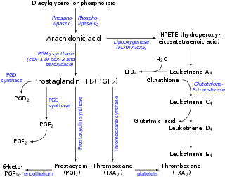
Eicosanoids are signaling molecules made by the enzymatic or non-enzymatic oxidation of arachidonic acid or other polyunsaturated fatty acids (PUFAs) that are, similar to arachidonic acid, around 20 carbon units in length. Eicosanoids are a sub-category of oxylipins, i.e. oxidized fatty acids of diverse carbon units in length, and are distinguished from other oxylipins by their overwhelming importance as cell signaling molecules. Eicosanoids function in diverse physiological systems and pathological processes such as: mounting or inhibiting inflammation, allergy, fever and other immune responses; regulating the abortion of pregnancy and normal childbirth; contributing to the perception of pain; regulating cell growth; controlling blood pressure; and modulating the regional flow of blood to tissues. In performing these roles, eicosanoids most often act as autocrine signaling agents to impact their cells of origin or as paracrine signaling agents to impact cells in the proximity of their cells of origin. Eicosanoids may also act as endocrine agents to control the function of distant cells.

<span class="mw-page-title-main">Leukotriene</span> Class of inflammation mediator molecules

Leukotrienes are a family of eicosanoid inflammatory mediators produced in leukocytes by the oxidation of arachidonic acid (AA) and the essential fatty acid eicosapentaenoic acid (EPA) by the enzyme arachidonate 5-lipoxygenase.

<span class="mw-page-title-main">Lipoxin</span> Acronym for lipoxygenase interaction product

A lipoxin (LX or Lx), an acronym for lipoxygenase interaction product, is a bioactive autacoid metabolite of arachidonic acid made by various cell types. They are categorized as nonclassic eicosanoids and members of the specialized pro-resolving mediators (SPMs) family of polyunsaturated fatty acid (PUFA) metabolites. Like other SPMs, LXs form during, and then act to resolve, inflammatory responses. Initially, two lipoxins were identified, lipoxin A4 (LXA4) and LXB4, but more recent studies have identified epimers of these two LXs: the epi-lipoxins, 15-epi-LXA4 and 15-epi-LXB4 respectively.

<span class="mw-page-title-main">Hepoxilin</span> Chemical compound

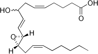
Hepoxilins (Hx) are a set of epoxyalcohol metabolites of polyunsaturated fatty acids (PUFA), i.e. they possess both an epoxide and an alcohol residue. HxA3, HxB3, and their non-enzymatically formed isomers are nonclassic eicosanoid derived from acid the (PUFA), arachidonic acid. A second group of less well studied hepoxilins, HxA4, HxB4, and their non-enzymatically formed isomers are nonclassical eicosanoids derived from the PUFA, eicosapentaenoic acid. Recently, 14,15-HxA3 and 14,15-HxB3 have been defined as arachidonic acid derivatives that are produced by a different metabolic pathway than HxA3, HxB3, HxA4, or HxB4 and differ from the aforementioned hepoxilins in the positions of their hydroxyl and epoxide residues. Finally, hepoxilin-like products of two other PUFAs, docosahexaenoic acid and linoleic acid, have been described. All of these epoxyalcohol metabolites are at least somewhat unstable and are readily enzymatically or non-enzymatically to their corresponding trihydroxy counterparts, the trioxilins (TrX). HxA3 and HxB3, in particular, are being rapidly metabolized to TrXA3, TrXB3, and TrXC3. Hepoxilins have various biological activities in animal models and/or cultured mammalian tissues and cells. The TrX metabolites of HxA3 and HxB3 have less or no activity in most of the systems studied but in some systems retain the activity of their precursor hepoxilins. Based on these studies, it has been proposed that the hepoxilins and trioxilins function in human physiology and pathology by, for example, promoting inflammation responses and dilating arteries to regulate regional blood flow and blood pressure.

Most of the eicosanoid receptors are integral membrane protein G protein-coupled receptors (GPCRs) that bind and respond to eicosanoid signaling molecules. Eicosanoids are rapidly metabolized to inactive products and therefore are short-lived. Accordingly, the eicosanoid-receptor interaction is typically limited to a local interaction: cells, upon stimulation, metabolize arachidonic acid to an eicosanoid which then binds cognate receptors on either its parent cell or on nearby cells to trigger functional responses within a restricted tissue area, e.g. an inflammatory response to an invading pathogen. In some cases, however, the synthesized eicosanoid travels through the blood to trigger systemic or coordinated tissue responses, e.g. prostaglandin (PG) E2 released locally travels to the hypothalamus to trigger a febrile reaction. An example of a non-GPCR receptor that binds many eicosanoids is the PPAR-γ nuclear receptor.

Arachidonate 5-lipoxygenase, also known as ALOX5, 5-lipoxygenase, 5-LOX, or 5-LO, is a non-heme iron-containing enzyme that in humans is encoded by the ALOX5 gene. Arachidonate 5-lipoxygenase is a member of the lipoxygenase family of enzymes. It transforms essential fatty acids (EFA) substrates into leukotrienes as well as a wide range of other biologically active products. ALOX5 is a current target for pharmaceutical intervention in a number of diseases.

<span class="mw-page-title-main">Mead acid</span> Chemical compound

Mead acid is an omega-9 fatty acid, first characterized by James F. Mead. As with some other omega-9 polyunsaturated fatty acids, animals can make Mead acid de novo. Its elevated presence in the blood is an indication of essential fatty acid deficiency. Mead acid is found in large quantities in cartilage.

The oxoeicosanoids are nonclassic eicosanoids, derived from arachidonic acid (AA). For example, Lipoxygenase produces 5-HETE from AA; a dehydrogenase then produces 5-oxo-eicosatetraenoic acid, an oxoeicosanoid, from 5-HETE.

<span class="mw-page-title-main">ALOX15</span> Lipoxygenase found in humans

ALOX15 is, like other lipoxygenases, a seminal enzyme in the metabolism of polyunsaturated fatty acids to a wide range of physiologically and pathologically important products. ▼ Gene Function

<span class="mw-page-title-main">ALOX12</span> Protein-coding gene in the species Homo sapiens

ALOX12, also known as arachidonate 12-lipoxygenase, 12-lipoxygenase, 12S-Lipoxygenase, 12-LOX, and 12S-LOX is a lipoxygenase-type enzyme that in humans is encoded by the ALOX12 gene which is located along with other lipoyxgenases on chromosome 17p13.3. ALOX12 is 75 kilodalton protein composed of 663 amino acids.

<span class="mw-page-title-main">GPR31</span> Protein in humans

G-protein coupled receptor 31 also known as 12-(S)-HETE receptor is a protein that in humans is encoded by the GPR31 gene. The human gene is located on chromosome 6q27 and encodes a G-protein coupled receptor protein composed of 319 amino acids.

<span class="mw-page-title-main">Oxoeicosanoid receptor 1</span> Protein-coding gene in the species Homo sapiens

Oxoeicosanoid receptor 1 (OXER1) also known as G-protein coupled receptor 170 (GPR170) is a protein that in humans is encoded by the OXER1 gene located on human chromosome 2p21; it is the principal receptor for the 5-Hydroxyicosatetraenoic acid family of carboxy fatty acid metabolites derived from arachidonic acid. The receptor has also been termed hGPCR48, HGPCR48, and R527 but OXER1 is now its preferred designation. OXER1 is a G protein-coupled receptor (GPCR) that is structurally related to the hydroxy-carboxylic acid (HCA) family of G protein-coupled receptors whose three members are HCA1 (GPR81), HCA2, and HCA3 ; OXER1 has 30.3%, 30.7%, and 30.7% amino acid sequence identity with these GPCRs, respectively. It is also related to the recently defined receptor, GPR31, for the hydroxyl-carboxy fatty acid 12-HETE.

<span class="mw-page-title-main">5-Hydroxyeicosatetraenoic acid</span> Chemical compound

5-Hydroxyeicosatetraenoic acid (5-HETE, 5(S)-HETE, or 5S-HETE) is an eicosanoid, i.e. a metabolite of arachidonic acid. It is produced by diverse cell types in humans and other animal species. These cells may then metabolize the formed 5(S)-HETE to 5-oxo-eicosatetraenoic acid (5-oxo-ETE), 5(S),15(S)-dihydroxyeicosatetraenoic acid (5(S),15(S)-diHETE), or 5-oxo-15-hydroxyeicosatetraenoic acid (5-oxo-15(S)-HETE).

<span class="mw-page-title-main">12-Hydroxyeicosatetraenoic acid</span> Chemical compound

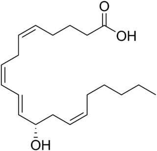
12-Hydroxyeicosatetraenoic acid (12-HETE) is a derivative of the 20 carbon polyunsaturated fatty acid, arachidonic acid, containing a hydroxyl residue at carbon 12 and a 5Z,8Z,10E,14Z Cis–trans isomerism configuration (Z=cis, E=trans) in its four double bonds. It was first found as a product of arachidonic acid metabolism made by human and bovine platelets through their 12S-lipoxygenase (i.e. ALOX12) enzyme(s). However, the term 12-HETE is ambiguous in that it has been used to indicate not only the initially detected "S" stereoisomer, 12S-hydroxy-5Z,8Z,10E,14Z-eicosatetraenoic acid (12(S)-HETE or 12S-HETE), made by platelets, but also the later detected "R" stereoisomer, 12(R)-hydroxy-5Z,8Z,10E,14Z-eicosatetraenoic acid (also termed 12(R)-HETE or 12R-HETE) made by other tissues through their 12R-lipoxygenase enzyme, ALOX12B. The two isomers, either directly or after being further metabolized, have been suggested to be involved in a variety of human physiological and pathological reactions. Unlike hormones which are secreted by cells, travel in the circulation to alter the behavior of distant cells, and thereby act as Endocrine signalling agents, these arachidonic acid metabolites act locally as Autocrine signalling and/or Paracrine signaling agents to regulate the behavior of their cells of origin or of nearby cells, respectively. In these roles, they may amplify or dampen, expand or contract cellular and tissue responses to disturbances.

<span class="mw-page-title-main">15-Hydroxyeicosatetraenoic acid</span> Chemical compound

15-Hydroxyeicosatetraenoic acid (also termed 15-HETE, 15(S)-HETE, and 15S-HETE) is an eicosanoid, i.e. a metabolite of arachidonic acid. Various cell types metabolize arachidonic acid to 15(S)-hydroperoxyeicosatetraenoic acid (15(S)-HpETE). This initial hydroperoxide product is extremely short-lived in cells: if not otherwise metabolized, it is rapidly reduced to 15(S)-HETE. Both of these metabolites, depending on the cell type which forms them, can be further metabolized to 15-oxo-eicosatetraenoic acid (15-oxo-ETE), 5(S),15(S)-dihydroxy-eicosatetraenoic acid (5(S),15(S)-diHETE), 5-oxo-15(S)-hydroxyeicosatetraenoic acid (5-oxo-15(S)-HETE), a subset of specialized pro-resolving mediators viz., the lipoxins, a class of pro-inflammatory mediators, the eoxins, and other products that have less well-defined activities and functions. Thus, 15(S)-HETE and 15(S)-HpETE, in addition to having intrinsic biological activities, are key precursors to numerous biologically active derivatives.

Eoxins are proposed to be a family of proinflammatory eicosanoids. They are produced by human eosinophils, mast cells, the L1236 Reed–Sternberg cell line derived from Hodgkin's lymphoma, and certain other tissues. These cells produce the eoxins by initially metabolizing arachidonic acid, an omega-6 (ω-6) fatty acid, via any enzyme possessing 15-lipoxygenase activity. The product of this initial metabolic step, 15(S)-hydroperoxyeicosatetraenoic acid, is then converted to a series of eoxins by the same enzymes that metabolize the 5-lipoxygenase product of arachidonic acid metabolism, i.e. 5-Hydroperoxy-eicosatetraenoic acid to a series of leukotrienes. That is, the eoxins are 14,15-disubstituted analogs of the 5,6-disubstituted leukotrienes.

<span class="mw-page-title-main">12-Hydroxyheptadecatrienoic acid</span> Chemical compound

12-Hydroxyheptadecatrienoic acid (also termed 12-HHT, 12(S)-hydroxyheptadeca-5Z,8E,10E-trienoic acid, or 12(S)-HHTrE) is a 17 carbon metabolite of the 20 carbon polyunsaturated fatty acid, arachidonic acid. It was discovered and structurally defined in 1973 by P. Wlodawer, Bengt I. Samuelsson, and M. Hamberg, as a product of arachidonic acid metabolism made by microsomes (i.e. endoplasmic reticulum) isolated from sheep seminal vesicle glands and by intact human platelets. 12-HHT is less ambiguously termed 12-(S)-hydroxy-5Z,8E,10E-heptadecatrienoic acid to indicate the S stereoisomerism of its 12-hydroxyl residue and the Z, E, and E cis-trans isomerism of its three double bonds. The metabolite was for many years thought to be merely a biologically inactive byproduct of prostaglandin synthesis. More recent studies, however, have attached potentially important activity to it.

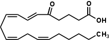
5-Hydroxyeicosanoid dehydrogenase (5-HEDH) or more formally, nicotinamide adenine dinucleotide phosphate (NADP+)-dependent dehydrogenase, is an enzyme that metabolizes an eicosanoid product of arachidonate 5-lipoxygenase (5-LOX), 5(S)-hydroxy-6S,8Z,11Z,14Z-eicosatetraenoic acid (i.e. 5-(S)-HETE; see 5-HETE) to its 5-keto analog, 5-oxo-eicosatetraenoic acid (i.e. 5-oxo-6S,8Z,11Z,14Z-eicosatetraenoic acid or 5-oxo-ETE). It also acts in the reverse direction, metabolizing 5-oxo-ETE to 5(S)-HETE. Since 5-oxo-ETE is 30–100-fold more potent than 5(S)-HETE in stimulating various cell types, 5-HEDH is regarded as a regulator and promoter of 5(S)HETE's and thereby 5-LOX's influences on cell function. Although 5-HEDH has been evaluated in a wide range of intact cells and in crude microsome preparations, it has not yet been evaluated for its structure, for its gene, of in pure form; furthermore, most studies on it have been conducted in human tissues.

Cytochrome P450 omega hydroxylases, also termed cytochrome P450 ω-hydroxylases, CYP450 omega hydroxylases, CYP450 ω-hydroxylases, CYP omega hydroxylase, CYP ω-hydroxylases, fatty acid omega hydroxylases, cytochrome P450 monooxygenases, and fatty acid monooxygenases, are a set of cytochrome P450-containing enzymes that catalyze the addition of a hydroxyl residue to a fatty acid substrate. The CYP omega hydroxylases are often referred to as monoxygenases; however, the monooxygenases are CYP450 enzymes that add a hydroxyl group to a wide range of xenobiotic and naturally occurring endobiotic substrates, most of which are not fatty acids. The CYP450 omega hydroxylases are accordingly better viewed as a subset of monooxygenases that have the ability to hydroxylate fatty acids. While once regarded as functioning mainly in the catabolism of dietary fatty acids, the omega oxygenases are now considered critical in the production or break-down of fatty acid-derived mediators which are made by cells and act within their cells of origin as autocrine signaling agents or on nearby cells as paracrine signaling agents to regulate various functions such as blood pressure control and inflammation.

Specialized pro-resolving mediators are a large and growing class of cell signaling molecules formed in cells by the metabolism of polyunsaturated fatty acids (PUFA) by one or a combination of lipoxygenase, cyclooxygenase, and cytochrome P450 monooxygenase enzymes. Pre-clinical studies, primarily in animal models and human tissues, implicate SPM in orchestrating the resolution of inflammation. Prominent members include the resolvins and protectins.

References

  1. 1 2 3 4 5 6 7 8 9 10 11 12 13 14 Powell WS, Rokach J (April 2015). "Biosynthesis, biological effects, and receptors of hydroxyeicosatetraenoic acids (HETEs) and oxoeicosatetraenoic acids (oxo-ETEs) derived from arachidonic acid". Biochimica et Biophysica Acta (BBA) - Molecular and Cell Biology of Lipids. 1851 (4): 340–55. doi:10.1016/j.bbalip.2014.10.008. PMC   5710736 . PMID   25449650.
  2. 1 2 Cossette C, Gravel S, Reddy CN, Gore V, Chourey S, Ye Q, Snyder NW, Mesaros CA, Blair IA, Lavoie JP, Reinero CR, Rokach J, Powell WS (August 2015). "Biosynthesis and actions of 5-oxoeicosatetraenoic acid (5-oxo-ETE) on feline granulocytes". Biochemical Pharmacology. 96 (3): 247–55. doi:10.1016/j.bcp.2015.05.009. PMC   4830392 . PMID   26032638.
  3. 1 2 3 4 5 6 7 8 9 10 11 12 13 14 15 16 17 18 Powell WS, Rokach J (October 2013). "The eosinophil chemoattractant 5-oxo-ETE and the OXE receptor". Progress in Lipid Research. 52 (4): 651–65. doi:10.1016/j.plipres.2013.09.001. PMC   5710732 . PMID   24056189.
  4. Bui P, Imaizumi S, Beedanagari SR, Reddy ST, Hankinson O (February 2011). "Human CYP2S1 metabolizes cyclooxygenase- and lipoxygenase-derived eicosanoids". Drug Metabolism and Disposition. 39 (2): 180–90. doi:10.1124/dmd.110.035121. PMC   3033693 . PMID   21068195.
  5. Zarini S, Murphy RC (March 2003). "Biosynthesis of 5-oxo-6,8,11,14-eicosatetraenoic acid from 5-hydroperoxyeicosatetraenoic acid in the murine macrophage". The Journal of Biological Chemistry. 278 (13): 11190–6. doi: 10.1074/jbc.M208496200 . PMID   12547823.
  6. Falgueyret J, Riendeau D (February 2000). "LTA(4)-derived 5-oxo-eicosatetraenoic acid: pH-dependent formation and interaction with the LTB(4) receptor of human polymorphonuclear leukocytes". Biochimica et Biophysica Acta (BBA) - Molecular and Cell Biology of Lipids. 1484 (1): 51–8. doi:10.1016/s1388-1981(99)00198-5. PMID   10685030.
  7. Zimpfer U, Dichmann S, Termeer CC, Simon JC, Schröder JM, Norgauer J (November 2000). "Human dendritic cells are a physiological source of the chemotactic arachidonic acid metabolite 5-oxo-eicosatetraenoic acid". Inflammation Research. 49 (11): 633–8. doi:10.1007/s000110050641. PMID   11131304. S2CID   21430700.
  8. 1 2 3 O'Flaherty JT, Taylor JS, Thomas MJ (December 1998). "Receptors for the 5-oxo class of eicosanoids in neutrophils". The Journal of Biological Chemistry. 273 (49): 32535–41. doi: 10.1074/jbc.273.49.32535 . PMID   9829988.
  9. Hevko JM, Bowers RC, Murphy RC (February 2001). "Synthesis of 5-oxo-6,8,11,14-eicosatetraenoic acid and identification of novel omega-oxidized metabolites in the mouse macrophage". The Journal of Pharmacology and Experimental Therapeutics. 296 (2): 293–305. PMID   11160610.
  10. Murphy RC, Zarini S (2002). "Glutathione adducts of oxyeicosanoids". Prostaglandins Other Lipid Mediat. 68–69: 471–82. doi:10.1016/s0090-6980(02)00049-7. PMID   12432937.
  11. Hevko JM, Bowers RC, Murphy RC (Feb 2001). "Synthesis of 5-oxo-6,8,11,14-eicosatetraenoic acid and identification of novel omega-oxidized metabolites in the mouse macrophage". J Pharmacol Exp Ther. 296 (2): 293–305. PMID   11160610.
  12. 1 2 3 4 Brink C, Dahlén SE, Drazen J, Evans JF, Hay DW, Rovati GE, Serhan CN, Shimizu T, Yokomizo T (March 2004). "International Union of Pharmacology XLIV. Nomenclature for the oxoeicosanoid receptor". Pharmacological Reviews. 56 (1): 149–57. doi:10.1124/pr.56.1.4. PMID   15001665. S2CID   7229884.W
  13. Konya V, Blättermann S, Jandl K, Platzer W, Ottersbach PA, Marsche G, Gütschow M, Kostenis E, Heinemann A (May 2014). "A biased non-Gαi OXE-R antagonist demonstrates that Gαi protein subunit is not directly involved in neutrophil, eosinophil, and monocyte activation by 5-oxo-ETE". Journal of Immunology. 192 (10): 4774–82. doi: 10.4049/jimmunol.1302013 . PMID   24733850.
  14. Rossi AG, O'Flaherty JT (1991). "Bioactions of 5-hydroxyicosatetraenoate and its interaction with platelet-activating factor". Lipids. 26 (12): 1184–8. doi:10.1007/bf02536528. PMID   1668115. S2CID   3964822.
  15. 1 2 Cooke M, Di Cónsoli H, Maloberti P, Cornejo Maciel F (May 2013). "Expression and function of OXE receptor, an eicosanoid receptor, in steroidogenic cells". Molecular and Cellular Endocrinology. 371 (1–2): 71–8. doi:10.1016/j.mce.2012.11.003. hdl: 11336/8381 . PMID   23159987. S2CID   8520991.
  16. 1 2 3 O'Flaherty JT, Rogers LC, Paumi CM, Hantgan RR, Thomas LR, Clay CE, High K, Chen YQ, Willingham MC, Smitherman PK, Kute TE, Rao A, Cramer SD, Morrow CS (October 2005). "5-Oxo-ETE analogs and the proliferation of cancer cells". Biochimica et Biophysica Acta (BBA) - Molecular and Cell Biology of Lipids. 1736 (3): 228–236. doi:10.1016/j.bbalip.2005.08.009. PMID   16154383.
  17. Morin C, Sirois M, Echave V, Gomes MM, Rousseau E (June 2007). "Relaxing effects of 5-oxo-ETE on human bronchi involve BK Ca channel activation". Prostaglandins & Other Lipid Mediators. 83 (4): 311–9. doi:10.1016/j.prostaglandins.2007.03.001. PMID   17499751.
  18. 1 2 Sozzani S, Zhou D, Locati M, Bernasconi S, Luini W, Mantovani A, O'Flaherty JT (November 1996). "Stimulating properties of 5-oxo-eicosanoids for human monocytes: synergism with monocyte chemotactic protein-1 and -3". Journal of Immunology. 157 (10): 4664–71. doi: 10.4049/jimmunol.157.10.4664 . PMID   8906847. S2CID   23499393.
  19. Muro S, Hamid Q, Olivenstein R, Taha R, Rokach J, Powell WS (October 2003). "5-oxo-6,8,11,14-eicosatetraenoic acid induces the infiltration of granulocytes into human skin". The Journal of Allergy and Clinical Immunology. 112 (4): 768–74. doi: 10.1016/S0091-6749(03)01888-8 . PMID   14564360.
  20. Morin C, Sirois M, Echave V, Gomes MM, Rousseau E (2007). "Relaxing effects of 5-oxo-ETE on human bronchi involve BK Ca channel activation". Prostaglandins & Other Lipid Mediators. 83 (4): 311–9. doi:10.1016/j.prostaglandins.2007.03.001. PMID   17499751.
  21. Grant GE, Rubino S, Gravel S, Wang X, Patel P, Rokach J, Powell WS (June 2011). "Enhanced formation of 5-oxo-6,8,11,14-eicosatetraenoic acid by cancer cells in response to oxidative stress, docosahexaenoic acid and neutrophil-derived 5-hydroxy-6,8,11,14-eicosatetraenoic acid". Carcinogenesis. 32 (6): 822–8. doi:10.1093/carcin/bgr044. PMC   3146358 . PMID   21393477.
  22. MacLeod RJ, Lembessis P, Hamilton JR, Powell WS (November 1999). "5-Oxo-6,8,11,14-eicosatetraenoic acid stimulates isotonic volume reduction of guinea pig jejunal crypt epithelial cells". The Journal of Pharmacology and Experimental Therapeutics. 291 (2): 511–6. PMID   10525065.
  23. 1 2 O'Flaherty JT, Kuroki M, Nixon AB, Wijkander J, Yee E, Lee SL, Smitherman PK, Wykle RL, Daniel LW (July 1996). "5-Oxo-eicosatetraenoate is a broadly active, eosinophil-selective stimulus for human granulocytes". Journal of Immunology. 157 (1): 336–42. doi: 10.4049/jimmunol.157.1.336 . PMID   8683135. S2CID   35264541.
  24. 1 2 O'Flaherty JT, Cordes JF, Lee SL, Samuel M, Thomas MJ (December 1994). "Chemical and biological characterization of oxo-eicosatetraenoic acids". Biochimica et Biophysica Acta (BBA) - General Subjects. 1201 (3): 505–15. doi:10.1016/0304-4165(94)90083-3. PMID   7803484.
  25. O'Flaherty JT, Kuroki M, Nixon AB, Wijkander J, Yee E, Lee SL, Smitherman PK, Wykle RL, Daniel LW (July 1996). "5-Oxo-eicosanoids and hematopoietic cytokines cooperate in stimulating neutrophil function and the mitogen-activated protein kinase pathway". The Journal of Biological Chemistry. 271 (30): 17821–8. doi: 10.1074/jbc.271.30.17821 . PMID   8663432.
  26. O'Flaherty JT, Cordes JF (June 1994). "Human neutrophil degranulation responses to nucleotides". Laboratory Investigation; A Journal of Technical Methods and Pathology. 70 (6): 816–21. PMID   8015286.
  27. Guilbert M, Ferland C, Bossé M, Flamand N, Lavigne S, Laviolette M (July 1999). "5-Oxo-6,8,11,14-eicosatetraenoic acid induces important eosinophil transmigration through basement membrane components: comparison of normal and asthmatic eosinophils". American Journal of Respiratory Cell and Molecular Biology. 21 (1): 97–104. CiteSeerX   10.1.1.326.2636 . doi:10.1165/ajrcmb.21.1.3517. PMID   10385597.
  28. Powell WS, Rokach J (March 2020). "Targeting the OXE receptor as a potential novel therapy for asthma". Biochemical Pharmacology. 179: 113930. doi:10.1016/j.bcp.2020.113930. PMC   10656995 . PMID   32240653. S2CID   214768793.