Adenine nucleotide translocator

Last updated
ADP/ATP translocases
ATP-ADP Translocase Top View.png
Cytoplasmic view of the binding pocket of ATP–ADP translocase 1, PDB: 1OKC .
Identifiers
SymbolAden_trnslctor
Pfam PF00153
InterPro IPR002113
TCDB 2.A.29.1.2
OPM superfamily 21
OPM protein 2c3e
Available protein structures:
Pfam   structures / ECOD  
PDB RCSB PDB; PDBe; PDBj
PDBsum structure summary
solute carrier family 25 (mitochondrial carrier; adenine nucleotide translocator), member 4
Identifiers
Symbol SLC25A4
Alt. symbolsPEO3, PEO2, ANT1
NCBI gene 291
HGNC 10990
OMIM 103220
RefSeq NM_001151
UniProt P12235
Other data
Locus Chr. 4 q35
Search for
Structures Swiss-model
Domains InterPro
solute carrier family 25 (mitochondrial carrier; adenine nucleotide translocator), member 5
Identifiers
SymbolSLC25A5
Alt. symbolsANT2
NCBI gene 292
HGNC 10991
OMIM 300150
RefSeq NM_001152
UniProt P05141
Other data
Locus Chr. X q24-q26
Search for
Structures Swiss-model
Domains InterPro
solute carrier family 25 (mitochondrial carrier; adenine nucleotide translocator), member 6
Identifiers
Symbol SLC25A6
Alt. symbolsANT3
NCBI gene 293
HGNC 10992
OMIM 403000
RefSeq NM_001636
UniProt P12236
Other data
Locus Chr. Y p
Search for
Structures Swiss-model
Domains InterPro

Adenine nucleotide translocator (ANT), also known as the ADP/ATP translocase (ANT), ADP/ATP carrier protein (AAC) or mitochondrial ADP/ATP carrier, exchanges free ATP with free ADP across the inner mitochondrial membrane. [1] [2] ANT is the most abundant protein in the inner mitochondrial membrane and belongs to mitochondrial carrier family. [3]

Contents

Free ADP is transported from the cytoplasm to the mitochondrial matrix, while ATP produced from oxidative phosphorylation is transported from the mitochondrial matrix to the cytoplasm, thus providing the cells with its main energy currency. [4] ADP/ATP translocases are exclusive to eukaryotes and are thought to have evolved during eukaryogenesis. [5] Human cells express four ADP/ATP translocases: SLC25A4, SLC25A5, SLC25A6 and SLC25A31, which constitute more than 10% of the protein in the inner mitochondrial membrane. [6] These proteins are classified under the mitochondrial carrier superfamily.

Types

In humans, there exist three paraologous ANT isoforms:

Structure

A side view of the translocase spanning the inner mitochondrial membrane. The six a-helices are denoted by different colors. The binding pocket is currently open to the cytoplasmic side and will bind to ADP, transporting it into the matrix. (From PDB: 1OKC ) ATP-ADP Translocase Side View (w) drawn over.png
A side view of the translocase spanning the inner mitochondrial membrane. The six α-helices are denoted by different colors. The binding pocket is currently open to the cytoplasmic side and will bind to ADP, transporting it into the matrix. (From PDB: 1OKC )
The translocase (as a molecular surface, green) viewed from both sides of a lipid bilayer representing the inner mitocondrial membrane. Left panel (IM): view from the intermembrane space. The protein is in the open conformation towards this side. Right panel (M): view from the matrix. The protein is closed towards this side. ATP-ADP translocase - top and bottom views.png
The translocase (as a molecular surface, green) viewed from both sides of a lipid bilayer representing the inner mitocondrial membrane. Left panel (IM): view from the intermembrane space. The protein is in the open conformation towards this side. Right panel (M): view from the matrix. The protein is closed towards this side.

ANT has long been thought to function as a homodimer, but this concept was challenged by the projection structure of the yeast Aac3p solved by electron crystallography, which showed that the protein was three-fold symmetric and monomeric, with the translocation pathway for the substrate through the centre. [7] The atomic structure of the bovine ANT confirmed this notion, and provided the first structural fold of a mitochondrial carrier. [8] Further work has demonstrated that ANT is a monomer in detergents [9] and functions as a monomer in mitochondrial membranes. [10] [11]

ADP/ATP translocase 1 is the major AAC in human cells and the archetypal protein of this family. It has a mass of approximately 30 kDa, consisting of 297 residues. [12] It forms six transmembrane α-helices that form a barrel that results in a deep cone-shaped depression accessible from the outside where the substrate binds. The binding pocket, conserved throughout most isoforms, mostly consists of basic residues that allow for strong binding to ATP or ADP and has a maximal diameter of 20 Å and a depth of 30 Å. [8] Indeed, arginine residues 96, 204, 252, 253, and 294, as well as lysine 38, have been shown to be essential for transporter activity. [13]

Function

ADP/ATP translocase transports ATP synthesized from oxidative phosphorylation into the cytoplasm, where it can be used as the principal energy currency of the cell to power thermodynamically unfavorable reactions. After the consequent hydrolysis of ATP into ADP, ADP is transported back into the mitochondrial matrix, where it can be rephosphorylated to ATP. Because a human typically exchanges the equivalent of his/her own mass of ATP on a daily basis, ADP/ATP translocase is an important transporter protein with major metabolic implications. [4] [8]

ANT transports the free, i.e. deprotonated, non-Magnesium, non-Calcium bound forms of ADP and ATP, in a 1:1 ratio. [1] Transport is fully reversible, and its directionality is governed by the concentrations of its substrates (ADP and ATP inside and outside mitochondria), the chelators of the adenine nucleotides, and the mitochondrial membrane potential. The relationship of these parameters can be expressed by an equation solving for the 'reversal potential of the ANT" (Erev_ANT), a value of the mitochondrial membrane potential at which no net transport of adenine nucleotides takes place by the ANT. [14] [15] [16] The ANT and the F0-F1 ATP synthase are not necessarily in directional synchrony. [14]

Apart from exchange of ADP and ATP across the inner mitochondrial membrane, the ANT also exhibits an intrinsic uncoupling activity [1] [17]

ANT is an important modulatory [18] and possible structural component of the Mitochondrial Permeability Transition Pore, a channel involved in various pathologies whose function still remains elusive. Karch et al. propose a "multi-pore model" in which ANT is at least one of the molecular components of the pore. [19]

Translocase mechanism

Under normal conditions, ATP and ADP cannot cross the inner mitochondrial membrane due to their high negative charges, but ADP/ATP translocase, an antiporter, couples the transport of the two molecules. The depression in ADP/ATP translocase alternatively faces the matrix and the cytoplasmic sides of the membrane. ADP in the intermembrane space, coming from the cytoplasm, binds the translocase and induces its eversion, resulting in the release of ADP into the matrix. Binding of ATP from the matrix induces eversion and results in the release of ATP into the intermembrane space, subsequently diffusing to the cytoplasm, and concomitantly brings the translocase back to its original conformation. [4] ATP and ADP are the only natural nucleotides recognized by the translocase. [8]

The net process is denoted by:

ADP3−cytoplasm + ATP4−matrix → ADP3−matrix + ATP4−cytoplasm

ADP/ATP exchange is energetically expensive: about 25% of the energy yielded from electron transfer by aerobic respiration, or one hydrogen ion, is consumed to regenerate the membrane potential that is tapped by ADP/ATP translocase. [4]

The translocator cycles between two states, called the cytoplasmic and matrix state, opening up to these compartments in an alternating way. [1] [2] There are structures available that show the translocator locked in a cytoplasmic state by the inhibitor carboxyatractyloside, [8] [20] or in the matrix state by the inhibitor bongkrekic acid. [21]

Alterations

Rare but severe diseases such as mitochondrial myopathies are associated with dysfunctional human ADP/ATP translocase. Mitochondrial myopathies (MM) refer to a group of clinically and biochemically heterogeneous disorders that share common features of major mitochondrial structural abnormalities in skeletal muscle. The major morphological hallmark of MM is ragged, red fibers containing peripheral and intermyofibrillar accumulations of abnormal mitochondria. [22] [23] In particular, autosomal dominant progressive external ophthalmoplegia (adPEO) is a common disorder associated with dysfunctional ADP/ATP translocase and can induce paralysis of muscles responsible for eye movements. General symptoms are not limited to the eyes and can include exercise intolerance, muscle weakness, hearing deficit, and more. adPEO shows Mendelian inheritance patterns but is characterized by large-scale mitochondrial DNA (mtDNA) deletions. mtDNA contains few introns, or non-coding regions of DNA, which increases the likelihood of deleterious mutations. Thus, any modification of ADP/ATP translocase mtDNA can lead to a dysfunctional transporter, [24] particularly residues involved in the binding pocket which will compromise translocase efficacy. [13] MM is commonly associated with dysfunctional ADP/ATP translocase, but MM can be induced through many different mitochondrial abnormalities.

Inhibition

Bongkrekic acid Bongkrekic acid.svg
Bongkrekic acid

ADP/ATP translocase is very specifically inhibited by two families of compounds. The first family, which includes atractyloside (ATR) and carboxyatractyloside (CATR), binds to the ADP/ATP translocase from the cytoplasmic side, locking it in a cytoplasmic side open conformation. In contrast, the second family, which includes bongkrekic acid (BA) and isobongkrekic acid (isoBA), binds the translocase from the matrix, locking it in a matrix side open conformation. [7] The negatively charged groups of the inhibitors bind strongly to the positively charged residues deep within the binding pocket. The high affinity (Kd in the nanomolar range) makes each inhibitor a deadly poison by obstructing cellular respiration/energy transfer to the rest of the cell. [8] There are structures available that show the translocator locked in a cytoplasmic state by the inhibitor carboxyatractyloside, [8] [20] or in the matrix state by the inhibitor bongkrekic acid. [21]

History

In 1955, Siekevitz and Potter demonstrated that adenine nucleotides were distributed in cells in two pools located in the mitochondrial and cytosolic compartments. [25] Shortly thereafter, Pressman hypothesized that the two pools could exchange nucleotides. [26] However, the existence of an ADP/ATP transporter was not postulated until 1964 when Bruni et al. uncovered an inhibitory effect of atractyloside on the energy-transfer system (oxidative phosphorylation) and ADP binding sites of rat liver mitochondria. [27]

Soon after, an overwhelming amount of research was done in proving the existence and elucidating the link between ADP/ATP translocase and energy transport. [28] [29] [30] cDNA of ADP/ATP translocase was sequenced for bovine in 1982 [31] and a yeast species Saccharomyces cerevisiae in 1986 [32] before finally Battini et al. sequenced a cDNA clone of the human transporter in 1989. The homology in the coding sequences between human and yeast ADP/ATP translocase was 47% while bovine and human sequences extended remarkable to 266 out of 297 residues, or 89.6%. In both cases, the most conserved residues lie in the ADP/ATP substrate binding pocket. [12]

See also

Related Research Articles

<span class="mw-page-title-main">Adenosine triphosphate</span> Energy-carrying molecule in living cells

Adenosine triphosphate (ATP) is a nucleotide that provides energy to drive and support many processes in living cells, such as muscle contraction, nerve impulse propagation, condensate dissolution, and chemical synthesis. Found in all known forms of life, it is often referred to as the "molecular unit of currency" of intracellular energy transfer.

<span class="mw-page-title-main">Cellular respiration</span> Process to convert glucose to ATP in cells

Cellular respiration is the process by which biological fuels are oxidized in the presence of an inorganic electron acceptor, such as oxygen, to drive the bulk production of adenosine triphosphate (ATP), which contains energy. Cellular respiration may be described as a set of metabolic reactions and processes that take place in the cells of organisms to convert chemical energy from nutrients into ATP, and then release waste products.

<span class="mw-page-title-main">Thermogenin</span> Mammalian protein found in Homo sapiens

Thermogenin is a mitochondrial carrier protein found in brown adipose tissue (BAT). It is used to generate heat by non-shivering thermogenesis, and makes a quantitatively important contribution to countering heat loss in babies which would otherwise occur due to their high surface area-volume ratio.

<span class="mw-page-title-main">Intermembrane space</span>

The intermembrane space (IMS) is the space occurring between or involving two or more membranes. In cell biology, it is most commonly described as the region between the inner membrane and the outer membrane of a mitochondrion or a chloroplast. It also refers to the space between the inner and outer nuclear membranes of the nuclear envelope, but is often called the perinuclear space. The IMS of mitochondria plays a crucial role in coordinating a variety of cellular activities, such as regulation of respiration and metabolic functions. Unlike the IMS of the mitochondria, the IMS of the chloroplast does not seem to have any obvious function.

Substrate-level phosphorylation is a metabolism reaction that results in the production of ATP or GTP supported by the energy released from another high-energy bond that leads to phosphorylation of ADP or GDP to ATP or GTP (note that the reaction catalyzed by creatine kinase is not considered as "substrate-level phosphorylation"). This process uses some of the released chemical energy, the Gibbs free energy, to transfer a phosphoryl (PO3) group to ADP or GDP. Occurs in glycolysis and in the citric acid cycle.

<span class="mw-page-title-main">Inner mitochondrial membrane</span>

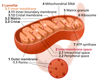
The inner mitochondrial membrane (IMM) is the mitochondrial membrane which separates the mitochondrial matrix from the intermembrane space.

The mitochondrial permeability transition pore is a protein that is formed in the inner membrane of the mitochondria under certain pathological conditions such as traumatic brain injury and stroke. Opening allows increase in the permeability of the mitochondrial membranes to molecules of less than 1500 daltons in molecular weight. Induction of the permeability transition pore, mitochondrial membrane permeability transition, can lead to mitochondrial swelling and cell death through apoptosis or necrosis depending on the particular biological setting.

<span class="mw-page-title-main">Bongkrek acid</span> Chemical compound

Bongkrek acid is a respiratory toxin produced in fermented coconut or corn contaminated by the bacterium Burkholderia gladioli pathovar cocovenenans. It is a highly toxic, heat-stable, colorless, odorless, and highly unsaturated tricarboxylic acid that inhibits the ADP/ATP translocase, also called the mitochondrial ADP/ATP carrier, preventing ATP from leaving the mitochondria to provide metabolic energy to the rest of the cell. Bongkrek acid, when consumed through contaminated foods, mainly targets the liver, brain, and kidneys along with symptoms that include vomiting, diarrhea, urinary retention, abdominal pain, and excessive sweating. Most of the outbreaks are found in Indonesia and China where fermented coconut and corn-based foods are consumed.

<span class="mw-page-title-main">TIM/TOM complex</span>

The TIM/TOM complex is a protein complex in cellular biochemistry which translocates proteins produced from nuclear DNA through the mitochondrial membrane for use in oxidative phosphorylation. In enzymology, the complex is described as an mitochondrial protein-transporting ATPase, or more systematically ATP phosphohydrolase , as the TIM part requires ATP hydrolysis to work.

<span class="mw-page-title-main">Mitochondrial membrane transport protein</span>

Mitochondrial membrane transport proteins, also known as mitochondrial carrier proteins, are proteins which exist in the membranes of mitochondria. They serve to transport molecules and other factors, such as ions, into or out of the organelles. Mitochondria contain both an inner and outer membrane, separated by the inter-membrane space, or inner boundary membrane. The outer membrane is porous, whereas the inner membrane restricts the movement of all molecules. The two membranes also vary in membrane potential and pH. These factors play a role in the function of mitochondrial membrane transport proteins. There are 53 discovered human mitochondrial membrane transporters, with many others that are known to still need discovered.

<span class="mw-page-title-main">Mitochondrial carrier</span>

Mitochondrial carriers are proteins from solute carrier family 25 which transfer molecules across the membranes of the mitochondria. Mitochondrial carriers are also classified in the Transporter Classification Database. The Mitochondrial Carrier (MC) Superfamily has been expanded to include both the original Mitochondrial Carrier (MC) family and the Mitochondrial Inner/Outer Membrane Fusion (MMF) family.

Translocase is a general term for a protein that assists in moving another molecule, usually across a cell membrane. These enzymes catalyze the movement of ions or molecules across membranes or their separation within membranes. The reaction is designated as a transfer from “side 1” to “side 2” because the designations “in” and “out”, which had previously been used, can be ambiguous. Translocases are the most common secretion system in Gram positive bacteria.

<span class="mw-page-title-main">Uncoupling protein</span> Mitochondrial protein

An uncoupling protein (UCP) is a mitochondrial inner membrane protein that is a regulated proton channel or transporter. An uncoupling protein is thus capable of dissipating the proton gradient generated by NADH-powered pumping of protons from the mitochondrial matrix to the mitochondrial intermembrane space. The energy lost in dissipating the proton gradient via UCPs is not used to do biochemical work. Instead, heat is generated. This is what links UCP to thermogenesis. However, not every type of UCPs are related to thermogenesis. Although UCP2 and UCP3 are closely related to UCP1, UCP2 and UCP3 do not affect thermoregulatory abilities of vertebrates. UCPs are positioned in the same membrane as the ATP synthase, which is also a proton channel. The two proteins thus work in parallel with one generating heat and the other generating ATP from ADP and inorganic phosphate, the last step in oxidative phosphorylation. Mitochondria respiration is coupled to ATP synthesis, but is regulated by UCPs. UCPs belong to the mitochondrial carrier (SLC25) family.

<span class="mw-page-title-main">ADP/ATP translocase 1</span> Protein-coding gene in the species Homo sapiens

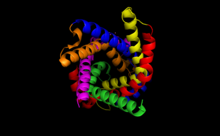
ADP/ATP translocase 1, or adenine nucleotide translocator 1 (ANT1), is an enzyme that in humans is encoded by the SLC25A4 gene.

<span class="mw-page-title-main">Tricarboxylate transport protein, mitochondrial</span> Mammalian protein found in Homo sapiens

Tricarboxylate transport protein, mitochondrial, also known as tricarboxylate carrier protein and citrate transport protein (CTP), is a protein that in humans is encoded by the SLC25A1 gene. SLC25A1 belongs to the mitochondrial carrier gene family SLC25. High levels of the tricarboxylate transport protein are found in the liver, pancreas and kidney. Lower or no levels are present in the brain, heart, skeletal muscle, placenta and lung.

<span class="mw-page-title-main">ADP/ATP translocase 4</span> Protein-coding gene in the species Homo sapiens

ADP/ATP translocase 4 (ANT4) is an enzyme that in humans is encoded by the SLC25A31 gene on chromosome 4. This enzyme inhibits apoptosis by catalyzing ADP/ATP exchange across the mitochondrial membranes and regulating membrane potential. In particular, ANT4 is essential to spermatogenesis, as it imports ATP into sperm mitochondria to support their development and survival. Outside this role, the SLC25AC31 gene has not been implicated in any human disease.

<span class="mw-page-title-main">ADP/ATP translocase 3</span> Protein-coding gene in humans

ADP/ATP translocase 3, also known as solute carrier family 25 member 6, is a protein that in humans is encoded by the SLC25A6 gene.

<span class="mw-page-title-main">ADP/ATP translocase 2</span> Protein-coding gene in humans

ADP/ATP translocase 2 is a protein that in humans is encoded by the SLC25A5 gene on the X chromosome.

The ATP:ADP Antiporter (AAA) Family is a member of the major facilitator superfamily. Members of the AAA family have been sequenced from bacteria and plants.

<span class="mw-page-title-main">Carboxyatractyloside</span> Chemical compound

Carboxyatractyloside (CATR) is a highly toxic diterpene glycoside that inhibits the ADP/ATP translocase. It is about 10 times more potent than its analog atractyloside. While atractyloside is effective in the inhibition of oxidative phosphorylation, carboxyatractyloside is considered to be more effective. The effects of carboxyatractyloside on the ADP/ATP translocase are not reversed by increasing the concentration of adenine nucleotides, unlike its counterpart atractyloside. Carboxyatractyloside behavior resembles bongkrekic acid while in the mitochondria. Carboxyatractyloside is poisonous to humans as well as livestock, including cows and horses.

References

  1. 1 2 3 4 Klingenberg M (October 2008). "The ADP and ATP transport in mitochondria and its carrier". Biochimica et Biophysica Acta (BBA) - Biomembranes. 1778 (10): 1978–2021. doi:10.1016/j.bbamem.2008.04.011. PMID   18510943.
  2. 1 2 Kunji ER, Aleksandrova A, King MS, Majd H, Ashton VL, Cerson E, Springett R, Kibalchenko M, Tavoulari S, Crichton PG, Ruprecht JJ (October 2016). "The transport mechanism of the mitochondrial ADP/ATP carrier". Biochimica et Biophysica Acta (BBA) - Molecular Cell Research. Channels and transporters in cell metabolism. 1863 (10): 2379–93. doi: 10.1016/j.bbamcr.2016.03.015 . PMID   27001633.
  3. Palmieri F, Monné M (October 2016). "Discoveries, metabolic roles and diseases of mitochondrial carriers: A review". Biochimica et Biophysica Acta (BBA) - Molecular Cell Research. Channels and transporters in cell metabolism. 1863 (10): 2362–78. doi: 10.1016/j.bbamcr.2016.03.007 . hdl: 11563/126168 . PMID   26968366.
  4. 1 2 3 4 Stryer L, Berg JM, Tymoczko JL (2007). Biochemistry. San Francisco: W.H. Freeman. p. 553. ISBN   978-0-7167-8724-2.
  5. Radzvilavicius AL, Blackstone NW (October 2015). "Conflict and cooperation in eukaryogenesis: implications for the timing of endosymbiosis and the evolution of sex". Journal of the Royal Society, Interface. 12 (111): 20150584. doi:10.1098/rsif.2015.0584. PMC   4614496 . PMID   26468067.
  6. Brandolin G, Dupont Y, Vignais PV (April 1985). "Substrate-induced modifications of the intrinsic fluorescence of the isolated adenine nucleotide carrier protein: demonstration of distinct conformational states". Biochemistry. 24 (8): 1991–7. doi:10.1021/bi00329a029. PMID   2990548.
  7. 1 2 Kunji ER, Harding M (September 2003). "Projection structure of the atractyloside-inhibited mitochondrial ADP/ATP carrier of Saccharomyces cerevisiae". The Journal of Biological Chemistry. 278 (39): 36985–8. doi: 10.1074/jbc.C300304200 . PMID   12893834.
  8. 1 2 3 4 5 6 7 Pebay-Peyroula E, Dahout-Gonzalez C, Kahn R, Trézéguet V, Lauquin GJ, Brandolin G (November 2003). "Structure of mitochondrial ADP/ATP carrier in complex with carboxyatractyloside". Nature. 426 (6962): 39–44. Bibcode:2003Natur.426...39P. doi:10.1038/nature02056. PMID   14603310. S2CID   4338748.
  9. Bamber L, Slotboom DJ, Kunji ER (August 2007). "Yeast mitochondrial ADP/ATP carriers are monomeric in detergents as demonstrated by differential affinity purification". Journal of Molecular Biology. 371 (2): 388–95. doi:10.1016/j.jmb.2007.05.072. PMID   17572439. S2CID   6153793.
  10. Bamber L, Harding M, Monné M, Slotboom DJ, Kunji ER (June 2007). "The yeast mitochondrial ADP/ATP carrier functions as a monomer in mitochondrial membranes". Proceedings of the National Academy of Sciences of the United States of America. 104 (26): 10830–4. Bibcode:2007PNAS..10410830B. doi: 10.1073/pnas.0703969104 . PMC   1891095 . PMID   17566106.
  11. Kunji ER, Crichton PG (March 2010). "Mitochondrial carriers function as monomers". Biochimica et Biophysica Acta (BBA) - Bioenergetics. 1797 (6–7): 817–31. doi:10.1016/j.bbabio.2010.03.023. PMID   20362544.
  12. 1 2 Battini R, Ferrari S, Kaczmarek L, Calabretta B, Chen ST, Baserga R (March 1987). "Molecular cloning of a cDNA for a human ADP/ATP carrier which is growth-regulated". The Journal of Biological Chemistry. 262 (9): 4355–9. doi: 10.1016/S0021-9258(18)61355-9 . hdl: 11380/811305 . PMID   3031073.
  13. 1 2 Nelson DR, Lawson JE, Klingenberg M, Douglas MG (April 1993). "Site-directed mutagenesis of the yeast mitochondrial ADP/ATP translocator. Six arginines and one lysine are essential". Journal of Molecular Biology. 230 (4): 1159–70. doi:10.1006/jmbi.1993.1233. PMID   8487299.
  14. 1 2 Chinopoulos C, Gerencser AA, Mandi M, Mathe K, Töröcsik B, Doczi J, Turiak L, Kiss G, Konràd C, Vajda S, Vereczki V, Oh RJ, Adam-Vizi V (July 2010). "Forward operation of adenine nucleotide translocase during FOF1-ATPase reversal: critical role of matrix substrate-level phosphorylation". FASEB Journal. 24 (7): 2405–16. doi: 10.1096/fj.09-149898 . PMC   2887268 . PMID   20207940.
  15. Chinopoulos C (May 2011). "Mitochondrial consumption of cytosolic ATP: not so fast". FEBS Letters. 585 (9): 1255–9. doi: 10.1016/j.febslet.2011.04.004 . PMID   21486564. S2CID   24773903.
  16. Chinopoulos C (December 2011). "The "B space" of mitochondrial phosphorylation". Journal of Neuroscience Research. 89 (12): 1897–904. doi: 10.1002/jnr.22659 . PMID   21541983. S2CID   6721812.
  17. Brustovetsky N, Klingenberg M (November 1994). "The reconstituted ADP/ATP carrier can mediate H+ transport by free fatty acids, which is further stimulated by mersalyl". The Journal of Biological Chemistry. 269 (44): 27329–36. doi: 10.1016/S0021-9258(18)46989-X . PMID   7961643.
  18. Doczi J, Torocsik B, Echaniz-Laguna A, Mousson de Camaret B, Starkov A, Starkova N, Gál A, Molnár MJ, Kawamata H, Manfredi G, Adam-Vizi V, Chinopoulos C (May 2016). "Alterations in voltage-sensing of the mitochondrial permeability transition pore in ANT1-deficient cells". Scientific Reports. 6: 26700. Bibcode:2016NatSR...626700D. doi:10.1038/srep26700. PMC   4879635 . PMID   27221760.
  19. Karch J, Bround MJ, Khalil H, et al. Inhibition of mitochondrial permeability transition by deletion of the ANT family and CypD. Sci Adv. 2019;5(8):eaaw4597. Published 2019 Aug 28. doi:10.1126/sciadv.aaw4597
  20. 1 2 Ruprecht JJ, Hellawell AM, Harding M, Crichton PG, McCoy AJ, Kunji ER (January 2014). "Structures of yeast mitochondrial ADP/ATP carriers support a domain-based alternating-access transport mechanism". Proceedings of the National Academy of Sciences of the United States of America. 111 (4): E426-34. Bibcode:2014PNAS..111E.426R. doi: 10.1073/pnas.1320692111 . PMC   3910652 . PMID   24474793.
  21. 1 2 Ruprecht JJ, King MS, Zögg T, Aleksandrova AA, Pardon E, Crichton PG, Steyaert J, Kunji ER (January 2019). "The Molecular Mechanism of Transport by the Mitochondrial ADP/ATP Carrier". Cell. 176 (3): 435–447.e15. doi:10.1016/j.cell.2018.11.025. PMC   6349463 . PMID   30611538.
  22. Harding AE, Petty RK, Morgan-Hughes JA (August 1988). "Mitochondrial myopathy: a genetic study of 71 cases". Journal of Medical Genetics. 25 (8): 528–35. doi:10.1136/jmg.25.8.528. PMC   1080029 . PMID   3050098.
  23. Rose MR (January 1998). "Mitochondrial myopathies: genetic mechanisms". Archives of Neurology. 55 (1): 17–24. doi:10.1001/archneur.55.1.17. PMID   9443707. S2CID   36345010.
  24. Kaukonen J, Juselius JK, Tiranti V, Kyttälä A, Zeviani M, Comi GP, Keränen S, Peltonen L, Suomalainen A (August 2000). "Role of adenine nucleotide translocator 1 in mtDNA maintenance". Science. 289 (5480): 782–5. Bibcode:2000Sci...289..782K. doi:10.1126/science.289.5480.782. PMID   10926541.
  25. Siekevitz P, Potter VR (July 1955). "Biochemical structure of mitochondria. II. Radioactive labeling of intra-mitochondrial nucleotides during oxidative phosphorylation". The Journal of Biological Chemistry. 215 (1): 237–55. doi: 10.1016/S0021-9258(18)66032-6 . PMID   14392158.
  26. Pressman BC (June 1958). "Intramitochondrial nucleotides. I. Some factors affecting net interconversions of adenine nucleotides". The Journal of Biological Chemistry. 232 (2): 967–78. doi: 10.1016/S0021-9258(19)77415-8 . PMID   13549480.
  27. Bruni A, Luciani S, Contessa AR (March 1964). "Inhibition by atractyloside of the binding of adenine-nucleotides to rat-liver mitochondria". Nature. 201 (1): 1219–20. Bibcode:1964Natur.201.1219B. doi:10.1038/2011219a0. PMID   14151375. S2CID   4170544.
  28. Duee ED, Vignais PV (August 1965). "[Exchange between extra- and intramitochondrial adenine nucleotides]". Biochimica et Biophysica Acta. 107 (1): 184–8. doi:10.1016/0304-4165(65)90419-8. PMID   5857365.
  29. Pfaff E, Klingenberg M, Heldt HW (June 1965). "Unspecific permeation and specific exchange of adenine nucleotides in liver mitochondria". Biochimica et Biophysica Acta (BBA) - General Subjects. 104 (1): 312–5. doi:10.1016/0304-4165(65)90258-8. PMID   5840415.
  30. Saks VA, Lipina NV, Smirnov VN, Chazov EI (March 1976). "Studies of energy transport in heart cells. The functional coupling between mitochondrial creatine phosphokinase and ATP ADP translocase: kinetic evidence". Archives of Biochemistry and Biophysics. 173 (1): 34–41. doi:10.1016/0003-9861(76)90231-9. PMID   1259440.
  31. Aquila H, Misra D, Eulitz M, Klingenberg M (March 1982). "Complete amino acid sequence of the ADP/ATP carrier from beef heart mitochondria". Hoppe-Seyler's Zeitschrift für Physiologische Chemie. 363 (3): 345–9. doi:10.1515/bchm2.1982.363.1.345. PMID   7076130.
  32. Adrian GS, McCammon MT, Montgomery DL, Douglas MG (February 1986). "Sequences required for delivery and localization of the ADP/ATP translocator to the mitochondrial inner membrane". Molecular and Cellular Biology. 6 (2): 626–34. doi:10.1128/mcb.6.2.626. PMC   367554 . PMID   3023860.