Anion-conducting channelrhodopsin

Last updated
Figure 1: It took 5 point mutations to create iChloC from cation-conducting Channelrhodopsin-2. ChloC Scheme 2.gif
Figure 1: It took 5 point mutations to create iChloC from cation-conducting Channelrhodopsin-2.

Anion-conducting channelrhodopsins are light-gated ion channels that open in response to light and let negatively charged ions (such as chloride) enter a cell. All channelrhodopsins use retinal as light-sensitive pigment, but they differ in their ion selectivity. Anion-conducting channelrhodopsins are used as tools to manipulate brain activity in mice, fruit flies and other model organisms (Optogenetics). Neurons expressing anion-conducting channelrhodopsins are silenced when illuminated with light, an effect that has been used to investigate information processing in the brain. For example, suppressing dendritic calcium spikes in specific neurons with light reduced the ability of mice to perceive a light touch to a whisker. [2] Studying how the behavior of an animal changes when specific neurons are silenced allows scientists to determine the role of these neurons in the complex circuits controlling behavior.

Contents

The first anion-conducting channelrhodopsins were engineered from the cation-conducting light-gated channel Channelrhodopsin-2 by removing negatively charged amino acids from the channel pore (Fig. 1). [3] As the main anion of extracellular fluid is chloride (Cl), anion-conducting channelrhodopsins are also known as “chloride-conducting channelrhodopsins” (ChloCs). Naturally occurring anion-conducting channelrhodopsins (ACRs) were subsequently identified in cryptophyte algae. [4] [5] [6] The crystal structure of the natural GtACR1 has recently been solved, paving the way for further protein engineering. [7] [8]

Structure of bromide-bound GtACR1 (PDB: 7LE1). The two gray planes indicate the hydrocarbon boundaries of the lipid bilayer and were calculated with the ANVIL algorithm. 7LE1.gif
Structure of bromide-bound GtACR1 (PDB: 7LE1). The two gray planes indicate the hydrocarbon boundaries of the lipid bilayer and were calculated with the ANVIL algorithm.

Variants

namespecies of originabsorptionreferenceproperties, applications
slowChloC Chlamydomonas reinhardtii blueWietek et al. 2014 [3] first generation, mixed conductance
iC1C2 Chlamydomonas reinhardtii blueBerndt et al. 2014 [10] first generation, mixed conductance
iChloC Chlamydomonas reinhardtii blueWietek et al. 2015 [1] inhibition of perception in mice [2]
iC++ Chlamydomonas reinhardtii blueBerndt et al. 2016 [11] inhibition of sleep in mice [12]
GtACR1 Guillardia theta greenGovorunova et al. 2015 [4] inhibition of behavior in Drosophila [13] [14] inhibition of rat heart muscle cells [15] holographic spike suppression in mouse cortex [16]
GtACR1(C102A) Guillardia theta green on

red off

Govorunova et al. 2018 [6] bistable
GtACR1(R83Q/N239Q) FLASH Guillardia theta green onKato et al. 2018 [7] very fast closing, large currents

inhibition of swimming in C. elegans, inhibition of spiking in mouse [7]

GtACR2 Guillardia theta blueGovorunova et al. 2015 [4] inhibition of behavior in Drosophila [13] inhibition of fear extinction in mice [17]
PsACR1 Proteomonas sulcata greenWietek et al. 2016, [18] Govorunova et al. 2016 [19] large currents
ZipACR Proteomonas sulcata greenGovorunova et al. 2017 [5] very fast
RapACR Rhodomonas salina greenGovorunova et al. 2018 [6] very fast, large currents
SwiChR++ Chlamydomonas reinhardtii blue on

red off

Berndt et al. 2016 [11] bistable
Phobos CA Chlamydomonas reinhardtii blue on

red off

Wietek et al. 2017 [20] bistable
Aurora Chlamydomonas reinhardtii orange-redWietek et al. 2017 [20] stop locomotion of Drosophila larvae
MerMAIDsunknowngreenOppermann et al. 2019 [21] rapidly inactivating

Applications

Anion-conducting channelrhodopsins (ACRs) have been used as optogenetic tools to inhibit neuronal activation. When expressed in nerve cells, ACRs act as light-gated chloride channels. Their effect on the activity of the neuron is comparable to GABAA receptors, ligand-gated chloride channels found in inhibitory synapses: As the chloride concentration in mature neurons is very low, illumination results in an inward flux of negatively charged ions, clamping the neuron at the chloride reversal potential (- 65 mV). Under these conditions, excitatory synaptic inputs are not able to efficiently depolarize the neuron. This effect is known as shunting inhibition (as opposed to inhibition by hyperpolarization). Illuminating the dendrite prevents the generation of dendritic calcium spikes while illumination of the entire neuron blocks action potential initiation in response to sensory stimulation. [2] [1] Axon terminals, however, have a higher chloride concentration and are therefore excited by ACRs. [22] To inhibit neurons with wide-field illumination, it has proven useful to restrict ACRs to the somatic compartment (ST variants). [17] [16]

Due to their high light sensitivity, ACRs can be activated with dim light which does not interfere with visual stimulation, even in very small animals like the fruit fly Drosophila . [14] When combined with a red-light sensitive cation-conducting channelrhodopsin, ACRs allow for bidirectional control of neurons: Silencing with blue light, activation with red light ('Bipoles'). [23]

Further reading

Neuron Review (2017): Silencing neurons: Tools, Applications, and Experimental Constraints [24]

Research highlight: A better way to turn off neurons [25]

Perspective: Expanding the optogenetics toolkit [26]

Related: Halorhodopsin, a light-driven chloride pump

Related Research Articles

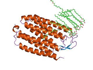
Channelrhodopsins are a subfamily of retinylidene proteins (rhodopsins) that function as light-gated ion channels. They serve as sensory photoreceptors in unicellular green algae, controlling phototaxis: movement in response to light. Expressed in cells of other organisms, they enable light to control electrical excitability, intracellular acidity, calcium influx, and other cellular processes. Channelrhodopsin-1 (ChR1) and Channelrhodopsin-2 (ChR2) from the model organism Chlamydomonas reinhardtii are the first discovered channelrhodopsins. Variants that are sensitive to different colors of light or selective for specific ions have been cloned from other species of algae and protists.

<span class="mw-page-title-main">Photostimulation</span>

Photostimulation is the use of light to artificially activate biological compounds, cells, tissues, or even whole organisms. Photostimulation can be used to noninvasively probe various relationships between different biological processes, using only light. In the long run, photostimulation has the potential for use in different types of therapy, such as migraine headache. Additionally, photostimulation may be used for the mapping of neuronal connections between different areas of the brain by “uncaging” signaling biomolecules with light. Therapy with photostimulation has been called light therapy, phototherapy, or photobiomodulation.

<span class="mw-page-title-main">Halorhodopsin</span> Family of transmembrane proteins


Halorhodopsin is a seven-transmembrane retinylidene protein from microbial rhodopsin family. It is a chloride-specific light-gated ion pump found in archaea known as halobacteria. It is activated by green light wavelengths of approximately 578nm. Halorhodopsin also shares sequence similarity to channelrhodopsin, another light-driven ion channel.

Retinylidene proteins, or rhodopsins in a broad sense, are proteins that use retinal as a chromophore for light reception. They are the molecular basis for a variety of light-sensing systems from phototaxis in flagellates to eyesight in animals. Retinylidene proteins include all forms of opsin and rhodopsin. While rhodopsin in the narrow sense refers to a dim-light visual pigment found in vertebrates, usually on rod cells, rhodopsin in the broad sense refers to any molecule consisting of an opsin and a retinal chromophore in the ground state. When activated by light, the chromophore is isomerized, at which point the molecule as a whole is no longer rhodopsin, but a related molecule such as metarhodopsin. However, it remains a retinylidene protein. The chromophore then separates from the opsin, at which point the bare opsin is a retinylidene protein. Thus, the molecule remains a retinylidene protein throughout the phototransduction cycle.

Light-gated ion channels are a family of ion channels regulated by electromagnetic radiation. Other gating mechanisms for ion channels include voltage-gated ion channels, ligand-gated ion channels, mechanosensitive ion channels, and temperature-gated ion channels. Most light-gated ion channels have been synthesized in the laboratory for study, although two naturally occurring examples, channelrhodopsin and anion-conducting channelrhodopsin, are currently known. Photoreceptor proteins, which act in a similar manner to light-gated ion channels, are generally classified instead as G protein-coupled receptors.

Optogenetics is a biological technique to control the activity of neurons or other cell types with light. This is achieved by expression of light-sensitive ion channels, pumps or enzymes specifically in the target cells. On the level of individual cells, light-activated enzymes and transcription factors allow precise control of biochemical signaling pathways. In systems neuroscience, the ability to control the activity of a genetically defined set of neurons has been used to understand their contribution to decision making, learning, fear memory, mating, addiction, feeding, and locomotion. In a first medical application of optogenetic technology, vision was partially restored in a blind patient.

<span class="mw-page-title-main">Axon terminal</span>

Axon terminals are distal terminations of the branches of an axon. An axon, also called a nerve fiber, is a long, slender projection of a nerve cell that conducts electrical impulses called action potentials away from the neuron's cell body in order to transmit those impulses to other neurons, muscle cells or glands. In the central nervous system, most presynaptic terminals are actually formed along the axons, not at their ends.

Guillardia is a genus of marine biflagellate cryptomonad algae with a plastid obtained through secondary endosymbiosis of a red alga.

<span class="mw-page-title-main">Edward Boyden</span> American neuroscientist

Edward S. Boyden is an American neuroscientist at MIT. He is the Y. Eva Tan Professor in Neurotechnology, a faculty member in the MIT Media Lab and an associate member of the McGovern Institute for Brain Research. In 2018 he was named a Howard Hughes Medical Institute Investigator. He is recognized for his work on optogenetics. In this technology, a light-sensitive ion channel such as channelrhodopsin-2 is genetically expressed in neurons, allowing neuronal activity to be controlled by light. There were early efforts to achieve targeted optical control dating back to 2002 that did not involve a directly light-activated ion channel, but it was the method based on directly light-activated channels from microbes, such as channelrhodopsin, emerging in 2005 that turned out to be broadly useful. Optogenetics in this way has been widely adopted by neuroscientists as a research tool, and it is also thought to have potential therapeutic applications. Boyden joined the MIT faculty in 2007, and continues to develop new optogenetic tools as well as other technologies for the manipulation of brain activity. Previously, Boyden received degrees in electrical engineering, computer science, and physics from MIT. During high school, Boyden attended the Texas Academy of Mathematics and Science.

<span class="mw-page-title-main">Karl Deisseroth</span> American optogeneticist (born 1971)

Karl Alexander Deisseroth is an American scientist. He is the D.H. Chen Professor of Bioengineering and of psychiatry and behavioral sciences at Stanford University.

A Light-oxygen-voltage-sensing domain is a protein sensor used by a large variety of higher plants, microalgae, fungi and bacteria to sense environmental conditions. In higher plants, they are used to control phototropism, chloroplast relocation, and stomatal opening, whereas in fungal organisms, they are used for adjusting the circadian temporal organization of the cells to the daily and seasonal periods. They are a subset of PAS domains.

<span class="mw-page-title-main">Peter Hegemann</span> German biophysicist

Peter Hegemann is a Hertie Senior Research Chair for Neurosciences and a Professor of Experimental Biophysics at the Department of Biology, Faculty of Life Sciences, Humboldt University of Berlin, Germany. He is known for his discovery of channelrhodopsin, a type of ion channels regulated by light, thereby serving as a light sensor. This created the field of optogenetics, a technique that controls the activities of specific neurons by applying light. He has received numerous accolades, including the Rumford Prize, the Shaw Prize in Life Science and Medicine, and the Albert Lasker Award for Basic Medical Research.

<span class="mw-page-title-main">Center for Molecular Neurobiology Hamburg</span> Molecular neuroscience research center

The Center for Molecular Neurobiology Hamburg (ZMNH), founded in 1988, is an internationally recognized molecular neuroscience research center, part of the University Medical Center Hamburg-Eppendorf (UKE), Germany. Headed by Matthias Kneussel, the ZMNH is currently home to 190 scientists and staff from 20 different countries (2022).

<span class="mw-page-title-main">Microbial rhodopsin</span> Retinal-binding proteins

Microbial rhodopsins, also known as bacterial rhodopsins, are retinal-binding proteins that provide light-dependent ion transport and sensory functions in halophilic and other bacteria. They are integral membrane proteins with seven transmembrane helices, the last of which contains the attachment point for retinal.

<span class="mw-page-title-main">Georg Nagel</span>

Georg Nagel is a biophysicist and professor at the Department for Neurophysiology at the University of Würzburg in Germany. His research is focused on microbial photoreceptors and the development of optogenetic tools.

Ernst Bamberg is a German biophysicist and director emeritus of the Department of Biophysical Chemistry at the Max Planck Institute of Biophysics.

<span class="mw-page-title-main">Photoactivated adenylyl cyclase</span>

Photoactivated adenylyl cyclase (PAC) is a protein consisting of an adenylyl cyclase enzyme domain directly linked to a BLUF type light sensor domain. When illuminated with blue light, the enzyme domain becomes active and converts ATP to cAMP, an important second messenger in many cells. In the unicellular flagellate Euglena gracilis, PACα and PACβ (euPACs) serve as a photoreceptor complex that senses light for photophobic responses and phototaxis. Small but potent PACs were identified in the genome of the bacteria Beggiatoa (bPAC) and Oscillatoria acuminata (OaPAC). While natural bPAC has some enzymatic activity in the absence of light, variants with no dark activity have been engineered (PACmn).

Lisa Gunaydin is an American neuroscientist and assistant professor at the Weill Institute for Neurosciences at the University of California San Francisco. Gunaydin helped discover optogenetics in the lab of Karl Deisseroth and now uses this technique in combination with neural and behavioral recordings to probe the neural circuits underlying emotional behaviors.

Fiber photometry is a calcium imaging technique that captures 'bulk' or population-level calcium (Ca2+) activity from specific cell-types within a brain region or functional network in order to study neural circuits Population-level calcium activity can be correlated with behavioral tasks, such as spatial learning, memory recall and goal-directed behaviors. The technique involves the surgical implantation of fiber optics into the brains of living animals. The benefits to researchers are that optical fibers are simpler to implant, less invasive and less expensive than other calcium methods, and there is less weight and stress on the animal, as compared to miniscopes. It also allows for imaging of multiple interacting brain regions and integration with other neuroscience techniques. The limitations of fiber photometry are low cellular and spatial resolution, and the fact that animals must be securely tethered to a rigid fiber bundle, which may impact the naturalistic behavior of smaller mammals such as mice.

Optogenetics began with methods to alter neuronal activity with light, using e.g. channelrhodopsins. In a broader sense, optogenetic approaches also include the use of genetically encoded biosensors to monitor the activity of neurons or other cell types by measuring fluorescence or bioluminescence. Genetically encoded calcium indicators (GECIs) are used frequently to monitor neuronal activity, but other cellular parameters such as membrane voltage or second messenger activity can also be recorded optically. The use of optogenetic sensors is not restricted to neuroscience, but plays increasingly important roles in immunology, cardiology and cancer research.

References

  1. 1 2 3 Wietek, Jonas; Beltramo, Riccardo; Scanziani, Massimo; Hegemann, Peter; Oertner, Thomas G.; Wiegert, J. Simon (2015-10-07). "An improved chloride-conducting channelrhodopsin for light-induced inhibition of neuronal activity in vivo". Scientific Reports. 5: 14807. doi:10.1038/srep14807. ISSN   2045-2322. PMC   4595828 . PMID   26443033.
  2. 1 2 3 Takahashi, Naoya; Oertner, Thomas G.; Hegemann, Peter; Larkum, Matthew E. (2016-12-23). "Active cortical dendrites modulate perception". Science. 354 (6319): 1587–1590. doi:10.1126/science.aah6066. ISSN   0036-8075. PMID   28008068. S2CID   28317052.
  3. 1 2 Wietek, Jonas; Wiegert, J. Simon; Adeishvili, Nona; Schneider, Franziska; Watanabe, Hiroshi; Tsunoda, Satoshi P.; Vogt, Arend; Elstner, Marcus; Oertner, Thomas G.; Hegemann, Peter (2014-04-25). "Conversion of Channelrhodopsin into a Light-Gated Chloride Channel". Science. 344 (6182): 409–412. doi: 10.1126/science.1249375 . ISSN   0036-8075. PMID   24674867. S2CID   206554245.
  4. 1 2 3 Govorunova, Elena G.; Sineshchekov, Oleg A.; Janz, Roger; Liu, Xiaoqin; Spudich, John L. (2015-08-07). "Natural light-gated anion channels: A family of microbial rhodopsins for advanced optogenetics". Science. 349 (6248): 647–650. doi:10.1126/science.aaa7484. ISSN   0036-8075. PMC   4764398 . PMID   26113638.
  5. 1 2 Govorunova, Elena G.; Sineshchekov, Oleg A.; Rodarte, Elsa M.; Janz, Roger; Morelle, Olivier; Melkonian, Michael; Wong, Gane K.-S.; Spudich, John L. (2017-03-03). "The Expanding Family of Natural Anion Channelrhodopsins Reveals Large Variations in Kinetics, Conductance, and Spectral Sensitivity". Scientific Reports. 7: 43358. doi:10.1038/srep43358. ISSN   2045-2322. PMC   5335703 . PMID   28256618.
  6. 1 2 3 Govorunova, Elena G.; Sineshchekov, Oleg A.; Hemmati, Raheleh; Janz, Roger; Morelle, Olivier; Melkonian, Michael; Wong, Gane K.-S.; Spudich, John L. (2018-05-01). "Extending the Time Domain of Neuronal Silencing with Cryptophyte Anion Channelrhodopsins". eNeuro. 5 (3): ENEURO.0174–18.2018. doi:10.1523/ENEURO.0174-18.2018. ISSN   2373-2822. PMC   6051594 . PMID   30027111.
  7. 1 2 3 Kato, Hideaki E.; Kim, Yoon Seok; Paggi, Joseph M.; Evans, Kathryn E.; Allen, William E.; Richardson, Claire; Inoue, Keiichi; Ito, Shota; Ramakrishnan, Charu (2018-08-29). "Structural mechanisms of selectivity and gating in anion channelrhodopsins". Nature. 561 (7723): 349–354. doi:10.1038/s41586-018-0504-5. ISSN   0028-0836. PMC   6317992 . PMID   30158697.
  8. Kim, Yoon Seok; Kato, Hideaki E.; Yamashita, Keitaro; Ito, Shota; Inoue, Keiichi; Ramakrishnan, Charu; Fenno, Lief E.; Evans, Kathryn E.; Paggi, Joseph M. (2018-08-29). "Crystal structure of the natural anion-conducting channelrhodopsin GtACR1". Nature. 561 (7723): 343–348. doi:10.1038/s41586-018-0511-6. ISSN   0028-0836. PMC   6340299 . PMID   30158696.
  9. Postic, Guillaume; Ghouzam, Yassine; Guiraud, Vincent; Gelly, Jean-Christophe (2016). "Membrane positioning for high- and low-resolution protein structures through a binary classification approach". Protein Engineering, Design and Selection. 29 (3): 87–91. doi: 10.1093/protein/gzv063 . PMID   26685702.
  10. Berndt, Andre; Lee, Soo Yeun; Ramakrishnan, Charu; Deisseroth, Karl (2014-04-25). "Structure-Guided Transformation of Channelrhodopsin into a Light-Activated Chloride Channel". Science. 344 (6182): 420–424. doi:10.1126/science.1252367. ISSN   0036-8075. PMC   4096039 . PMID   24763591.
  11. 1 2 Berndt, Andre; Lee, Soo Yeun; Wietek, Jonas; Ramakrishnan, Charu; Steinberg, Elizabeth E.; Rashid, Asim J.; Kim, Hoseok; Park, Sungmo; Santoro, Adam (2016-01-26). "Structural foundations of optogenetics: Determinants of channelrhodopsin ion selectivity". Proceedings of the National Academy of Sciences. 113 (4): 822–829. doi: 10.1073/pnas.1523341113 . ISSN   0027-8424. PMC   4743797 . PMID   26699459.
  12. Chung, Shinjae; Weber, Franz; Zhong, Peng; Tan, Chan Lek; Nguyen, Thuc Nghi; Beier, Kevin T.; Hörmann, Nikolai; Chang, Wei-Cheng; Zhang, Zhe (2017). "Identification of preoptic sleep neurons using retrograde labelling and gene profiling". Nature. 545 (7655): 477–481. doi:10.1038/nature22350. PMC   5554302 . PMID   28514446.
  13. 1 2 Mohammad, Farhan; Stewart, James C; Ott, Stanislav; Chlebikova, Katarina; Chua, Jia Yi; Koh, Tong-Wey; Ho, Joses; Claridge-Chang, Adam (2017). "Optogenetic inhibition of behavior with anion channelrhodopsins". Nature Methods. 14 (3): 271–274. doi:10.1038/nmeth.4148. PMID   28114289. S2CID   4133602.
  14. 1 2 Mauss, Alex S.; Busch, Christian; Borst, Alexander (2017-10-23). "Optogenetic Neuronal Silencing in Drosophila during Visual Processing". Scientific Reports. 7 (1): 13823. doi:10.1038/s41598-017-14076-7. ISSN   2045-2322. PMC   5653863 . PMID   29061981.
  15. Govorunova, Elena G.; Cunha, Shane R.; Sineshchekov, Oleg A.; Spudich, John L. (2016-09-15). "Anion channelrhodopsins for inhibitory cardiac optogenetics". Scientific Reports. 6 (1): 33530. doi:10.1038/srep33530. ISSN   2045-2322. PMC   5024162 . PMID   27628215.
  16. 1 2 Mardinly, Alan R.; Oldenburg, Ian Antón; Pégard, Nicolas C.; Sridharan, Savitha; Lyall, Evan H.; Chesnov, Kirill; Brohawn, Stephen G.; Waller, Laura; Adesnik, Hillel (2018-04-30). "Precise multimodal optical control of neural ensemble activity". Nature Neuroscience. 21 (6): 881–893. doi:10.1038/s41593-018-0139-8. ISSN   1097-6256. PMC   5970968 . PMID   29713079.
  17. 1 2 Mahn, Mathias; Gibor, Lihi; Malina, Katayun Cohen-Kashi; Patil, Pritish; Printz, Yoav; Oring, Shir; Levy, Rivka; Lampl, Ilan; Yizhar, Ofer (2018). "High-efficiency optogenetic silencing with soma-targeted anion-conducting channelrhodopsins". Nature Communications. 9 (1): 4125. doi: 10.1038/s41467-018-06511-8 . PMC   6175909 . PMID   30297821.
  18. Wietek, Jonas; Broser, Matthias; Krause, Benjamin S.; Hegemann, Peter (2016-02-19). "Identification of a Natural Green Light Absorbing Chloride Conducting Channelrhodopsin from Proteomonas sulcata". Journal of Biological Chemistry. 291 (8): 4121–4127. doi: 10.1074/jbc.M115.699637 . ISSN   0021-9258. PMC   4759187 . PMID   26740624.
  19. Govorunova, Elena G.; Sineschekov, Oleg A.; Spudich, John L. (2016-02-01). "Proteomonas sulcata ACR1: A Fast Anion Channelrhodopsin". Photochemistry and Photobiology. 92 (2): 257–263. doi:10.1111/php.12558. PMC   4914479 . PMID   26686819.
  20. 1 2 Wietek, Jonas; Rodriguez-Rozada, Silvia; Tutas, Janine; Tenedini, Federico; Grimm, Christiane; Oertner, Thomas G.; Soba, Peter; Hegemann, Peter; Wiegert, J. Simon (Nov 2017). "Anion-conducting channelrhodopsins with tuned spectra and modified kinetics engineered for optogenetic manipulation of behavior". Scientific Reports. 7 (1): 14957. doi:10.1038/s41598-017-14330-y. ISSN   2045-2322. PMC   5668261 . PMID   29097684.
  21. Oppermann, Johannes; Fischer, Paul; Silapetere, Arita; Liepe, Bernhard; Rodriguez-Rozada, Silvia; Flores-Uribe, José; Peter, Enrico; Keidel, Anke; Vierock, Johannes; Kaufmann, Joel; Broser, Matthias (2019-07-25). "MerMAIDs: a family of metagenomically discovered marine anion-conducting and intensely desensitizing channelrhodopsins". Nature Communications. 10 (1): 3315. doi:10.1038/s41467-019-11322-6. ISSN   2041-1723. PMC   6658528 . PMID   31346176.
  22. Mahn, Mathias; Prigge, Matthias; Ron, Shiri; Levy, Rivka; Yizhar, Ofer (2016). "Biophysical constraints of optogenetic inhibition at presynaptic terminals". Nature Neuroscience. 19 (4): 554–556. doi:10.1038/nn.4266. PMC   4926958 . PMID   26950004.
  23. Vierock, Johannes; Rodriguez-Rozada, Silvia; Dieter, Alexander; Pieper, Florian; Sims, Ruth; Tenedini, Federico; Bergs, Amelie C. F.; Bendifallah, Imane; Zhou, Fangmin; Zeitzschel, Nadja; Ahlbeck, Joachim (2021-07-26). "BiPOLES is an optogenetic tool developed for bidirectional dual-color control of neurons". Nature Communications. 12 (1): 4527. doi:10.1038/s41467-021-24759-5. ISSN   2041-1723. PMC   8313717 . PMID   34312384.
  24. Wiegert, J. Simon; Mahn, Mathias; Prigge, Matthias; Printz, Yoav; Yizhar, Ofer (2017). "Silencing Neurons: Tools, Applications, and Experimental Constraints". Neuron. 95 (3): 504–529. doi:10.1016/j.neuron.2017.06.050. PMC   5830081 . PMID   28772120.
  25. Evanko, Daniel (2014). "Neuroscience: A better way to turn off neurons". Nature Methods. 11 (6): 608. doi: 10.1038/nmeth.2988 . S2CID   1699434.
  26. Berndt, Andre; Deisseroth, Karl (2015-08-07). "Expanding the optogenetics toolkit". Science. 349 (6248): 590–591. doi:10.1126/science.aac7889. ISSN   0036-8075. PMC   4776750 . PMID   26250674.