
The human leg, in the general word sense, is the entire lower limb of the human body, including the foot, thigh and even the hip or gluteal region. However, the definition in human anatomy refers only to the section of the lower limb extending from the knee to the ankle, also known as the crus or, especially in non-technical use, the shank. Legs are used for standing, and all forms of locomotion including recreational such as dancing, and constitute a significant portion of a person's mass. Female legs generally have greater hip anteversion and tibiofemoral angles, but shorter femur and tibial lengths than those in males.
In human anatomy, the thigh is the area between the hip (pelvis) and the knee. Anatomically, it is part of the lower limb.

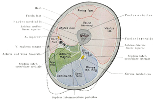
The femoral triangle is an anatomical region of the upper third of the thigh. It is a subfascial space which appears as a triangular depression below the inguinal ligament when the thigh is flexed, abducted and laterally rotated.

The deep artery of the thigh, is a large branch of the femoral artery. It travels more deeply (posteriorly) than the rest of the femoral artery.

The pectineus muscle is a flat, quadrangular muscle, situated at the anterior (front) part of the upper and medial (inner) aspect of the thigh. The pectineus muscle is the most anterior adductor of the hip. The muscle does adduct and internally rotate the thigh but its primary function is hip flexion.

In human anatomy, the groin is the junctional area between the abdomen and the thigh on either side of the pubic bone. This is also known as the medial compartment of the thigh that consists of the adductor muscles of the hip or the groin muscles.

In vertebrate anatomy, hip refers to either an anatomical region or a joint.

The external obturator muscle, obturator externus muscle is a flat, triangular muscle, which covers the outer surface of the anterior wall of the pelvis.
The adductor brevis is a muscle in the thigh situated immediately deep to the pectineus and adductor longus. It belongs to the adductor muscle group. The main function of the adductor brevis is to pull the thigh medially. The adductor brevis and the rest of the adductor muscle group is also used to stabilize left to right movements of the trunk, when standing on both feet, or to balance when standing on a moving surface. The adductor muscle group is used pressing the thighs together to ride a horse, and kicking with the inside of the foot in soccer or swimming. Last, they contribute to flexion of the thigh when running or against resistance.
In the human body, the adductor longus is a skeletal muscle located in the thigh. One of the adductor muscles of the hip, its main function is to adduct the thigh and it is innervated by the obturator nerve. It forms the medial wall of the femoral triangle.
The adductor magnus is a large triangular muscle, situated on the medial side of the thigh.

The iliopsoas muscle refers to the joined psoas and the iliacus muscles. The two muscles are separate in the abdomen, but usually merge in the thigh. They are usually given the common name iliopsoas. The iliopsoas muscle joins to the femur at the lesser trochanter. It acts as the strongest flexor of the hip.
The adductor muscles of the hip are a group of muscles mostly used for bringing the thighs together.

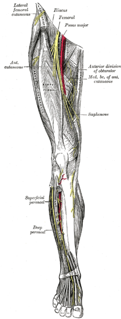
The lumbar plexus is a web of nerves in the lumbar region of the body which forms part of the larger lumbosacral plexus. It is formed by the divisions of the first four lumbar nerves (L1-L4) and from contributions of the subcostal nerve (T12), which is the last thoracic nerve. Additionally, the ventral rami of the fourth lumbar nerve pass communicating branches, the lumbosacral trunk, to the sacral plexus. The nerves of the lumbar plexus pass in front of the hip joint and mainly support the anterior part of the thigh.
The obturator nerve in human anatomy arises from the ventral divisions of the second, third, and fourth lumbar nerves in the lumbar plexus; the branch from the third is the largest, while that from the second is often very small.

The obturator artery is a branch of the internal iliac artery that passes antero-inferiorly on the lateral wall of the pelvis, to the upper part of the obturator foramen, and, escaping from the pelvic cavity through the obturator canal, it divides into both an anterior and a posterior branch.

The medial compartment of thigh is one of the fascial compartments of the thigh and contains the hip adductor muscles and the gracilis muscle.
The posterior branch of the obturator nerve pierces the anterior part of the obturator externus, and supplies this muscle; it then passes behind the adductor brevis on the front of the adductor magnus, where it divides into numerous muscular branches which are distributed to the adductor magnus and the adductor brevis.
Occasionally the communicating branch to the anterior cutaneous and saphenous branches of the femoral is continued down, as a cutaneous branch, to the thigh and leg, as the cutaneous branch of the obturator nerve.
The subsartorial plexus is a plexus of nerves that is located under the sartorius muscle.