The common plantar digital nerves of medial plantar nerve are nerves of the foot. The three common digital nerves (nn. digitales plantares communes) pass between the divisions of the plantar aponeurosis, and each splits into two proper digital nerves:
- Those of the first common digital nerve supply the adjacent sides of the great and second toes;
- Those of the second, the adjacent sides of the second and third toes; and those of the third, the adjacent sides of the third and fourth toes.
- The third common digital nerve receives a communicating branch from the lateral plantar nerve; the first gives a twig to the first Lumbricalis.

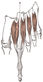
The lumbricals are four small skeletal muscles, accessory to the tendons of the flexor digitorum longus and numbered from the medial side of the foot; they arise from these tendons, as far back as their angles of division, each springing from two tendons, except the first.So the first lumbricle is unipenate and second, third and fourth are bipenate.
Each proper digital nerve gives off cutaneous and articular filaments; and opposite the last phalanx sends upward a dorsal branch, which supplies the structures around the nail, the continuation of the nerve being distributed to the ball of the toe.
It will be observed that these digital nerves are similar in their distribution to those of the median nerve in the hand.

The median nerve is a nerve in humans and other animals in the upper limb. It is one of the five main nerves originating from the brachial plexus.
This page is based on this
Wikipedia article Text is available under the
CC BY-SA 4.0 license; additional terms may apply.
Images, videos and audio are available under their respective licenses.

The human leg, in the general meaning, is the entire lower limb of the human body, including the foot, thigh and even the hip or gluteal region. However, the definition in human anatomy refers only to the section of the lower limb extending from the knee to the ankle, also known as the crus.
Legs are used for standing, and all forms of locomotion including recreational such as dancing, and constitute a significant portion of a person's mass. Female legs generally have greater hip anteversion and tibiofemoral angles, but shorter femur and tibial lengths than those in males.

Toes are the digits of the foot of a tetrapod. Animal species such as cats that walk on their toes are described as being digitigrade. Humans, and other animals that walk on the soles of their feet, are described as being plantigrade; unguligrade animals are those that walk on hooves at the tips of their toes.

The tibial nerve is a branch of the sciatic nerve. The tibial nerve passes through the popliteal fossa to pass below the arch of soleus.
In human anatomy, the dorsal interossei of the foot are four muscles situated between the metatarsal bones.
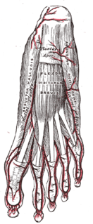
The flexor digitorum brevis lies in the middle of the sole of the foot, immediately above the central part of the plantar aponeurosis, with which it is firmly united.

The superficial peroneal nerve or superior fibular nerve, innervates the peroneus longus and peroneus brevis muscles and the skin over the antero-lateral aspect of the leg along with the greater part of the dorsum of the foot.
The deep peroneal nerve begins at the bifurcation of the common peroneal nerve between the fibula and upper part of the peroneus longus, passes infero-medially, deep to extensor digitorum longus, to the anterior surface of the interosseous membrane, and comes into relation with the anterior tibial artery above the middle of the leg; it then descends with the artery to the front of the ankle-joint, where it divides into a lateral and a medial terminal branch.
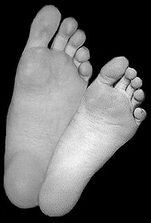
The sole is the bottom of the foot.
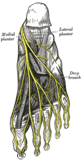
The medial plantar nerve is the larger of the two terminal divisions of the tibial nerve, which accompanies the medial plantar artery.

In the palm of the hand the median nerve is covered by the skin and the palmar aponeurosis, and rests on the tendons of the Flexor muscles. Immediately after emerging from under the transverse carpal ligament the median nerve becomes enlarged and flattened and splits into a smaller, lateral, and a larger, medial portion.
In the palm of the hand the median nerve is covered by the skin and the palmar aponeurosis, and rests on the tendons of the Flexor muscles. Immediately after emerging from under the transverse carpal ligament the median nerve becomes enlarged and flattened and splits into a smaller, lateral, and a larger, medial portion.
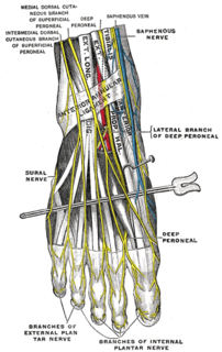
The medial dorsal cutaneous nerve passes in front of the ankle-joint, and divides into two dorsal digital branches, one of which supplies the medial side of the great toe, the other, the adjacent side of the second and third toes.
The superficial branch of the lateral plantar nerve splits into a proper and a common plantar digital nerve:
Dorsal digital nerves of foot are branches of the intermediate dorsal cutaneous nerve, medial dorsal cutaneous nerve, sural nerve and deep fibular nerve. There are 10 total dorsal digital branches.
The proper plantar digital nerves of lateral plantar nerve are nerves of the foot that arise from the superficial branch of the lateral plantar nerve. The superficial branch splits into a proper digital nerve and a common digital nerve:
The common plantar digital nerves of lateral plantar nerve are nerves of the foot. The common digital nerve communicates with the third common digital branch of the medial plantar nerve and divides into two proper digital nerves which supply the adjoining sides of the fourth and fifth toes.
The proper plantar digital nerves of medial plantar nerve are nerves of the foot. They primarily arise from the medial plantar nerve's superficial and deep branches. The superficial branch of the medial plantar nerve turns into a proper digital nerve and is responsible for supplies the medial side of the great toe.
The proper palmar digital nerves of the ulnar nerve are nerves of the hand.
Plantar digital nerves may refer to;
