Assay

Last updated

An assay is an investigative (analytic) procedure in laboratory medicine, mining, pharmacology, environmental biology and molecular biology for qualitatively assessing or quantitatively measuring the presence, amount, or functional activity of a target entity. The measured entity is often called the analyte, the measurand, or the target of the assay. The analyte can be a drug, biochemical substance, chemical element or compound, or cell in an organism or organic sample. [1] [2] An assay usually aims to measure an analyte's intensive property and express it in the relevant measurement unit (e.g. molarity, density, functional activity in enzyme international units, degree of effect in comparison to a standard, etc.).

Contents

If the assay involves exogenous reactants (the reagents), then their quantities are kept fixed (or in excess) so that the quantity and quality of the target are the only limiting factors. The difference in the assay outcome is used to deduce the unknown quality or quantity of the target in question. Some assays (e.g., biochemical assays) may be similar to chemical analysis and titration. However, assays typically involve biological material or phenomena that are intrinsically more complex in composition or behavior, or both. Thus, reading of an assay may be noisy and involve greater difficulties in interpretation than an accurate chemical titration. On the other hand, older generation qualitative assays, especially bioassays, may be much more gross and less quantitative (e.g., counting death or dysfunction of an organism or cells in a population, or some descriptive change in some body part of a group of animals).

Assays have become a routine part of modern medical, environmental, pharmaceutical, and forensic technology. Other businesses may also employ them at the industrial, curbside, or field levels. Assays in high commercial demand have been well investigated in research and development sectors of professional industries. They have also undergone generations of development and sophistication. In some cases, they are protected by intellectual property regulations such as patents granted for inventions. Such industrial-scale assays are often performed in well-equipped laboratories and with automated organization of the procedure, from ordering an assay to pre-analytic sample processing (sample collection, necessary manipulations e.g. spinning for separation, aliquoting if necessary, storage, retrieval, pipetting, aspiration, etc.). Analytes are generally tested in high-throughput autoanalyzers, and the results are verified and automatically returned to ordering service providers and end-users. These are made possible through the use of an advanced laboratory informatics system that interfaces with multiple computer terminals with end-users, central servers, the physical autoanalyzer instruments, and other automata.[ clarification needed ]

Etymology

According to Etymology Online, [3] the verb assay means "to try, endeavor, strive, test the quality of"; from Anglo-French assaier, from assai (noun), from Old French essai, "trial". Thus the noun assay means "trial, test of quality, test of character" (from mid-14th century), from Anglo-French assai; and its meaning "analysis" is from the late 14th century.

For assay of currency coins this literally meant analysis of the purity of the gold or silver (or whatever the precious component) that represented the true value of the coin. This might have translated later (possibly after the 14th century) into a broader usage of "analysis",[ citation needed ] e.g., in pharmacology, analysis for an important component of a target inside a mixture—such as the active ingredient of a drug inside the inert excipients in a formulation that previously was measured only grossly by its observable action on an organism (e.g., a lethal dose or inhibitory dose).

General steps

An assay (analysis) is never an isolated process, as it must be accompanied with pre- and post-analytic procedures. Both the communication order (the request to perform an assay plus related information) and the handling of the specimen itself (the collecting, documenting, transporting, and processing done before beginning the assay) are pre-analytic steps. Similarly, after the assay is completed the results must be documented, verified and communicated—the post-analytic steps. As with any multi-step information handling and transmission system, the variation and errors in reporting final results entail not only those intrinsic to the assay itself but also those occurring in the pre-analytic and post-analytic procedures.

While the analytic steps of the assay itself get much attention, [4] it is those that get less attention of the chain of users—the pre-analytic and post-analytic procedures—that typically accumulate the most errors; e.g., pre-analytic steps in medical laboratory assays may contribute 32–75% of all lab errors. [5]

Assays can be very diverse, but generally involve the following general steps:

  1. Sample processing and manipulation in order to selectively present the target in a discernible or measurable form to a discrimination/identification/detection system. It might involve a simple centrifugal separation or washing or filtration or capture by some form of selective binding or it may even involve modifying the target e.g. epitope retrieval in immunological assays or cutting down the target into pieces e.g. in Mass Spectrometry. Generally there are multiple separate steps done before an assay and are called preanalytic processing. But some of the manipulations may be inseparable part of the assay itself and will not thus be considered pre-analytic.
  2. Target-specific discrimination/identification principle: to discriminate from background (noise) of similar components and specifically identify a particular target component ("analyte") in a biological material by its specific attributes. (e.g. in a PCR assay a specific oligonucleotide primer identifies the target by base pairing based on the specific nucleotide sequence unique to the target).
  3. Signal (or target) amplification system: The presence and quantity of that analyte is converted into a detectable signal generally involving some method of signal amplification, so that it can be easily discriminated from noise and measured - e.g. in a PCR assay among a mixture of DNA sequences only the specific target is amplified into millions of copies by a DNA polymerase enzyme so that it can be discerned as a more prominent component compared to any other potential components. Sometimes the concentration of the analyte is too large and in that case the assay may involve sample dilution or some sort of signal diminution system which is a negative amplification.
  4. Signal detection (and interpretation) system: A system of deciphering the amplified signal into an interpretable output that can be quantitative or qualitative. It can be visual or manual very crude methods or can be very sophisticated electronic digital or analog detectors.
  5. Signal enhancement and noise filtering may be done at any or all of the steps above. Since the more downstream a step/process during an assay, the higher the chance of carrying over noise from the previous process and amplifying it, multiple steps in a sophisticated assay might involve various means of signal-specific sharpening/enhancement arrangements and noise reduction or filtering arrangements. These may simply be in the form of a narrow band-pass optical filter, or a blocking reagent in a binding reaction that prevents nonspecific binding or a quenching reagent in a fluorescence detection system that prevents "autofluorescence" of background objects. [ citation needed ]

Assay types based on the nature of the assay process

Time and number of measurements taken

Depending on whether an assay just looks at a single time point or timed readings taken at multiple time points, an assay may be:

  1. An end point assay, in which a single measurement is performed after a fixed incubation period; or
  2. A kinetic assay, in which measurements are performed multiple times over a fixed time interval. Kinetic assay results may be visualized numerically (for example, as a slope parameter representing the rate of signal change over time), or graphically (for example, as a plot of the signal measured at each time point). For kinetic assays, both the magnitude and shape of the measured response over time provide important information.
  3. A high throughput assay can be either an endpoint or a kinetic assay usually done on an automated platform in 96-, 384- or 1536-well microplate formats (High Throughput Screening). Such assays are able to test large number of compounds or analytes or make functional biological readouts in response to a stimuli and/or compounds being tested. [6]

Number of analytes detected

Depending on how many targets or analytes are being measured:

  1. Usual assays are simple or single target assays which is usually the default unless it is called multiplex.
  2. Multiplex assays are used to simultaneously measure the presence, concentration, activity, or quality of multiple analytes in a single test. The advent of multiplexing enabled rapid, efficient sample testing in many fields, including immunology, cytochemistry, genetics/genomics, pharmacokinetics, and toxicology. [7]

Result type

Depending on the quality of the result produced, assays may be classified into:

  1. Qualitative assays, i.e. assays which generally give just a pass or fail, or positive or negative or some such sort of only small number of qualitative gradation rather than an exact quantity.
  2. Semi-quantitative assays, i.e. assays that give the read-out in an approximate fashion rather than an exact number for the quantity of the substance. Generally they have a few more gradations than just two outcomes, positive or negative, e.g. scoring on a scale of 1+ to 4+ as used for blood grouping tests based on RBC agglutination in response to grouping reagents (antibody against blood group antigens).
  3. Quantitative assays, i.e. assays that give accurate and exact numeric quantitative measure of the amount of a substance in a sample. An example of such an assay used in coagulation testing laboratories for the most common inherited bleeding disease - Von Willebrand disease is VWF antigen assay where the amount of VWF present in a blood sample is measured by an immunoassay.
  4. Functional assays, i.e. an assay that tries to quantify functioning of an active substance rather than just its quantity. The functional counterpart of the VWF antigen assay is Ristocetin Cofactor assay, which measures the functional activity of the VWF present in a patient's plasma by adding exogenous formalin-fixed platelets and gradually increasing quantities of drug named ristocetin while measuring agglutination of the fixed platelets. A similar assay but used for a different purpose is called Ristocetin Induced Platelet Aggregation or RIPA, which tests response of endogenous live platelets from a patient in response to Ristocetin (exogenous) & VWF (usually endogenous).

Sample type and method

Depending on the general substrate on which the assay principle is applied:

  1. Bioassay: when the response is biological activity of live objects. Examples include
    1. in vivo, whole organism (e.g. mouse or other subject injected with a drug)
    2. ex vivo body part (e.g. leg of a frog)
    3. ex vivo organ (e.g. heart of a dog)
    4. ex vivo part of an organ (e.g. a segment of an intestine).
    5. tissue (e.g. limulus lysate)
    6. cell (e.g. platelets)
  2. Ligand binding assay when a ligand (usually a small molecule) binds a receptor (usually a large protein).
  3. Immunoassay when the response is an antigen antibody binding type reaction.

Signal amplification

Depending on the nature of the signal amplification system assays may be of numerous types, to name a few:

  1. Enzyme assay : Enzymes may be tested by their highly repeating activity on a large number of substrates when loss of a substrate or the making of a product may have a measurable attribute like color or absorbance at a particular wavelength or light or Electrochemiluminescence or electrical/redox activity.
  2. Light detection systems that may use amplification e.g. by a photodiode or a photomultiplier tube or a cooled charge coupled device.
  3. Radioisotope labeled substrates as used in radioimmunoassays and equilibrium dialysis assays and can be detected by the amplification in Gamma counters or X-ray plates, or phosphorimager
  4. Polymerase Chain Reaction Assays that amplify a DNA (or RNA) target rather than the signal
  5. Combination Methods Assays may utilize a combination of the above and other amplification methods to improve sensitivity. e.g. Enzyme-linked immunoassay or EIA, enzyme linked immunosorbent assay.

Detection method or technology

Depending on the nature of the Detection system assays can be based on:

  1. Colony forming or virtual colony count: e.g. by multiplying bacteria or proliferating cells.
  2. Photometry / spectrophotometry When the absorbance of a specific wavelength of light while passing through a fixed path-length through a cuvette of liquid test sample is measured and the absorbance is compared with a blank and standards with graded amounts of the target compound. If the emitted light is of a specific visible wavelength it may be called colorimetry , or it may involve specific wavelength of light e.g. by use of laser and emission of fluorescent signals of another specific wavelength which is detected via very specific wavelength optical filters.
  3. Transmittance of light may be used to measure e.g. clearing of opacity of a liquid created by suspended particles due to decrease in number of clumps during a platelet agglutination reaction.
  4. Turbidimetry when the opacity of straight-transmitted light passing through a liquid sample is measured by detectors placed straight across the light source.
  5. Nephelometry where a measurement of the amount of light scattering that occurs when a beam of light is passed through the solution is used to determine size and/or concentration and/or size distribution of particles in the sample. [8]
  6. Reflectometry When color of light reflected from a (usually dry) sample or reactant is assessed e.g. the automated readings of the strip urine dipstick assays.
  7. Viscoelastic measurements e.g. viscometry, elastography (e.g. thromboelastography)
  8. Counting assays: e.g. optic Flow cytometric cell or particle counters, or coulter/impedance principle based cell counters
  9. Imaging assays, that involve image analysis manually or by software:
    1. Cytometry : When the size statistics of cells is assessed by an image processor.
  10. Electric detection e.g. involving amperometry, Voltammetry, coulometry may be used directly or indirectly for many types of quantitative measurements.
  11. Other physical property based assays may use
    1. Osmometer
    2. Viscometer
    3. Ion Selective electrodes
    4. Syndromic testing

Assay types based on the targets being measured

DNA

Assays for studying interactions of proteins with DNA include:

Protein

RNA

Cell counting, viability, proliferation or cytotoxicity assays

A cell-counting assay may determine the number of living cells, the number of dead cells, or the ratio of one cell type to another, such as enumerating and typing red versus different types of white blood cells. This is measured by different physical methods (light transmission, electric current change). But other methods use biochemical probing cell structure or physiology (stains). Another application is to monitor cell culture (assays of cell proliferation or cytotoxicity). A cytotoxicity assay measures how toxic a chemical compound is to cells.

Environmental or food contaminants

Surfactants

Other cell assays

Many cell assays have been developed to assess specific parameters or response of cells (biomarkers, cell physiology). Techniques used to study cells include :

Metastasis Assay

Petrochemistry

Virology

The HPCE-based viral titer assay uses a proprietary, high-performance capillary electrophoresis system to determine baculovirus titer.

The Trofile assay is used to determine HIV tropism.

The viral plaque assay is to calculate the number of viruses present in a sample. In this technique the number of viral plaques formed by a viral inoculum is counted, from which the actual virus concentration can be determined.

Cellular secretions

A wide range of cellular secretions (say, a specific antibody or cytokine) can be detected using the ELISA technique. The number of cells which secrete those particular substances can be determined using a related technique, the ELISPOT assay.

Drugs

Quality

When multiple assays measure the same target their results and utility may or may not be comparable depending on the natures of the assay and their methodology, reliability etc. Such comparisons are possible through study of general quality attributes of the assays e.g. principles of measurement (including identification, amplification and detection), dynamic range of detection (usually the range of linearity of the standard curve), analytic sensitivity, functional sensitivity, analytic specificity, positive, negative predictive values, turn around time i.e. time taken to finish a whole cycle from the preanalytic steps till the end of the last post analytic step (report dispatch/transmission), throughput i.e. number of assays done per unit time (usually expressed as per hour) etc. Organizations or laboratories that perform Assays for professional purposes e.g. medical diagnosis and prognostics, environmental analysis, forensic proceeding, pharmaceutical research and development must undergo well regulated quality assurance procedures including method validation, regular calibration, analytical quality control, proficiency testing, test accreditation, test licensing and must document appropriate certifications from the relevant regulating bodies in order to establish the reliability of their assays, especially to remain legally acceptable and accountable for the quality of the assay results and also to convince customers to use their assay commercially/professionally.

List of BioAssay databases

Bioactivity databases

Bioactivity databases correlate structures or other chemical information to bioactivity results taken from bioassays in literature, patents, and screening programs.

NameDeveloper(s)Initial release
ScrubChem Jason Bret Harris 2016 [10]
PubChem-BioAssay NIH 2004 [11]
ChEMBL EMBL-EBI 2009

Protocol databases

Protocol databases correlate results from bioassays to their metadata about experimental conditions and protocol designs.

NameDeveloper(s)Initial release
BioMetaData or BioAssay Express Collaborative Drug Discovery 2016 [12]
PubChem-BioAssay NIH 2004 [11]

See also

Related Research Articles

<span class="mw-page-title-main">ELISA</span> Method to detect an antigen using an antibody and enzyme

The enzyme-linked immunosorbent assay (ELISA) is a commonly used analytical biochemistry assay, first described by Eva Engvall and Peter Perlmann in 1971. The assay uses a solid-phase type of enzyme immunoassay (EIA) to detect the presence of a ligand in a liquid sample using antibodies directed against the protein to be measured. ELISA has been used as a diagnostic tool in medicine, plant pathology, and biotechnology, as well as a quality control check in various industries.

Viral load, also known as viral burden, is a numerical expression of the quantity of virus in a given volume of fluid, including biological and environmental specimens. It is not to be confused with viral titre or viral titer, which depends on the assay. When an assay for measuring the infective virus particle is done, viral titre often refers to the concentration of infectious viral particles, which is different from the total viral particles. Viral load is measured using body fluids sputum and blood plasma. As an example of environmental specimens, the viral load of norovirus can be determined from run-off water on garden produce. Norovirus has not only prolonged viral shedding and has the ability to survive in the environment but a minuscule infectious dose is required to produce infection in humans: less than 100 viral particles.

<span class="mw-page-title-main">Reverse transcription polymerase chain reaction</span> Laboratory technique to multiply an RNA sample for study

Reverse transcription polymerase chain reaction (RT-PCR) is a laboratory technique combining reverse transcription of RNA into DNA and amplification of specific DNA targets using polymerase chain reaction (PCR). It is primarily used to measure the amount of a specific RNA. This is achieved by monitoring the amplification reaction using fluorescence, a technique called real-time PCR or quantitative PCR (qPCR). Combined RT-PCR and qPCR are routinely used for analysis of gene expression and quantification of viral RNA in research and clinical settings.

von Willebrand disease Medical condition

Von Willebrand disease (VWD) is the most common hereditary blood-clotting disorder in humans. An acquired form can sometimes result from other medical conditions. It arises from a deficiency in the quality or quantity of von Willebrand factor (VWF), a multimeric protein that is required for platelet adhesion. It is known to affect several breeds of dogs as well as humans. The three forms of VWD are hereditary, acquired, and pseudo or platelet type. The three types of hereditary VWD are VWD type 1, VWD type 2, and VWD type 3. Type 2 contains various subtypes. Platelet type VWD is also an inherited condition.

<span class="mw-page-title-main">Automated analyser</span>

An automated analyser is a medical laboratory instrument designed to measure various substances and other characteristics in a number of biological samples quickly, with minimal human assistance. These measured properties of blood and other fluids may be useful in the diagnosis of disease.

A biosensor is an analytical device, used for the detection of a chemical substance, that combines a biological component with a physicochemical detector. The sensitive biological element, e.g. tissue, microorganisms, organelles, cell receptors, enzymes, antibodies, nucleic acids, etc., is a biologically derived material or biomimetic component that interacts with, binds with, or recognizes the analyte under study. The biologically sensitive elements can also be created by biological engineering. The transducer or the detector element, which transforms one signal into another one, works in a physicochemical way: optical, piezoelectric, electrochemical, electrochemiluminescence etc., resulting from the interaction of the analyte with the biological element, to easily measure and quantify. The biosensor reader device connects with the associated electronics or signal processors that are primarily responsible for the display of the results in a user-friendly way. This sometimes accounts for the most expensive part of the sensor device, however it is possible to generate a user friendly display that includes transducer and sensitive element. The readers are usually custom-designed and manufactured to suit the different working principles of biosensors.

<span class="mw-page-title-main">Calibration curve</span> Method for determining the concentration of a substance in an unknown sample

In analytical chemistry, a calibration curve, also known as a standard curve, is a general method for determining the concentration of a substance in an unknown sample by comparing the unknown to a set of standard samples of known concentration. A calibration curve is one approach to the problem of instrument calibration; other standard approaches may mix the standard into the unknown, giving an internal standard. The calibration curve is a plot of how the instrumental response, the so-called analytical signal, changes with the concentration of the analyte.

<span class="mw-page-title-main">Biochip</span> Substrates performing biochemical reactions

In molecular biology, biochips are engineered substrates that can host large numbers of simultaneous biochemical reactions. One of the goals of biochip technology is to efficiently screen large numbers of biological analytes, with potential applications ranging from disease diagnosis to detection of bioterrorism agents. For example, digital microfluidic biochips are under investigation for applications in biomedical fields. In a digital microfluidic biochip, a group of (adjacent) cells in the microfluidic array can be configured to work as storage, functional operations, as well as for transporting fluid droplets dynamically.

<span class="mw-page-title-main">Radioimmunoassay</span> Immunoassay that uses radiolabeled molecules

A radioimmunoassay (RIA) is an immunoassay that uses radiolabeled molecules in a stepwise formation of immune complexes. A RIA is a very sensitive in vitro assay technique used to measure concentrations of substances, usually measuring antigen concentrations by use of antibodies.

<span class="mw-page-title-main">Immunoassay</span> Biochemical test for a protein or other molecule using an antibody

An immunoassay (IA) is a biochemical test that measures the presence or concentration of a macromolecule or a small molecule in a solution through the use of an antibody (usually) or an antigen (sometimes). The molecule detected by the immunoassay is often referred to as an "analyte" and is in many cases a protein, although it may be other kinds of molecules, of different sizes and types, as long as the proper antibodies that have the required properties for the assay are developed. Analytes in biological liquids such as serum or urine are frequently measured using immunoassays for medical and research purposes.

The limit of detection is the lowest signal, or the lowest corresponding quantity to be determined from the signal, that can be observed with a sufficient degree of confidence or statistical significance. However, the exact threshold used to decide when a signal significantly emerges above the continuously fluctuating background noise remains arbitrary and is a matter of policy and often of debate among scientists, statisticians and regulators depending on the stakes in different fields.

<span class="mw-page-title-main">Real-time polymerase chain reaction</span> Laboratory technique of molecular biology

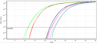
A real-time polymerase chain reaction is a laboratory technique of molecular biology based on the polymerase chain reaction (PCR). It monitors the amplification of a targeted DNA molecule during the PCR, not at its end, as in conventional PCR. Real-time PCR can be used quantitatively and semi-quantitatively.

<span class="mw-page-title-main">Lateral flow test</span> Immunochromatographic testing devices

A lateral flow test (LFT), is an assay also known as a lateral flow device (LFD), lateral flow immunochromatographic assay, or rapid test. It is a simple device intended to detect the presence of a target substance in a liquid sample without the need for specialized and costly equipment. LFTs are widely used in medical diagnostics in the home, at the point of care, and in the laboratory. For instance, the home pregnancy test is an LFT that detects a specific hormone. These tests are simple and economical and generally show results in around five to thirty minutes. Many lab-based applications increase the sensitivity of simple LFTs by employing additional dedicated equipment. Because the target substance is often a biological antigen, many lateral flow tests are rapid antigen tests.

Virus quantification is counting or calculating the number of virus particles (virions) in a sample to determine the virus concentration. It is used in both research and development (R&D) in academic and commercial laboratories as well as in production situations where the quantity of virus at various steps is an important variable that must be monitored. For example, the production of virus-based vaccines, recombinant proteins using viral vectors, and viral antigens all require virus quantification to continually monitor and/or modify the process in order to optimize product quality and production yields and to respond to ever changing demands and applications. Other examples of specific instances where viruses need to be quantified include clone screening, multiplicity of infection (MOI) optimization, and adaptation of methods to cell culture.

There are many methods to investigate protein–protein interactions which are the physical contacts of high specificity established between two or more protein molecules involving electrostatic forces and hydrophobic effects. Each of the approaches has its own strengths and weaknesses, especially with regard to the sensitivity and specificity of the method. A high sensitivity means that many of the interactions that occur are detected by the screen. A high specificity indicates that most of the interactions detected by the screen are occurring in reality.

<span class="mw-page-title-main">Surround optical-fiber immunoassay</span>

Surround optical-fiber immunoassay (SOFIA) is an ultrasensitive, in vitro diagnostic platform incorporating a surround optical-fiber assembly that captures fluorescence emissions from an entire sample. The technology's defining characteristics are its extremely high limit of detection, sensitivity, and dynamic range. SOFIA's sensitivity is measured at the attogram level (10−18 g), making it about one billion times more sensitive than conventional diagnostic techniques. Based on its enhanced dynamic range, SOFIA is able to discriminate levels of analyte in a sample over 10 orders of magnitude, facilitating accurate titering.

A ligand binding assay (LBA) is an assay, or an analytic procedure, which relies on the binding of ligand molecules to receptors, antibodies or other macromolecules. A detection method is used to determine the presence and extent of the ligand-receptor complexes formed, and this is usually determined electrochemically or through a fluorescence detection method. This type of analytic test can be used to test for the presence of target molecules in a sample that are known to bind to the receptor.

<span class="mw-page-title-main">CD/DVD based immunoassay</span>

A compact disk/digital versatile disk (CD/DVD) based immunoassay is a method for determining the concentration of a compound in research and diagnostic laboratories by performing the test on an adapted CD/DVD surface using an adapted optical disc drive; these methods have been discussed and prototyped in research labs since 1991.

<span class="mw-page-title-main">Fluorescence polarization immunoassay</span> Class of invitro biochemical test

Fluorescence polarization immunoassay (FPIA) is a class of in vitro biochemical test used for rapid detection of antibody or antigen in sample. FPIA is a competitive homogenous assay, that consists of a simple prepare and read method, without the requirement of separation or washing steps.

The enzyme-linked immunosorbent spot (ELISpot) is a type of assay that focuses on quantitatively measuring the frequency of cytokine secretion for a single cell. The ELISpot Assay is also a form of immunostaining since it is classified as a technique that uses antibodies to detect a protein analyte, with the word analyte referring to any biological or chemical substance being identified or measured.

References

  1. The American heritage dictionary of the English language (4th ed.). Boston, MA: Houghton Mifflin. 2006. ISBN   9780618701735.
  2. Abate, Frank (2001). J. Jewell, Elizabeth (ed.). The new Oxford American dictionary (2nd ed.). Oxford: Oxford University Press. ISBN   9780195112276.
  3. "Online Etymology Dictionary - Assay". etymonline. Douglas Harper. 2016. Retrieved 13 August 2016.
  4. Bonini, P; Plebani, M; Ceriotti, F; Rubboli, F (May 2002). "Errors in laboratory medicine". Clinical Chemistry. 48 (5): 691–8. doi: 10.1093/clinchem/48.5.691 . PMID   11978595.
  5. Hammerling, Julie A. (1 February 2012). "A Review of Medical Errors in Laboratory Diagnostics and Where We Are Today: Table 1". Laboratory Medicine. 43 (2): 41–44. doi: 10.1309/LM6ER9WJR1IHQAUY .
  6. Sittampalam, GS (2004). "Assay Guidance Manual [Internet]". ncbi.nlm.com. Eli Lilly & Company and the National Center for Advancing Translational Sciences . Retrieved 12 August 2016.
  7. Banks, Peter (7 June 2010). "Multiplexed Assays in the Life Sciences". biotek.com. BioTek Instruments Inc. Retrieved 13 August 2016.
  8. "Nephelometry". The Free Dictionary. Farlex. 2016. Retrieved 9 September 2016.
  9. Lowry OH, Rosebrough NJ, Farr AL, Randall RJ (November 1951). "Protein measurement with the Folin phenol reagent". J. Biol. Chem. 193 (1): 265–75. doi: 10.1016/S0021-9258(19)52451-6 . PMID   14907713.
  10. Harris, JB (2019). Post-processing of Large Bioactivity Data. Methods in Molecular Biology. Vol. 1939. pp. 37–47. doi:10.1007/978-1-4939-9089-4_3. ISBN   978-1-4939-9088-7. PMID   30848455. S2CID   73493315.
  11. 1 2 Wang, Yanli; Bryant, Stephen H.; Cheng, Tiejun; Wang, Jiyao; Gindulyte, Asta; Shoemaker, Benjamin A.; Thiessen, Paul A.; He, Siqian; Zhang, Jian (4 January 2017). "PubChem BioAssay: 2017 update". Nucleic Acids Research. 45 (D1): D955–D963. doi:10.1093/nar/gkw1118. PMC   5210581 . PMID   27899599.
  12. "Home". assay.biometadata.com.