Blastic plasmacytoid dendritic cell neoplasm

Last updated
Blastic plasmacytoid dendritic cell neoplasm
PMC4314974 OL-09-03-1388-g02.png
CD4+ CD56+ lymphoma

Blastic plasmacytoid dendritic cell neoplasm (BPDCN) is a rare hematologic malignancy. It was initially regarded as a form of lymphocyte-derived cutaneous lymphoma and alternatively named CD4+CD56+ hematodermic tumor, blastic NK cell lymphoma, [1] and agranular CD4+ NK cell leukemia. [2] Later, however, the disease was determined to be a malignancy of plasmacytoid dendritic cells rather than lymphocytes and therefore termed blastic plasmacytoid dendritic cell neoplasm. In 2016, the World Health Organization designated BPDCN to be in its own separate category within the myeloid class of neoplasms. [3] It is estimated that BPDCN constitutes 0.44% of all hematological malignancies. [4]

Contents

Blastic plasmacytoid dendritic cell neoplasm is an aggressive malignancy with features of cutaneous lymphoma (e.g. malignant plasmacytoid dendritic cell infiltrations into the skin to form single or multiple lesions) and/or leukemia (i.e. malignant plasmacytoid dendritic cells in blood and bone marrow). [2] While commonly presenting with these clinical features, BPDCN, particularly in its more advanced stages, may also involve malignant plasmacytoid dendritic cell infiltrations in and thereby injury to the liver, spleen, lymph nodes, central nervous system, or other tissues. The neoplasm occurs in individuals of all ages but predominates in the elderly; in children, it afflicts males and females equally but in adults is far more common (~75% of cases) in males. [5]

Blastic plasmacytoid dendritic cell neoplasm typically responds to chemotherapy regimens used to treat hematological malignancies. All too often, however, the disease rapidly recurs and does so in a more drug-resistant form. [5] Furthermore, the disease may occur in association with the myelodysplastic syndrome or transform to acute myeloid leukemia. [4] Consequently, BPDCN has a very low 5 year survival rate. [5] Current translational research studies on treating BPDCN have therefore focused on non-chemotherapeutic regimens that target the molecular pathways which may promote the disease. [6]

Presentation

Blastic plasmacytoid dendritic cell neoplasm occurs in children, [5] including neonates, [7] but is more common in adults, particularly those between the ages 60–80. [5] BPDCN usually (i.e. 61% [5] to 90% [8] of cases) presents with skin lesions, i.e. nodules, tumors, red or purple papules, bruise-like patches, and/or ulcers that most often occur on the head, face, and upper torso. [2] The lesions are due to diffuse infiltrations of the skin by malignant pDC. In one large study, this presentation was accompanied by swollen lymph nodes, usually in the neck, due to malignant pDC infiltrations (~50% of cases); enlarged liver (~16% of cases) and/or spleen (26% of cases), also due to malignant pDC infiltrations; [5] increased levels of malignant pDC in blood (i.e. >2% of nucleated cells) (~40% of cases), bone marrow (~65% of cases) and cerebrospinal fluid (47% of childhood cases but less often detected in adult cases). [5] More advanced or severe cases may present with extreme organ and/or lymph node enlargements, skin lesions in virtually any site, and clinical evidence of malignant pDC infiltrations in the breasts, eyes, kidneys, lungs, gastrointestinal tract, bone, sinuses, ears, or testes. [5] About 10% of individuals with BPDCN present with a leukemia-like disease, [4] i.e. they exhibit circulating malignant pDC, anemia, thrombocytopenia, and/or leukopenia due to extensive malignant pDC infiltrations in the bone marrow. [4] A leukemic phase of the disease is a common feature of end stage and post-therapy relapsing BPDCN. [2]

Pathophysiology

There are three types of dendritic cells, plasmacytic dendritic cells (pDC) and two types of conventional dendritic cells (cDC), myeloid cDC1 and myeloid cDC2. [9] pDC circulate in the blood, representing <0.4% of all nucleated blood cells, and are present in various hematological tissues such as lymph nodes and spleen. [2] Their major function is to detect and then initiate immune responses to intracellular pathogens, particularly viruses such as the cold sore-causing Herpes simplex viruses, HIV, and hepatitis viruses but also bacteria such as the tuberculosis-causing Mycobacterium tuberculosis , fungi such as the aspergillosis-causing Aspergillus fumigatus and parasites such as malaria-causing Plasmodium falciparum . Following detection of these intracellular pathogens, pCD initiate immune responses by producing massive amounts of type I [10] and type III [9] interferons as well as by differentiating (i.e. maturing) into conventional dendritic cells that further promote immune responses by, e.g. functioning as antigen-presenting cells. [10] The malignant pDC in BPDCN have the appearance of immature plasmacytoid dendritic cells. They are distinguished from other dendritic, myeloid, lymphoid and NK cell types by exhibiting at least several of the following properties: 1) plasmacytoid morphology; 2) production of large amounts of type I interferons when properly stimulated; 3) ability to differentiate into conventional dendritic cells when properly stimulated; 4) the expression of key marker proteins such as granzyme B, [10] TCF4, [11] interleukin-3 receptor (i.e. CD123), CLEC4C, and Neuropilin, [9] and 5) failure to express certain marker proteins that are commonly expressed by myeloid, lymphoid, and NK cell lineages. [11]

Blastic plasmacytoid dendritic cell neoplasm typically arises after the serial acquisition of multiple genetic abnormalities in pDC or their precursor cells. Inactivating mutations (i.e. mutations which cause the gene to make no or a less active product) in the TET2 gene are the most common genetic abnormality in the disease, [11] occurring in 32–67% of all BPDCN cases and often accompanied by mutations in either the NPM1 or SRSF2 gene. Numerous other genetic abnormalities are associated with the disease: 1) mutations in NRAS, ASXL1, and TP53; 2) deletions of the CDKN2A-ARF-CDKN2B locus on the short arm of chromosome 9, CDKN1B locus on the short arm of chromosome 12, RB1 locus on the long arm of chromosome 13, or NRC1 locus on the long arm of chromosome 5; 3) fusions of KMT2A on the long arm of chromosome 11 with MLLT1 on the short arm of chromosome 10, SUPT3H on the short arm of chromosome with MYC on the long arm of chromosome 8, or KMT2A on the long arm of chromosome 11 with MLLT1 on the long arm of chromosome 19; [12] and 4) duplication or loss of entire chromosomes, particularly chromosomes 9, 13, or 15. [4] Laboratory studies indicate that malignant pDC have a pathologically overactive NF-κB pathway that promotes their survival and production of various cytokines) that stimulate their own proliferation. [8] Presumably, these genetic abnormalities lead to the activation of the NF-κB pathway and/or other cellular activation pathways which promote the survival, proliferation, and/or other malignant phenotypic traits in pDC and thereby cause BPDCN. [12]

Diagnosis

BPDCN is suggested by a biopsy of skin lesions which reveals the infiltration by medium-sized blast (i.e. immature) cells into the dermis while sparing the epidermis. [4] These cells exhibit irregular nuclei, fine chromatin, and at least one small nucleolus. [8] Such blast cells may also be observed in the circulation, bone marrow, or other tissues and suggest BPDCN. However, the diagnosis of this disease requires determination that these cells are pDC blast cells rather than AML, T-cell lymphoblastic lymphoma (TCLL), or aggressive NK-cell leukemia (NKL) blast cells. Various studies have offered similar but not identical criteria to make this determination. All studies agree that pDC should have a typical plasmacytoid morphology and express a particular profile of marker proteins as detected by immunoassay and/or flow cytometry. However, the studies disagree on which marker proteins to profile. One study's profile assayed 1) CD4, CD56, CD123 (i.e. Interleukin-3 receptor, and TLC1, which are expressed on 80–100% of pDC but uncommon on AML, TCLL, or NKL blasts); 2) CD2AP and CLEC4C which are unique to pDC; and 3) myeloperoxidase, lysozyme, CD34, CD14, CD11c, and CD163 which are unique to AML, TCLL, or NKL blasts. [4] Two other studies recommended assaying somewhat different sets of marker proteins. [2] [12]

Treatment

There have been no controlled studies to define the optimal treatment for BPDCN. [8] Studies on small numbers of individuals with the disease have found that the standard chemotherapy regimens used for the initial induction treatments of AML, acute lymphoblastic leukemia, and high-grade lymphoma give complete remission rates of 77%, 93%, and 80%, respectively, in childhood PBDN and 47%, 77%, and 53%, respectively, in adult PBDN. However, these remissions were short-lived: post-treatment mean times to relapse or death were 12 months for children and 6.8 months for adults. [5] Given these poor remission and survival rates, other treatments have been added to the initial treatment regimens. Studies have shown that the addition of intrathecally administered drugs (administered directly into the spinal canal) as prophylaxis prolongs the period of CNS-free disease and increases overall survival. Hematopoietic stem cell transplantation following initial chemotherapy-induced remission also prolongs these remissions and, it is suggested, offers potential for curing the disease. (A graft-versus-leukemia effect may have contributed to the benefits seen after transplantation.) [4] Studies have not yet determined whether allogenic (i.e. taken from others) or autologous (i.e. taken from self) stem cells achieve better results, although one retrospective study in Japan found that autologous stem cells gave significantly better overall and progression-free survival rates. [8] A phase I clinical research study to test the safety and efficacy of a combination chemotherapy regimen consisting of methotrexate, L-asparaginase, idarubicin, and dexamethasone followed by allogenic or autologous bone marrow transplantation in 26 participants newly diagnosed with BPDCN is planned but not yet in its recruiting phase. [13]

While few studies have reported on the treatment of BPDCN that has recurred following initial therapy, donor lymphocyte infusions coupled with alternative chemotherapy treatments have induced second complete or partial remissions in a few patients. [4]

Tagraxofusp-erzs

Tagraxofusp-erzs (trade name Elzonris; formerly SL-401 and DT388-IL3) was approved in the United States in December 2018 for the treatment of BPDCN. [14] Tagraxofusp-erzs is a fusion protein consisting of interleukin 3 (i.e. IL-3) fused to diphtheria toxin. The fusion protein readily kills cultured pDC by binding to their IL-3 receptors to thereby gain entrance to the cells and then blocking these cells' protein synthesis (due to diphtheria toxin-mediated inhibition of eukaryotic elongation factor 2).[ citation needed ]

Prognosis

Due to the high rates of recurrence following initial therapy and the short overall survival times of individuals with BPDCN, prognosis of the disease is poor. However, further study of treatment regimens that include intrathecal chemotherapy and hematological stem cell transplantation in initial treatment regimens (see previous section) and newer non-chemotherapeutic drug treatments (see next section) may improve this situation. [8]

Research

UCART123

UCART123 are chimeric T cell receptor-bearing cells, i.e. T lymphocytes engineered to bear a monoclonal antibody that directs them to attack and kill BPDCN cells. The intravenous infusion of these cells in patients with BPDCN is in phase 1 clinical trials [15] but in September 2017, the Federal Drug Administration suspended these because one patient developed a Grade 5 (i.e. lethal) cytokine release syndrome (see UCART123#CAR-T cancer treatment). [15] The suspension was lifted in November 2017 after the trial used reduced amounts of the cells and with additional conditions were applied. [16] A new phase 1 clinical trial is now recruiting 76 new patients to study the safety and efficacy of UCAR123 in treating BPDCN. The study began in June 2017 and is scheduled to end in December 2021. [17]

Venetoclax

BCL-2 is a cellular protein that can act to inhibit cell death due to apoptosis. The BCL-2 gene appears to be one of the most up-regulated (i.e. overactive) genes in BPDCN. Venetoclax inhibits the apoptosis-inhibiting action of BCL-2 and proved active in treating two patients with relapsed or refractory BPDCN. [8] A phase I clinical trial testing the safety and efficacy of the drug in BPDCN is planned but not yet in its recruiting phase. [18]

Related Research Articles

<span class="mw-page-title-main">Tumors of the hematopoietic and lymphoid tissues</span> Tumors that affect the blood, bone marrow, lymph, and lymphatic system

Tumors of the hematopoietic and lymphoid tissues or tumours of the haematopoietic and lymphoid tissues are tumors that affect the blood, bone marrow, lymph, and lymphatic system. Because these tissues are all intimately connected through both the circulatory system and the immune system, a disease affecting one will often affect the others as well, making aplasia, myeloproliferation and lymphoproliferation closely related and often overlapping problems. While uncommon in solid tumors, chromosomal translocations are a common cause of these diseases. This commonly leads to a different approach in diagnosis and treatment of hematological malignancies. Hematological malignancies are malignant neoplasms ("cancer"), and they are generally treated by specialists in hematology and/or oncology. In some centers "hematology/oncology" is a single subspecialty of internal medicine while in others they are considered separate divisions. Not all hematological disorders are malignant ("cancerous"); these other blood conditions may also be managed by a hematologist.

<span class="mw-page-title-main">Anaplastic large-cell lymphoma</span> Medical condition

Anaplastic large-cell lymphoma (ALCL) refers to a group of non-Hodgkin lymphomas in which aberrant T cells proliferate uncontrollably. Considered as a single entity, ALCL is the most common type of peripheral lymphoma and represents ~10% of all peripheral lymphomas in children. The incidence of ALCL is estimated to be 0.25 cases per 100,000 people in the United States of America. There are four distinct types of anaplastic large-cell lymphomas that on microscopic examination share certain key histopathological features and tumor marker proteins. However, the four types have very different clinical presentations, gene abnormalities, prognoses, and/or treatments.

<span class="mw-page-title-main">Myeloid sarcoma</span> Medical condition

A myeloid sarcoma is a solid tumor composed of immature white blood cells called myeloblasts. A chloroma is an extramedullary manifestation of acute myeloid leukemia; in other words, it is a solid collection of leukemic cells occurring outside of the bone marrow.

<span class="mw-page-title-main">Follicular lymphoma</span> Medical condition

Follicular lymphoma (FL) is a cancer that involves certain types of white blood cells known as lymphocytes. The cancer originates from the uncontrolled division of specific types of B-cells known as centrocytes and centroblasts. These cells normally occupy the follicles in the germinal centers of lymphoid tissues such as lymph nodes. The cancerous cells in FL typically form follicular or follicle-like structures in the tissues they invade. These structures are usually the dominant histological feature of this cancer.

<span class="mw-page-title-main">Primary effusion lymphoma</span> Medical condition

Primary effusion lymphoma (PEL) is classified as a diffuse large B cell lymphoma. It is a rare malignancy of plasmablastic cells that occurs in individuals that are infected with the Kaposi's sarcoma-associated herpesvirus. Plasmablasts are immature plasma cells, i.e. lymphocytes of the B-cell type that have differentiated into plasmablasts but because of their malignant nature do not differentiate into mature plasma cells but rather proliferate excessively and thereby cause life-threatening disease. In PEL, the proliferating plasmablastoid cells commonly accumulate within body cavities to produce effusions, primarily in the pleural, pericardial, or peritoneal cavities, without forming a contiguous tumor mass. In rare cases of these cavitary forms of PEL, the effusions develop in joints, the epidural space surrounding the brain and spinal cord, and underneath the capsule which forms around breast implants. Less frequently, individuals present with extracavitary primary effusion lymphomas, i.e., solid tumor masses not accompanied by effusions. The extracavitary tumors may develop in lymph nodes, bone, bone marrow, the gastrointestinal tract, skin, spleen, liver, lungs, central nervous system, testes, paranasal sinuses, muscle, and, rarely, inside the vasculature and sinuses of lymph nodes. As their disease progresses, however, individuals with the classical effusion-form of PEL may develop extracavitary tumors and individuals with extracavitary PEL may develop cavitary effusions.

<span class="mw-page-title-main">Acute myeloid leukemia</span> Cancer of the myeloid line of blood cells

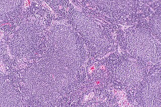
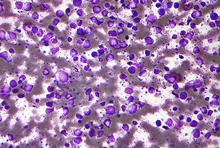
Acute myeloid leukemia (AML) is a cancer of the myeloid line of blood cells, characterized by the rapid growth of abnormal cells that build up in the bone marrow and blood and interfere with normal blood cell production. Symptoms may include feeling tired, shortness of breath, easy bruising and bleeding, and increased risk of infection. Occasionally, spread may occur to the brain, skin, or gums. As an acute leukemia, AML progresses rapidly, and is typically fatal within weeks or months if left untreated.

<span class="mw-page-title-main">Diffuse large B-cell lymphoma</span> Type of blood cancer

Diffuse large B-cell lymphoma (DLBCL) is a cancer of B cells, a type of lymphocyte that is responsible for producing antibodies. It is the most common form of non-Hodgkin lymphoma among adults, with an annual incidence of 7–8 cases per 100,000 people per year in the US and UK. This cancer occurs primarily in older individuals, with a median age of diagnosis at ~70 years, although it can occur in young adults and, in rare cases, children. DLBCL can arise in virtually any part of the body and, depending on various factors, is often a very aggressive malignancy. The first sign of this illness is typically the observation of a rapidly growing mass or tissue infiltration that is sometimes associated with systemic B symptoms, e.g. fever, weight loss, and night sweats.

Plasmacytoid dendritic cells (pDCs) are a rare type of immune cell that are known to secrete large quantities of type 1 interferon (IFNs) in response to a viral infection. They circulate in the blood and are found in peripheral lymphoid organs. They develop from bone marrow hematopoietic stem cells and constitute < 0.4% of peripheral blood mononuclear cells (PBMC). Other than conducting antiviral mechanisms, pDCs are considered to be key in linking the innate and adaptive immune systems. However, pDCs are also responsible for participating in and exacerbating certain autoimmune diseases like lupus. pDCs that undergo malignant transformation cause a rare hematologic disorder, blastic plasmacytoid dendritic cell neoplasm.

<span class="mw-page-title-main">ETV6</span> Protein-coding gene in the species Homo sapiens

ETV6 protein is a transcription factor that in humans is encoded by the ETV6 gene. The ETV6 protein regulates the development and growth of diverse cell types, particularly those of hematological tissues. However, its gene, ETV6 frequently suffers various mutations that lead to an array of potentially lethal cancers, i.e., ETV6 is a clinically significant proto-oncogene in that it can fuse with other genes to drive the development and/or progression of certain cancers. However, ETV6 is also an anti-oncogene or tumor suppressor gene in that mutations in it that encode for a truncated and therefore inactive protein are also associated with certain types of cancers.

Richter's transformation (RT), also known as Richter's syndrome, is the conversion of chronic lymphocytic leukemia (CLL) or its variant, small lymphocytic lymphoma (SLL), into a new and more aggressively malignant disease. CLL is the circulation of malignant B lymphocytes with or without the infiltration of these cells into lymphatic or other tissues while SLL is the infiltration of these malignant B lymphocytes into lymphatic and/or other tissues with little or no circulation of these cells in the blood. CLL along with its SLL variant are grouped together in the term CLL/SLL.

<span class="mw-page-title-main">PDGFRB</span> Protein-coding gene in the species Homo sapiens

Platelet-derived growth factor receptor beta is a protein that in humans is encoded by the PDGFRB gene. Mutations in PDGFRB are mainly associated with the clonal eosinophilia class of malignancies.

<span class="mw-page-title-main">Acute megakaryoblastic leukemia</span> Medical condition

Acute megakaryoblastic leukemia (AMKL) is life-threatening leukemia in which malignant megakaryoblasts proliferate abnormally and injure various tissues. Megakaryoblasts are the most immature precursor cells in a platelet-forming lineage; they mature to promegakaryocytes and, ultimately, megakaryocytes which cells shed membrane-enclosed particles, i.e. platelets, into the circulation. Platelets are critical for the normal clotting of blood. While malignant megakaryoblasts usually are the predominant proliferating and tissue-damaging cells, their similarly malignant descendants, promegakaryocytes and megakaryocytes, are variable contributors to the malignancy.

<span class="mw-page-title-main">Marginal zone B-cell lymphoma</span> Group of lymphomas

Marginal zone B-cell lymphomas, also known as marginal zone lymphomas (MZLs), are a heterogeneous group of lymphomas that derive from the malignant transformation of marginal zone B-cells. Marginal zone B cells are innate lymphoid cells that normally function by rapidly mounting IgM antibody immune responses to antigens such as those presented by infectious agents and damaged tissues. They are lymphocytes of the B-cell line that originate and mature in secondary lymphoid follicles and then move to the marginal zones of mucosa-associated lymphoid tissue, the spleen, or lymph nodes. Mucosa-associated lymphoid tissue is a diffuse system of small concentrations of lymphoid tissue found in various submucosal membrane sites of the body such as the gastrointestinal tract, mouth, nasal cavity, pharynx, thyroid gland, breast, lung, salivary glands, eye, skin and the human spleen.

<span class="mw-page-title-main">Extranodal NK/T-cell lymphoma, nasal type</span> Medical condition

Extranodal NK/T-cell lymphoma, nasal type (ENKTCL-NT) is a rare type of lymphoma that commonly involves midline areas of the nasal cavity, oral cavity, and/or pharynx At these sites, the disease often takes the form of massive, necrotic, and extremely disfiguring lesions. However, ENKTCL-NT can also involve the eye, larynx, lung, gastrointestinal tract, skin, and various other tissues. ENKTCL-NT mainly affects adults; it is relatively common in Asia and to lesser extents Mexico, Central America, and South America but is rare in Europe and North America. In Korea, ENKTCL-NT often involves the skin and is reported to be the most common form of cutaneous lymphoma after mycosis fungoides.

Myeloid leukemia is a type of leukemia affecting myeloid tissue.

Clonal hypereosinophilia, also termed primary hypereosinophilia or clonal eosinophilia, is a grouping of hematological disorders all of which are characterized by the development and growth of a pre-malignant or malignant population of eosinophils, a type of white blood cell that occupies the bone marrow, blood, and other tissues. This population consists of a clone of eosinophils, i.e. a group of genetically identical eosinophils derived from a sufficiently mutated ancestor cell.

GATA2 deficiency is a grouping of several disorders caused by common defect, namely, familial or sporadic inactivating mutations in one of the two parental GATA2 genes. Being the gene haploinsufficient, mutations that cause a reduction in the cellular levels of the gene's product, GATA2, are autosomal dominant. The GATA2 protein is a transcription factor critical for the embryonic development, maintenance, and functionality of blood-forming, lymphatic-forming, and other tissue-forming stem cells. In consequence of these mutations, cellular levels of GATA2 are deficient and individuals develop over time hematological, immunological, lymphatic, or other presentations that may begin as apparently benign abnormalities but commonly progress to severe organ failure, opportunistic infections, virus infection-induced cancers, the myelodysplastic syndrome, and/or leukemia. GATA2 deficiency is a life-threatening and precancerous condition.

Epstein–Barr virus–associated lymphoproliferative diseases are a group of disorders in which one or more types of lymphoid cells, i.e. B cells, T cells, NK cells, and histiocytic-dendritic cells, are infected with the Epstein–Barr virus (EBV). This causes the infected cells to divide excessively, and is associated with the development of various non-cancerous, pre-cancerous, and cancerous lymphoproliferative disorders (LPDs). These LPDs include the well-known disorder occurring during the initial infection with the EBV, infectious mononucleosis, and the large number of subsequent disorders that may occur thereafter. The virus is usually involved in the development and/or progression of these LPDs although in some cases it may be an "innocent" bystander, i.e. present in, but not contributing to, the disease.

Monomorphic epitheliotropic intestinal T cell lymphoma (MEITL) is an extremely rare peripheral T-cell lymphoma that involves the malignant proliferation of a type of lymphocyte, the T cell, in the gastrointestinal tract. Over time, these T cells commonly spread throughout the mucosal lining of a portion of the GI tract, lead to GI tract nodules and ulcerations, and cause symptoms such as abdominal pain, weight loss, diarrhea, obstruction, bleeding, and/or perforation.

T-cell/histiocyte-rich large B-cell lymphoma (THRLBCL) is a malignancy of B cells. B-cells are lymphocytes that normally function in the humoral immunity component of the adaptive immune system by secreting antibodies that, for example, bind to and neutralize invasive pathogens. Among the various forms of B-cell lymphomas, THRLBCL is a rarely occurring subtype of the diffuse large B-cell lymphomas (DLBCL). DLBCL are a large group of lymphomas that account for ~25% of all non-Hodgkin lymphomas worldwide. THRLBCL is distinguished from the other DLBCL subtypes by the predominance of non-malignant T-cell lymphocytes and histiocytes over malignant B-cells in its tumors and tissue infiltrates.

References

  1. Slater DN (November 2005). "The new World Health Organization-European Organization for Research and Treatment of Cancer classification for cutaneous lymphomas: a practical marriage of two giants". The British Journal of Dermatology. 153 (5): 874–80. doi: 10.1111/j.1365-2133.2005.06905.x . PMID   16225594. S2CID   23625855.
  2. 1 2 3 4 5 6 Owczarczyk-Saczonek A, Sokołowska-Wojdyło M, Olszewska B, Malek M, Znajewska-Pander A, Kowalczyk A, Biernat W, Poniatowska-Broniek G, Knopińska-Posłuszny W, Kozielec Z, Nowicki R, Placek W (April 2018). "Clinicopathologic retrospective analysis of blastic plasmacytoid dendritic cell neoplasms". Postepy Dermatologii I Alergologii. 35 (2): 128–138. doi:10.5114/ada.2017.72269. PMC   5949541 . PMID   29760611.
  3. Arber DA, Orazi A, Hasserjian R, Thiele J, Borowitz MJ, Le Beau MM, Bloomfield CD, Cazzola M, Vardiman JW (May 2016). "The 2016 revision to the World Health Organization classification of myeloid neoplasms and acute leukemia". Blood. 127 (20): 2391–405. doi: 10.1182/blood-2016-03-643544 . PMID   27069254.
  4. 1 2 3 4 5 6 7 8 9 Sullivan JM, Rizzieri DA (December 2016). "Treatment of blastic plasmacytoid dendritic cell neoplasm". Hematology. American Society of Hematology. Education Program. 2016 (1): 16–23. doi:10.1182/asheducation-2016.1.16. PMC   6142460 . PMID   27913457.
  5. 1 2 3 4 5 6 7 8 9 10 Kim MJ, Nasr A, Kabir B, de Nanassy J, Tang K, Menzies-Toman D, Johnston D, El Demellawy D (October 2017). "Pediatric Blastic Plasmacytoid Dendritic Cell Neoplasm: A Systematic Literature Review". Journal of Pediatric Hematology/Oncology. 39 (7): 528–537. doi:10.1097/MPH.0000000000000964. PMID   28906324. S2CID   11799428.
  6. Pemmaraju N (December 2017). "Novel Pathways and Potential Therapeutic Strategies for Blastic Plasmacytoid Dendritic Cell Neoplasm (BPDCN): CD123 and Beyond". Current Hematologic Malignancy Reports. 12 (6): 510–512. doi:10.1007/s11899-017-0425-7. PMID   29064022. S2CID   207330989.
  7. Roberts I, Fordham NJ, Rao A, Bain BJ (July 2018). "Neonatal leukaemia". British Journal of Haematology. 182 (2): 170–184. doi: 10.1111/bjh.15246 . hdl: 10044/1/59959 . PMID   29806701.
  8. 1 2 3 4 5 6 7 Wang S, Wang X, Liu M, Bai O (April 2018). "Blastic plasmacytoid dendritic cell neoplasm: update on therapy especially novel agents". Annals of Hematology. 97 (4): 563–572. doi:10.1007/s00277-018-3259-z. PMID   29455234. S2CID   3627886.
  9. 1 2 3 Collin M, Bigley V (May 2018). "Human dendritic cell subsets: an update". Immunology. 154 (1): 3–20. doi:10.1111/imm.12888. PMC   5904714 . PMID   29313948.
  10. 1 2 3 Alculumbre S, Raieli S, Hoffmann C, Chelbi R, Danlos FX, Soumelis V (February 2018). "Plasmacytoid pre-dendritic cells (pDC): from molecular pathways to function and disease association" (PDF). Seminars in Cell & Developmental Biology. 86: 24–35. doi: 10.1016/j.semcdb.2018.02.014 . PMID   29444460.
  11. 1 2 3 Sumarriva Lezama L, Chisholm KM, Carneal E, Nagy A, Cascio MJ, Yan J, Chang CC, Cherry A, George TI, Ohgami RS (June 2018). "An analysis of blastic plasmacytoid dendritic cell neoplasm with translocations involving the MYC locus identifies t(6;8)(p21;q24) as a recurrent cytogenetic abnormality". Histopathology. 73 (5): 767–776. doi:10.1111/his.13668. PMID   29884995. S2CID   47003308.
  12. 1 2 3 Suma S, Sakata-Yanagimoto M, Nguyen TB, Hattori K, Sato T, Noguchi M, Nannya Y, Ogawa S, Watanabe R, Fujimoto M, Nakamura N, Kusakabe M, Nishikii H, Kato T, Chiba S (April 2018). "Blastic plasmacytoid dendritic cell neoplasm arising from clonal hematopoiesis". International Journal of Hematology. 108 (4): 447–451. doi:10.1007/s12185-018-2461-z. PMID   29705980. S2CID   13993911.
  13. "Combination Chemotherapy (Methotrexate, L-asparaginase, Idarubicin and Dexamethasone) in Patients with Newly Diagnosed Blastic Plasmacytoid Dendritic Cell Neoplasm (BPDCN)". 19 November 2019.
  14. "FDA approves first treatment for rare blood disease" (Press release). Food and Drug Administration. December 21, 2018.
  15. 1 2 McKee, Selina (2017-09-05). "FDA holds trials of Cellectis' cell therapy after patient death". www.pharmatimes.com. Retrieved 2017-10-08.
  16. Melão, Alice (9 November 2017). "FDA Lifts Clinical Hold on Cellectis' UCART123 Trials in AML, BPDCN".
  17. "Phase 1, Open-label Dose-escalation and Dose-expansion Study to Evaluate the Safety, Expansion, Persistence and Clinical Activity of a Single Dose of UCART123 (Allogeneic Engineered T-cells Expressing Anti-CD123 Chimeric Antigen Receptor), Administered in Patients with Blastic Plasmacytoid Dendritic Cell Neoplasm (BPDCN)". 29 July 2019.
  18. Lane, Andrew (6 May 2021). "Phase 1 Study of Venetoclax, a BCL2 Antagonist, for Patients with Blastic Plasmacytoid Dendritic Cell Neoplasm (BPDCN)".