Coherent anti-Stokes Raman spectroscopy

Last updated

Coherent anti-Stokes Raman spectroscopy, also called Coherent anti-Stokes Raman scattering spectroscopy (CARS), is a form of spectroscopy used primarily in chemistry, physics and related fields. It is sensitive to the same vibrational signatures of molecules as seen in Raman spectroscopy, typically the nuclear vibrations of chemical bonds. Unlike Raman spectroscopy, CARS employs multiple photons to address the molecular vibrations, and produces a coherent signal. As a result, CARS is orders of magnitude stronger than spontaneous Raman emission. CARS is a third-order nonlinear optical process involving three laser beams: a pump beam of frequency ωp, a Stokes beam of frequency ωS and a probe beam at frequency ωpr. These beams interact with the sample and generate a coherent optical signal at the anti-Stokes frequency (ωprpS). The latter is resonantly enhanced when the frequency difference between the pump and the Stokes beams (ωpS) coincides with the frequency of a Raman resonance, which is the basis of the technique's intrinsic vibrational contrast mechanism. [1] [2]

Contents

Coherent Stokes Raman spectroscopy (CSRS pronounced as "scissors") is closely related to Raman spectroscopy and lasing processes. It is very similar to CARS except it uses an anti-Stokes frequency stimulation beam and a Stokes frequency beam is observed (the opposite of CARS).

History

In 1965, a paper was published by two researchers of the Scientific Laboratory at the Ford Motor Company, P. D. Maker and R. W. Terhune, in which the CARS phenomenon was reported for the first time. [3] Maker and Terhune used a pulsed ruby laser to investigate the third order response of several materials. They first passed the ruby beam of frequency ω through a Raman shifter to create a second beam at ω-ωv, and then directed the two beams simultaneously onto the sample. When the pulses from both beams overlapped in space and time, the Ford researchers observed a signal at ω+ωv, which is the blue-shifted CARS signal. They also demonstrated that the signal increases significantly when the difference frequency ωv between the incident beams matches a Raman frequency of the sample. Maker and Terhune called their technique simply 'three wave mixing experiments'. The name coherent anti-Stokes Raman spectroscopy was assigned almost ten years later, by Begley et al. at Stanford University in 1974. [4] Since then, this vibrationally sensitive nonlinear optical technique has been commonly known as CARS.

Principle

CARS energy diagram CARS diagram.png
CARS energy diagram

The CARS process can be physically explained by using either a classical oscillator model or by using a quantum mechanical model that incorporates the energy levels of the molecule. Classically, the Raman active vibrator is modeled as a (damped) harmonic oscillator with a characteristic frequency of ωv. In CARS, this oscillator is not driven by a single optical wave, but by the difference in frequency (ωpS) between the pump and the Stokes beams instead. This driving mechanism is similar to hearing the low combination tone when striking two different high tone piano keys: your ear is sensitive to the difference in the frequencies of the high tones. Similarly, the Raman oscillator is susceptible to the difference in frequency between two optical waves. When the difference of frequency ωpS approaches ωv, the oscillator is driven very efficiently. On a molecular level, this implies that the electron cloud surrounding the chemical bond is vigorously oscillating with the frequency ωpS. These electron motions alter the optical properties of the sample, i.e. there is a periodic modulation of the refractive index of the material. This periodic modulation can be probed by a third laser beam, the probe beam. When the probe beam is propagating through the periodically altered medium, it acquires the same modulation. Part of the probe, originally at ωpr will now get modified to ωprpS, which is the observed anti-Stokes emission. Under certain beam geometries, the anti-Stokes emission may diffract away from the probe beam, and can be detected in a separate direction.

While intuitive, this classical picture does not take into account the quantum mechanical energy levels of the molecule. Quantum mechanically, the CARS process can be understood as follows. Our molecule is initially in the ground state, the lowest energy state of the molecule. The pump beam excites the molecule to a virtual state. A virtual state is not an eigenstate of the molecule and it can not be occupied but it does allow for transitions between otherwise unoccupied real states. If a Stokes beam is simultaneously present along with the pump, the virtual state can be used as an instantaneous gateway to address a vibrational eigenstate of the molecule. The joint action of the pump and the Stokes has effectively established a coupling between the ground state and the vibrationally excited state of the molecule. The molecule is now in two states at the same time: it resides in a coherent superposition of states. This coherence between the states can be probed by the probe beam, which promotes the system to a virtual state. Again, the molecule cannot stay in the virtual state and will fall back instantaneously to the ground state under the emission of a photon at the anti-Stokes frequency. The molecule is no longer in a superposition, as it resides again in one state, the ground state. In the quantum mechanical model, no energy is deposited in the molecule during the CARS process. Instead, the molecule acts like a medium for converting the frequencies of the three incoming waves into a CARS signal (a parametric process). There are, however, related coherent Raman processes that occur simultaneously which do deposit energy into the molecule.

Comparison to Raman spectroscopy

CARS is often compared to Raman spectroscopy as both techniques probe the same Raman active modes. Raman can be done using a single continuous wave (CW) laser whereas CARS requires (generally) two pulsed laser sources. The Raman signal is detected on the red side of the incoming radiation where it might have to compete with other fluorescent processes. The CARS signal is detected on the blue side, which is free from fluorescence, but it comes with a non-resonant contribution. The differences between the signals from Raman and CARS (there are many variants of both techniques) stems largely from the fact that Raman relies on a spontaneous transition whereas CARS relies on a coherently driven transition. The total Raman signal collected from a sample is the incoherent addition of the signal from individual molecules. It is therefore linear in the concentration of those molecules and the signal is emitted in all directions. The total CARS signal comes from a coherent addition of the signal from individual molecules. For the coherent addition to be additive, phase-matching must be fulfilled. For tight focusing conditions this is generally not a restriction. Once phase-matching is fulfilled the signal amplitude grows linearly with distance so that the power grows quadratically. This signal forms a collimated beam that is therefore easily collected. The fact that the CARS signal is quadratic in the distance makes it quadratic with respect to the concentration and therefore especially sensitive to the majority constituent. The total CARS signal also contains an inherent non-resonant background. This non-resonant signal can be considered as the result of (several) far off-resonance transitions that also add coherently. The resonant amplitude contains a phase shift of π/2 radians over the resonance whereas the non-resonant part does not. The spectroscopic line shape of the CARS intensity therefore resembles a Fano profile which is shifted with respect to the Raman signal. To compare the spectra from multi-component compounds, the (resonant) CARS spectral amplitude should be compared to the Raman spectral intensity.

Coherent anti-Stokes Raman spectrum of microscopy oil.
Pump beam: 800 nm;
Stokes beam: broadband from 1000 nm to 1100 nm;
anti-Stokes emission: centered to -1250 cm (CH2 groups symmetric vibration). CARS spectrum.png
Coherent anti-Stokes Raman spectrum of microscopy oil.
Pump beam: 800 nm;
Stokes beam: broadband from 1000 nm to 1100 nm;
anti-Stokes emission: centered to -1250 cm (CH2 groups symmetric vibration).

Theoretically Raman spectroscopy and CARS spectroscopy are equally sensitive as they use the same molecular transitions. However, given the limits on input power (damage threshold) and detector noise (integration time), the signal from a single transition can be collected much faster in practical situations (a factor of 105) using CARS. Imaging of known substances (known spectra) is therefore often done using CARS. Given the fact that CARS is a higher order nonlinear process, the CARS signal from a single molecule is larger than the Raman signal from a single molecule for a sufficiently high driving intensity. However, at very low concentrations, the advantages of the coherent addition for the CARS signal are reduced and the presence of the incoherent background becomes an increasing problem.

Since CARS is such a nonlinear process there are not really any 'typical' experimental numbers. One example is given below under the explicit warning that just changing the pulse duration by one order of magnitude changes the CARS signal by three orders of magnitude. The comparison should only be used as an indication of the order of magnitude of the signals. 200 mW average power input (CW for the Raman), in a 0.9NA objective with a center wavelength around 800 nm, constitutes a power density of 26 MW/cm,2 (focus length = 1.5 micrometre, focus volume = 1.16 micrometre3, photon energy = 2.31×10⁻¹⁹ J or 1.44 eV). The Raman cross section for the vibration of the aromatic ring in toluene around 1000 cm−1 is on the order of 10−29cm2/molecule·steradian. Therefore, the Raman signal is around 26×10−23 W/molecule·steradian or 3.3×10⁻²¹ W/molecule (over 4π steradians). That is 0.014 photon/sec·molecule. The density of toluene = 0.8668×10³ kg/m3, molecular mass = 92.14×10⁻³ kg/mol. Therefore, the focal volume (~1 cubic micrometre) contains 6×10⁹ molecules. Those molecules together generate a Raman signal in the order of 2×10⁻¹¹ W (20 pW) or roughly one hundred million photons/sec (over 4π steradians). A CARS experiment with similar parameters (150 mW at 1064 nm, 200 mW at 803.5 nm, 15ps pulses at 80 MHz repetition frequency, same objective lens) yields roughly 17.5×10⁻⁶ W (on the 3000 cm−1 line, which has 1/3 of the strength and roughly 3 times the width). This CARS power is roughly 106 higher than the Raman but since there are 6×10⁹ molecules, the signal per molecule from CARS is only 4×10⁻²⁵ W/molecule·s or 1.7×10⁻⁶ photons/molecule·s. If we allow two factors of three (line strength and line width) then the spontaneous Raman signal per molecule still exceeds the CARS per molecule by more than two orders of magnitude. The coherent addition of the CARS signal from the molecules however yields a total signal that is much higher than the Raman.

The sensitivity in many CARS experiments is not limited by the detection of CARS photons but rather by the distinction between the resonant and non-resonant part of the CARS signal.

Coherent Stokes Raman spectroscopy

Coherent Stokes Raman spectroscopy (CSRS pronounced as "scissors") is a form of spectroscopy used primarily in chemistry, physics and related fields. It is closely related to Raman spectroscopy and lasing processes. It is very similar to Raman spectroscopy, but involves a lasing process that dramatically improves the signal.

It is very similar to the more common CARS except it uses an anti-Stokes frequency stimulation beam and a Stokes frequency beam is observed (the opposite of CARS). [2] This is disadvantageous because anti-stokes processes must start in a less populated excited state.

Applications

CARS Microscopy

CARS is used for species selective microscopy and combustion diagnostics. The first exploits the selectivity of vibrational spectroscopy. More recently, CARS microscopy has been utilized as a method for non-invasive imaging of lipids in biological samples, both in vivo and in vitro. Moreover, RP-CARS, a particular implementation of the Coherent anti-Stokes Raman spectroscopy microscopy, is used to study myelin and myelopathies. In 2020, Scully and team used femtosecond adaptive spectroscopic techniques via CARS to identify individual virus particles. [5]

Combustion diagnostics

CARS spectroscopy can be used for temperature measurements; because the CARS signal is temperature dependent. The strength of the signal scales (non-linearly) with the difference in the ground state population and the vibrationally excited state population. Since the population of states follows the temperature dependent Boltzmann distribution, the CARS signal carries an intrinsic temperature dependence as well. This temperature dependence makes CARS a popular technique for monitoring the temperature of hot gases and flames.

Other applications

CARS-based detectors for roadside bombs are under development. [6] [7]

See also

Related Research Articles

<span class="mw-page-title-main">Spectroscopy</span> Study involving matter and electromagnetic radiation

Spectroscopy is the field of study that measures and interprets electromagnetic spectra. In narrower contexts, spectroscopy is the precise study of color as generalized from visible light to all bands of the electromagnetic spectrum.

<span class="mw-page-title-main">Raman spectroscopy</span> Spectroscopic technique

Raman spectroscopy is a spectroscopic technique typically used to determine vibrational modes of molecules, although rotational and other low-frequency modes of systems may also be observed. Raman spectroscopy is commonly used in chemistry to provide a structural fingerprint by which molecules can be identified.

<span class="mw-page-title-main">Laser cooling</span> Class of methods for cooling atoms to very low temperatures

Laser cooling includes several techniques where atoms, molecules, and small mechanical systems are cooled with laser light. The directed energy of lasers is often associated with heating materials, e.g. laser cutting, so it can be counterintuitive that laser cooling often results in sample temperatures approaching absolute zero. Laser cooling relies on the change in momentum when an object, such as an atom, absorbs and re-emits a photon. For example, if laser light illuminates a warm cloud of atoms from all directions and the laser's frequency is tuned below an atomic resonance, the atoms will be cooled. This common type of laser cooling relies on the Doppler effect where individual atoms will preferentially absorb laser light from the direction opposite to the atom's motion. The absorbed light is re-emitted by the atom in a random direction. After repeated emission and absorption of light the net effect on the cloud of atoms is that they will expand more slowly. The slower expansion reflects a decrease in the velocity distribution of the atoms, which corresponds to a lower temperature and therefore the atoms have been cooled. For an ensemble of particles, their thermodynamic temperature is proportional to the variance in their velocity. More homogeneous velocities between particles corresponds to a lower temperature. Laser cooling techniques combine atomic spectroscopy with the aforementioned mechanical effect of light to compress the velocity distribution of an ensemble of particles, thereby cooling the particles.

In physics and physical chemistry, time-resolved spectroscopy is the study of dynamic processes in materials or chemical compounds by means of spectroscopic techniques. Most often, processes are studied after the illumination of a material occurs, but in principle, the technique can be applied to any process that leads to a change in properties of a material. With the help of pulsed lasers, it is possible to study processes that occur on time scales as short as 10−16 seconds. All time-resolved spectra are suitable to be analyzed using the two-dimensional correlation method for a correlation map between the peaks.

In electromagnetism, Brillouin scattering, named after Léon Brillouin, refers to the interaction of light with the material waves in a medium. It is mediated by the refractive index dependence on the material properties of the medium; as described in optics, the index of refraction of a transparent material changes under deformation.

<span class="mw-page-title-main">Raman scattering</span> Inelastic scattering of photons by matter

In physics, Raman scattering or the Raman effect is the inelastic scattering of photons by matter, meaning that there is both an exchange of energy and a change in the light's direction. Typically this effect involves vibrational energy being gained by a molecule as incident photons from a visible laser are shifted to lower energy. This is called normal Stokes-Raman scattering.

<span class="mw-page-title-main">Optical parametric amplifier</span>

An optical parametric amplifier, abbreviated OPA, is a laser light source that emits light of variable wavelengths by an optical parametric amplification process. It is essentially the same as an optical parametric oscillator, but without the optical cavity.

<span class="mw-page-title-main">Optical parametric oscillator</span>

An optical parametric oscillator (OPO) is a parametric oscillator that oscillates at optical frequencies. It converts an input laser wave with frequency into two output waves of lower frequency by means of second-order nonlinear optical interaction. The sum of the output waves' frequencies is equal to the input wave frequency: . For historical reasons, the two output waves are called "signal" and "idler", where the output wave with higher frequency is the "signal". A special case is the degenerate OPO, when the output frequency is one-half the pump frequency, , which can result in half-harmonic generation when signal and idler have the same polarization.

Ultrafast laser spectroscopy is a category of spectroscopic techniques using ultrashort pulse lasers for the study of dynamics on extremely short time scales. Different methods are used to examine the dynamics of charge carriers, atoms, and molecules. Many different procedures have been developed spanning different time scales and photon energy ranges; some common methods are listed below.

<span class="mw-page-title-main">Resonance-enhanced multiphoton ionization</span> Spectroscopy technique

Resonance-enhanced multiphoton ionization (REMPI) is a technique applied to the spectroscopy of atoms and small molecules. In practice, a tunable laser can be used to access an excited intermediate state. The selection rules associated with a two-photon or other multiphoton photoabsorption are different from the selection rules for a single photon transition. The REMPI technique typically involves a resonant single or multiple photon absorption to an electronically excited intermediate state followed by another photon which ionizes the atom or molecule. The light intensity to achieve a typical multiphoton transition is generally significantly larger than the light intensity to achieve a single photon photoabsorption. Because of this, subsequent photoabsorption is often very likely. An ion and a free electron will result if the photons have imparted enough energy to exceed the ionization threshold energy of the system. In many cases, REMPI provides spectroscopic information that can be unavailable to single photon spectroscopic methods, for example rotational structure in molecules is easily seen with this technique.

Sum frequency generation spectroscopy (SFG) is a nonlinear laser spectroscopy technique used to analyze surfaces and interfaces. It can be expressed as a sum of a series of Lorentz oscillators. In a typical SFG setup, two laser beams mix at an interface and generate an output beam with a frequency equal to the sum of the two input frequencies, traveling in a direction allegedly given by the sum of the incident beams' wavevectors. The technique was developed in 1987 by Yuen-Ron Shen and his students as an extension of second harmonic generation spectroscopy and rapidly applied to deduce the composition, orientation distributions, and structural information of molecules at gas–solid, gas–liquid and liquid–solid interfaces. Soon after its invention, Philippe Guyot-Sionnest extended the technique to obtain the first measurements of electronic and vibrational dynamics at surfaces. SFG has advantages in its ability to be monolayer surface sensitive, ability to be performed in situ, and its capability to provide ultrafast time resolution. SFG gives information complementary to infrared and Raman spectroscopy.

The technique of vibrational analysis with scanning probe microscopy allows probing vibrational properties of materials at the submicrometer scale, and even of individual molecules. This is accomplished by integrating scanning probe microscopy (SPM) and vibrational spectroscopy. This combination allows for much higher spatial resolution than can be achieved with conventional Raman/FTIR instrumentation. The technique is also nondestructive, requires non-extensive sample preparation, and provides more contrast such as intensity contrast, polarization contrast and wavelength contrast, as well as providing specific chemical information and topography images simultaneously.

Rotating-polarization coherent anti-Stokes Raman spectroscopy, (RP-CARS) is a particular implementation of the coherent anti-Stokes Raman spectroscopy (CARS). RP-CARS takes advantage of polarization-dependent selection rules in order to gain information about molecule orientation anisotropy and direction within the optical point spread function.

Brillouin spectroscopy is an empirical spectroscopy technique which allows the determination of elastic moduli of materials. The technique uses inelastic scattering of light when it encounters acoustic phonons in a crystal, a process known as Brillouin scattering, to determine phonon energies and therefore interatomic potentials of a material. The scattering occurs when an electromagnetic wave interacts with a density wave, photon-phonon scattering.

<span class="mw-page-title-main">Infrared Nanospectroscopy (AFM-IR)</span> Infrared microscopy technique

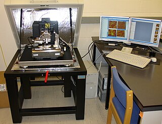
AFM-IR or infrared nanospectroscopy is one of a family of techniques that are derived from a combination of two parent instrumental techniques. AFM-IR combines the chemical analysis power of infrared spectroscopy and the high-spatial resolution of scanning probe microscopy (SPM). The term was first used to denote a method that combined a tuneable free electron laser with an atomic force microscope equipped with a sharp probe that measured the local absorption of infrared light by a sample with nanoscale spatial resolution.

<span class="mw-page-title-main">Cho Minhaeng</span> South Korean scientist (born 1965)

Cho Minhaeng is a South Korean scientist in researching physical chemistry, spectroscopy, and microscopy. He was director of the National Creative Research Initiative Center for Coherent Multidimensional Spectroscopy and is founding director of the Center for Molecular Spectroscopy and Dynamics in the Institute for Basic Science (IBS), located in Korea University.

<span class="mw-page-title-main">Cavity optomechanics</span>

Cavity optomechanics is a branch of physics which focuses on the interaction between light and mechanical objects on low-energy scales. It is a cross field of optics, quantum optics, solid-state physics and materials science. The motivation for research on cavity optomechanics comes from fundamental effects of quantum theory and gravity, as well as technological applications.

Stimulated Raman spectroscopy, also referred to as stimulated Raman scattering (SRS) is a form of spectroscopy employed in physics, chemistry, biology, and other fields. The basic mechanism resembles that of spontaneous Raman spectroscopy: a pump photon, of the angular frequency , which is scattered by a molecule has some small probability of inducing some vibrational transition, as opposed to inducing a simple Rayleigh transition. This makes the molecule emit a photon at a shifted frequency. However, SRS, as opposed to spontaneous Raman spectroscopy, is a third-order non-linear phenomenon involving a second photon—the Stokes photon of angular frequency —which stimulates a specific transition. When the difference in frequency between both photons resembles that of a specific vibrational transition the occurrence of this transition is resonantly enhanced. In SRS, the signal is equivalent to changes in the intensity of the pump and Stokes beams. The signals are typically rather low, of the order of a part in 10^5, thus calling for modulation-transfer techniques: one beam is modulated in amplitude and the signal is detected on the other beam via a lock-in amplifier. Employing a pump laser beam of a constant frequency and a Stokes laser beam of a scanned frequency allows for the unraveling of the spectral fingerprint of the molecule. This spectral fingerprint differs from those obtained by other spectroscopy methods such as Rayleigh scattering as the Raman transitions confer to different exclusion rules than those that apply for Rayleigh transitions.

<span class="mw-page-title-main">Coherent Raman scattering microscopy</span> Multi-photon microscopy technique

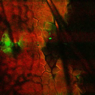
Coherent Raman scattering (CRS) microscopy is a multi-photon microscopy technique based on Raman-active vibrational modes of molecules. The two major techniques in CRS microscopy are stimulated Raman scattering (SRS) and coherent anti-Stokes Raman scattering (CARS). SRS and CARS were theoretically predicted and experimentally realized in the 1960s. In 1982 the first CARS microscope was demonstrated. In 1999, CARS microscopy using a collinear geometry and high numerical aperture objective were developed in Xiaoliang Sunney Xie's lab at Harvard University. This advancement made the technique more compatible with modern laser scanning microscopes. Since then, CRS's popularity in biomedical research started to grow. CRS is mainly used to image lipid, protein, and other bio-molecules in live or fixed cells or tissues without labeling or staining. CRS can also be used to image samples labeled with Raman tags, which can avoid interference from other molecules and normally allows for stronger CRS signals than would normally be obtained for common biomolecules. CRS also finds application in other fields, such as material science and environmental science.

References

  1. Tolles, W.M.; Nibler, J.W.; McDonald, J.R.3; Harvey, A.B. (1977). "A Review of the Theory and Application of Coherent Anti-Stokes Raman Spectroscopy (CARS)". Applied Spectroscopy. 31 (4): 253–271. Bibcode:1977ApSpe..31..253T. doi:10.1366/000370277774463625. S2CID   98395453.{{cite journal}}: CS1 maint: numeric names: authors list (link)
  2. 1 2 Zheltikov, A.M. (August–September 2000). "Coherent anti-Stokes Raman scattering: from proof-of-the-principle experiments to femtosecond CARS and higher order wave-mixing generalizations". Journal of Raman Spectroscopy. 31 (8–9): 653–667. Bibcode:2000JRSp...31..653Z. doi:10.1002/1097-4555(200008/09)31:8/9<653::AID-JRS597>3.0.CO;2-W.[ dead link ]
  3. Maker, P.D.; Terhune, R.W. (1965). "Study of Optical Effects Due to an Induced Polarization Third Order in the Electric Field Strength". Physical Review. 137 (3A): 801–818. Bibcode:1965PhRv..137..801M. doi:10.1103/PhysRev.137.A801.
  4. Begley, R.F.; Harvey, A.B.; Byer, R.L. (1974). "Coherent anti-Stokes Raman spectroscopy". Applied Physics Letters. 25 (7): 387–390. Bibcode:1974ApPhL..25..387B. doi:10.1063/1.1655519.
  5. Deckert, V.; Deckert-Gaudig, T.; Cialla-May, D.; Popp, J.; Zell, R.; Deinhard-Emmer, S.; Sokolov, A.V.; Yi, Z.; Scully, M.O. (2020). "Laser spectroscopic technique for direct identification of a single virus : FASTER CARS" (PDF). Proceedings of the National Academy of Sciences of the United States of America. 117 (45): 27820–27824. arXiv: 2003.07951 . Bibcode:2020PNAS..11727820D. doi: 10.1073/pnas.2013169117 . PMC   7668096 . PMID   33093197.
  6. Ori Katz; Adi Natan; Salman Rosenwaks; Yaron Silberberg (December 2008). "Shaped Femtosecond Pulses for Remote Chemical Detection" (PDF). OPN. Archived from the original (PDF) on 2013-09-28.
  7. "Laser beam 'kicks' molecules to detect roadside bombs". BBC . 2011-09-19.

Further reading