Congenital vertebral anomaly

Last updated
Congenital anomalies of spine
Specialty Medical genetics, orthopedics

Congenital vertebral anomalies are a collection of malformations of the spine. Most, around 85%, are not clinically significant, but they can cause compression of the spinal cord by deforming the vertebral canal or causing instability. This condition occurs in the womb. Congenital vertebral anomalies include alterations of the shape and number of vertebrae.

Contents

Lumbarization and sacralization

Lumbarization of sacral vertebra 1, seen as 6 vertebrae that do not connect to ribs. Lumbarization of S1.jpg
Lumbarization of sacral vertebra 1, seen as 6 vertebrae that do not connect to ribs.

Lumbarization is an anomaly in the spine. It is defined by the nonfusion of the first and second segments of the sacrum. The lumbar spine subsequently appears to have six vertebrae or segments, not five. This sixth lumbar vertebra is known as a transitional vertebra. Conversely the sacrum appears to have only four segments instead of its designated five segments. Lumbosacral transitional vertebrae consist of the process of the last lumbar vertebra fusing with the first sacral segment. [1] While only around 10 percent of adults have a spinal abnormality due to genetics, a sixth lumbar vertebra is one of the more common abnormalities. [2]

Sacralization of the L5 vertebra is seen at the lower right of the image. Sacralization of the fifth lumbar vertebra.jpg
Sacralization of the L5 vertebra is seen at the lower right of the image.

Sacralization of the fifth lumbar vertebra (or sacralization) is a congenital anomaly, in which the transverse process of the last lumbar vertebra (L5) fuses to the sacrum on one side or both, or to ilium, or both. These anomalies are observed in about 3.5 percent of people, and it is usually bilateral but can be unilateral or incomplete (ipsilateral or contralateral rudimentary facets) as well. Although sacralization may be a cause of low back pain, it is asymptomatic in many cases (especially bilateral type). Low back pain in these cases most likely occurs due to biomechanics. In sacralization, the L5-S1 intervertebral disc may be thin and narrow. This abnormality is found by X-ray.[ citation needed ]

Sacralization of L6 means L6 attaches to S1 via a rudimentary joint. This L6-S1 joint creates additional motion, increasing the potential for motion-related stress and lower back pain/conditions. This condition can usually be treated without surgery, injecting steroid medication at the pseudoarticulation instead. Additionally, if L6 fuses to another vertebra this is increasingly likely to cause lower back pain. [3] The presence of a sixth vertebra in the space where five vertebrae normally reside also decreases the flexibility of the spine and increases the likelihood of injury. [4]

Hemivertebrae

Hemivertebrae are wedge-shaped vertebrae and therefore can cause an angle in the spine (such as kyphosis, scoliosis, and lordosis). Among the congenital vertebral anomalies, hemivertebrae are the most likely to cause neurologic problems. [5] The most common location is the midthoracic vertebrae, especially the eighth (T8). [6] Neurologic signs result from severe angulation of the spine, narrowing of the spinal canal, instability of the spine, and luxation or fracture of the vertebrae. Signs include rear limb weakness or paralysis, urinary or fecal incontinence, and spinal pain. [5] Most cases of hemivertebrae have no or mild symptoms, so treatment is usually conservative. Severe cases may respond to surgical spinal cord decompression and vertebral stabilization. [6]

Recognised associations are many and include: Aicardi syndrome, cleidocranial dysostosis, gastroschisis 3, Gorlin syndrome, fetal pyelectasis 3, Jarcho-Levin syndrome, OEIS complex, VACTERL association. [7]

The probable cause of hemivertebrae is a lack of blood supply causing part of the vertebrae not to form. Hemivertebrae in dogs are most common in the tail, resulting in a screw shape.[ citation needed ]

Block vertebrae

Block vertebrae occur when there is improper segmentation of the vertebrae, leading to parts of or the entire vertebrae being fused. The adjacent vertebrae fuse through their intervertebral discs and also through other intervertebral joints so that it can lead to blocking or stretching of the exiting nerve roots from that segment. It may lead to certain neurological problems depending on the severity of the block. It can increase stress on the inferior and the superior intervertebral joints. It can lead to an abnormal angle in the spine, there are certain syndromes associated with block vertebrae; for example, Klippel–Feil syndrome. The sacrum is a normal block vertebra.[ citation needed ]

Fossil record

Evidence for block vertebrae found in the fossil record is studied by paleopathologists, specialists in ancient disease and injury. A block vertebra has been documented in T. rex. This suggests that the basic development pattern of vertebrae goes at least as far back as the most recent common ancestor of archosaurs and mammals. The tyrannosaur's block vertebra was probably caused by a "failure of the resegmentation of the sclerotomes". [8]

Butterfly vertebrae

Butterfly vertebrae have a sagittal cleft through the body of the vertebrae and a funnel shape at the ends. This gives the appearance of a butterfly on an x-ray. It is caused by persistence of the notochord (which usually only remains as the center of the intervertebral disc) during vertebrae formation. There are usually no symptoms. There are also coronal clefts mainly in skeletal dysplasias such as chondrodysplasia punctata. In dogs, butterfly vertebrae occur most often in Bulldogs, Pugs, and Boston Terriers. [9]

Transitional vertebrae

Short ribs at the first lumbar vertebra, which is thus a transitional vertebra, since lumbar vertebrae normally do not have ribs attached to them. Stummelrippe LWK1.jpg
Short ribs at the first lumbar vertebra, which is thus a transitional vertebra, since lumbar vertebrae normally do not have ribs attached to them.

Transitional vertebrae have the characteristics of two types of vertebra. The condition usually involves the vertebral arch or transverse processes. It occurs at the cervicothoracic, thoracolumbar, or lumbosacral junction. For instance, the transverse process of the last cervical vertebra may resemble a rib. A transitional vertebra at the lumbosacral junction can cause arthritis, disk changes, or thecal sac compression. Back pain associated with lumbosacral transitional vertebrae (LSTV) is known as Bertolotti's syndrome. One study found that male German Shepherd Dogs with a lumbosacral transitional vertebra are at greater risk for cauda equina syndrome, which can cause rear limb weakness and incontinence. [10]

The significance of transitional vertebrae has been questioned by one study finding similar prevalence in the general population as those with low back pain, [11] but more recent study found a large difference. [12]

Spina bifida

Spina bifida is characterized by a midline cleft in the vertebral arch. It usually causes no symptoms in dogs. It is seen most commonly in Bulldogs and Manx cats. [5] In Manx it accompanies a condition known as sacrocaudal dysgenesis that gives these cats their characteristic tailless or stumpy tail appearance. It is inherited in Manx as an autosomal dominant trait. [13]

Associations

Vertebral anomalies is associated with an increased incidence of some other specific anomalies as well, together being called the VACTERL association: [14]

Related Research Articles

<span class="mw-page-title-main">Intervertebral disc</span> Cartilage of spine

An intervertebral disc lies between adjacent vertebrae in the vertebral column. Each disc forms a fibrocartilaginous joint, to allow slight movement of the vertebrae, to act as a ligament to hold the vertebrae together, and to function as a shock absorber for the spine.

<span class="mw-page-title-main">Sacrum</span> Triangular-shaped bone at the bottom of the spine

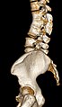
The sacrum, in human anatomy, is a large, triangular bone at the base of the spine that forms by the fusing of the sacral vertebrae (S1–S5) between ages 18 and 30.

<span class="mw-page-title-main">Spinal nerve</span> Nerve that carries signals between the spinal cord and the body

A spinal nerve is a mixed nerve, which carries motor, sensory, and autonomic signals between the spinal cord and the body. In the human body there are 31 pairs of spinal nerves, one on each side of the vertebral column. These are grouped into the corresponding cervical, thoracic, lumbar, sacral and coccygeal regions of the spine. There are eight pairs of cervical nerves, twelve pairs of thoracic nerves, five pairs of lumbar nerves, five pairs of sacral nerves, and one pair of coccygeal nerves. The spinal nerves are part of the peripheral nervous system.

<span class="mw-page-title-main">Lumbar vertebrae</span> Five vertebrae between the pelvis and the rib cage

The lumbar vertebrae are, in human anatomy, the five vertebrae between the rib cage and the pelvis. They are the largest segments of the vertebral column and are characterized by the absence of the foramen transversarium within the transverse process and by the absence of facets on the sides of the body. They are designated L1 to L5, starting at the top. The lumbar vertebrae help support the weight of the body, and permit movement.

<span class="mw-page-title-main">Kyphosis</span> Medical condition

Kyphosis is an abnormally excessive convex curvature of the spine as it occurs in the thoracic and sacral regions. Abnormal inward concave lordotic curving of the cervical and lumbar regions of the spine is called lordosis. It can result from degenerative disc disease; developmental abnormalities, most commonly Scheuermann's disease; Copenhagen disease, osteoporosis with compression fractures of the vertebra; multiple myeloma; or trauma. A normal thoracic spine extends from the 1st thoracic to the 12th thoracic vertebra and should have a slight kyphotic angle, ranging from 20° to 45°. When the "roundness" of the upper spine increases past 45° it is called kyphosis or "hyperkyphosis". Scheuermann's kyphosis is the most classic form of hyperkyphosis and is the result of wedged vertebrae that develop during adolescence. The cause is not currently known and the condition appears to be multifactorial and is seen more frequently in males than females.

<span class="mw-page-title-main">Spondylosis</span> Degeneration of the vertebral column

Spondylosis is the degeneration of the vertebral column from any cause. In the more narrow sense it refers to spinal osteoarthritis, the age-related degeneration of the spinal column, which is the most common cause of spondylosis. The degenerative process in osteoarthritis chiefly affects the vertebral bodies, the neural foramina and the facet joints. If severe, it may cause pressure on the spinal cord or nerve roots with subsequent sensory or motor disturbances, such as pain, paresthesia, imbalance, and muscle weakness in the limbs.

<span class="mw-page-title-main">Neuromere</span>

Neuromeres are distinct groups of neural crest cells, forming segments in the neural tube of the early embryonic development of the brain. There are three classes of neuromeres in the central nervous system – prosomeres, mesomeres and rhombomeres that will develop the forebrain, midbrain, and hindbrain respectively.

<span class="mw-page-title-main">Degenerative disc disease</span> Medical condition

Degenerative disc disease (DDD) is a medical condition typically brought on by the normal aging process in which there are anatomic changes and possibly a loss of function of one or more intervertebral discs of the spine. DDD can take place with or without symptoms, but is typically identified once symptoms arise. The root cause is thought to be loss of soluble proteins within the fluid contained in the disc with resultant reduction of the oncotic pressure, which in turn causes loss of fluid volume. Normal downward forces cause the affected disc to lose height, and the distance between vertebrae is reduced. The anulus fibrosus, the tough outer layers of a disc, also weakens. This loss of height causes laxity of the longitudinal ligaments, which may allow anterior, posterior, or lateral shifting of the vertebral bodies, causing facet joint malalignment and arthritis; scoliosis; cervical hyperlordosis; thoracic hyperkyphosis; lumbar hyperlordosis; narrowing of the space available for the spinal tract within the vertebra ; or narrowing of the space through which a spinal nerve exits with resultant inflammation and impingement of a spinal nerve, causing a radiculopathy.

<span class="mw-page-title-main">Retrolisthesis</span> Posterior displacement (but not dislocation) of a vertebra relative to the one below it

A retrolisthesis is a posterior displacement of one vertebral body with respect to the subjacent vertebra to a degree less than a luxation (dislocation). Retrolistheses are most easily diagnosed on lateral x-ray views of the spine. Views where care has been taken to expose for a true lateral view without any rotation offer the best diagnostic quality.

<span class="mw-page-title-main">Cauda equina syndrome</span> Nerve damage at the end of the spinal cord

Cauda equina syndrome (CES) is a condition that occurs when the bundle of nerves below the end of the spinal cord known as the cauda equina is damaged. Signs and symptoms include low back pain, pain that radiates down the leg, numbness around the anus, and loss of bowel or bladder control. Onset may be rapid or gradual.

<span class="mw-page-title-main">Anterior longitudinal ligament</span> Ligament running down the front of the vertebral column

The anterior longitudinal ligament is a ligament that extends across the anterior/ventral aspect of the vertebral bodies and intervertebral discs the spine.

<span class="mw-page-title-main">Lumbar nerves</span>

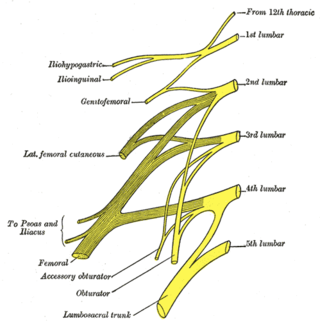
The lumbar nerves are the five pairs of spinal nerves emerging from the lumbar vertebrae. They are divided into posterior and anterior divisions.

<span class="mw-page-title-main">Bertolotti's syndrome</span> Medical condition

Bertolotti's syndrome is a commonly missed cause of back pain which occurs due to lumbosacral transitional vertebrae (LSTV). It is a congenital condition but is not usually symptomatic until one's later twenties or early thirties. However, there are a few cases of Bertolotti's that become symptomatic at a much earlier age.

<span class="mw-page-title-main">Diastematomyelia</span> Medical condition

Diastematomyelia is a congenital disorder in which a part of the spinal cord is split, usually at the level of the upper lumbar vertebra in the longitudinal (sagittal) direction. Females are affected much more commonly than males. This condition occurs in the presence of an osseous, cartilaginous or fibrous septum in the central portion of the spinal canal which then produces a complete or incomplete sagittal division of the spinal cord into two hemicords. When the split does not reunite distally to the spur, the condition is referred to as diplomyelia, which is true duplication of the spinal cord.

<span class="mw-page-title-main">Laminotomy</span> Surgical procedure

A laminotomy is an orthopaedic neurosurgical procedure that removes part of the lamina of a vertebral arch in order to relieve pressure in the vertebral canal. A laminotomy is less invasive than conventional vertebral column surgery techniques, such as laminectomy because it leaves more ligaments and muscles attached to the spinous process intact and it requires removing less bone from the vertebra. As a result, laminotomies typically have a faster recovery time and result in fewer postoperative complications. Nevertheless, possible risks can occur during or after the procedure like infection, hematomas, and dural tears. Laminotomies are commonly performed as treatment for lumbar spinal stenosis and herniated disks. MRI and CT scans are often used pre- and post surgery to determine if the procedure was successful.

<span class="mw-page-title-main">Spinal cord</span> Long, tubular central nervous system structure in the vertebral column

The spinal cord is a long, thin, tubular structure made up of nervous tissue that extends from the medulla oblongata in the brainstem to the lumbar region of the vertebral column (backbone) of vertebrate animals. The center of the spinal cord is hollow and contains a structure called central canal, which contains cerebrospinal fluid. The spinal cord is also covered by meninges and enclosed by the neural arches. Together, the brain and spinal cord make up the central nervous system (CNS).

<span class="mw-page-title-main">Facet syndrome</span> Medical condition

Facet syndrome is a syndrome in which the facet joints cause painful symptoms. In conjunction with degenerative disc disease, a distinct but functionally related condition, facet arthropathy is believed to be one of the most common causes of lower back pain.

<span class="mw-page-title-main">Vertebral column</span> Bony structure found in vertebrates

The vertebral column, also known as the backbone or spine, is the core part of the axial skeleton in vertebrate animals. The vertebral column is the defining characteristic of vertebrate endoskeleton in which the notochord found in all chordates has been replaced by a segmented series of mineralized irregular bones called vertebrae, separated by fibrocartilaginous intervertebral discs. The dorsal portion of the vertebral column houses the spinal canal, a cavity formed by alignment of the neural arches that encloses and protects the spinal cord.

Williams flexion exercises (WFE) – also called Williams lumbar flexion exercises – are a set of related physical exercises intended to enhance lumbar flexion, avoid lumbar extension, and strengthen the abdominal and gluteal musculature in an effort to manage low back pain non-surgically. The system was first devised in 1937 by Dallas orthopedic surgeon Dr. Paul C. Williams.

<span class="mw-page-title-main">Vertebra</span> Bone in the vertebral column

Each vertebra is an irregular bone with a complex structure composed of bone and some hyaline cartilage, that make up the vertebral column or spine, of vertebrates. The proportions of the vertebrae differ according to their spinal segment and the particular species.

References

  1. Jancuska, JM; Spivak, JM; Bendo, JA (2015). "A Review of Symptomatic Lumbosacral Transitional Vertebrae: Bertolotti's Syndrome". Int J Spine Surg. 9: 42. doi:10.14444/2042. PMC   4603258 . PMID   26484005.
  2. Dorland's Medical Dictionary
  3. "Spinal Cord, Inc". Spinalcord.com. Retrieved November 27, 2016.
  4. "Laser Spine Institute". Archived from the original on November 28, 2016. Retrieved November 27, 2016.
  5. 1 2 3 Braund, K.G. (2003). "Developmental Disorders". Clinical Neurology in Small Animals: Localization, Diagnosis and Treatment. Retrieved 2007-02-04.
  6. 1 2 Jeffery N, Smith P, Talbot C (2007). "Imaging findings and surgical treatment of hemivertebrae in three dogs". J Am Vet Med Assoc. 230 (4): 532–6. doi: 10.2460/javma.230.4.532 . PMID   17302550.
  7. Weerakkody, Yuranga. "Hemivertebra | Radiology Reference Article". Radiopaedia.org. Retrieved 26 April 2022.
  8. Molnar, R. E., 2001, Theropod paleopathology: a literature survey: In: Mesozoic Vertebrate Life, edited by Tanke, D. H., and Carpenter, K., Indiana University Press, p. 337-363.
  9. Ettinger, Stephen J.; Feldman, Edward C. (1995). Textbook of Veterinary Internal Medicine (4th ed.). W.B. Saunders Company. ISBN   0-7216-6795-3.
  10. Flückiger M, Damur-Djuric N, Hässig M, Morgan J, Steffen F (2006). "A lumbosacral transitional vertebra in the dog predisposes to cauda equina syndrome". Vet Radiol Ultrasound. 47 (1): 39–44. doi:10.1111/j.1740-8261.2005.00103.x. PMID   16429983.
  11. Apazidis A, Ricart PA, Diefenbach CM, Spivak JM (September 2011). "The prevalence of transitional vertebrae in the lumbar spine". Spine J. 11 (9): 858–62. doi:10.1016/j.spinee.2011.08.005. PMID   21951610.
  12. Jin L, Yin Y, Chen W, Zhang R, Guo J, Tao S, Guo Z, Hou Z, Zhang Y (December 2021). "Role of the Lumbosacral Transition Vertebra and Vertebral Lamina in the Pathogenesis of Lumbar Disc Herniation". Orthop Surg. 13 (8): 2355–2362. doi:10.1111/os.13122. PMC   8654657 . PMID   34791784.
  13. "Congenital and Inherited Anomalies of the Nervous System: Small Animals". The Merck Veterinary Manual. 2006. Retrieved 2007-02-04.
  14. Solomon, Benjamin D (2011). "VACTERL/VATER Association". Orphanet Journal of Rare Diseases. Springer Science and Business Media LLC. 6 (1): 56. doi: 10.1186/1750-1172-6-56 . ISSN   1750-1172. PMC   3169446 . PMID   21846383.