Axial skeleton

Last updated
Axial skeleton
Axial skeleton diagram.svg
Diagram of the axial skeleton
Details
Identifiers
Latin skeleton axiale
TA98 A02.0.00.009
TA2 353
Anatomical terminology
3D medical animation still shot of human skull 3D Medical Animation Axial Skeleton.jpg
3D medical animation still shot of human skull

The axial skeleton is the part of the skeleton that consists of the bones of the head and trunk of a vertebrate. In the human skeleton, it consists of 80 bones and is composed of six parts; the skull (22 bones), also the ossicles of the middle ear, the hyoid bone, the rib cage, sternum and the vertebral column. The axial skeleton together with the appendicular skeleton form the complete skeleton. Another definition of axial skeleton is the bones including the vertebrae, sacrum, coccyx, skull, ribs, and sternum. [1]

Contents

Structure

Sobo 1909 35.png
Sobo 1909 36.png
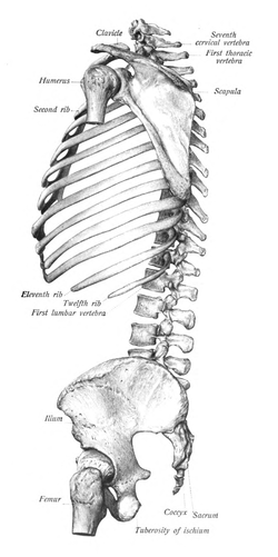
View from both directions of a axial skeleton cut in half. Shows attachments to pelvis and does not show skull. From Sobotta's atlas of human anatomy 1909.

Flat bones house the brain and other vital organs. This article mainly deals with the axial skeletons of humans; however, it is important to understand the evolutionary lineage of the axial skeleton. The human axial skeleton consists of 81 different bones. It is the medial core of the body and connects the pelvis to the body, where the appendix skeleton attaches. As the skeleton grows older the bones get weaker with the exception of the skull. The skull remains strong to protect the brain from injury.

In humans, the axial skeleton serves to protect the brain, spinal cord, heart, and lungs. It also serves as the attachment site for muscles that move the head, neck, and back, and for muscles that act across the shoulder and hip joints to move their corresponding limbs. [2]

Human skull

The human skull consists of the cranium and the facial bones. The cranium holds and protects the brain in a large space called the cranial vault. The cranium is formed from eight plate-shaped bones which fit together at meeting points (joints) called sutures. In addition there are 14 facial bones which form the lower front part of the skull. Together the 22 bones that compose the skull form additional, smaller spaces besides the cranial vault, such as the cavities for the eyes, the internal ear, the nose, and the mouth. The most important facial bones include the jaw or mandible, the upper jaw or maxilla, the zygomatic or cheek bone, and the nasal bone. [3]

Humans are born with separate plates which later fuse to allow flexibility as the skull passes through the pelvis and birth canal during birth. During development the eight separate plates of the immature bones fuse into one single structure known as the Skull. The only bone that remains separate from the rest of the skull is the mandible. [4]

Rib cage

The rib cages are composed of 12 pairs of ribs plus the sternum for a total of 25 separate bones. The rib cage functions as protection for the vital organs such as the heart and lungs. The ribs are shaped like crescents, with one end flattened and the other end rounded. The rounded ends are attached at joints to the thoracic vertebrae at the back and the flattened ends come together at the sternum, in the front. [5]

The upper seven pairs of ribs attach to the sternum with costal cartilage and are known as "true ribs". The 8th through 10th ribs have non-costal cartilage which connects them to the ribs above, and for this they are known as "false ribs". The last two ribs are called "floating ribs" because they do not attach to the sternum or to other ribs and simply "hang free". The length of each rib increases from number one to seven and then decreases until rib pair number 12. The first rib is the shortest, broadest, flattest, and most curved.[ medical citation needed ]

Vertebral column

At birth the majority of humans have 33 separate vertebrae. However, during normal development several vertebrae fuse, leaving a total of 24, in most cases. The confusion about whether or not there are 32-34 vertebrae stems from the fact that the two lowest vertebrae, the sacrum and the coccyx, are single bones made up of several smaller bones which have fused together. This is how the vertebrae are counted: 24 separate vertebrae and the sacrum, formed from 5 fused vertebrae, and the coccyx, formed from 3-5 fused vertebrae. If you count the coccyx and sacrum each as one vertebra, then there are 26 vertebrae. If the fused vertebrae are all counted separately, then the total number of vertebrae comes to between 32 and 34 (due to the number making up the coccyx varying between 3 and 5).

The vertebral column consists of 5 parts. The most cranial (uppermost) part is made up by the cervical vertebrae (7), followed by thoracic vertebrae (12), lumbar (5), sacral (5) and coccygeal vertebrae (3-5).

Cervical vertebrae make up the junction between the vertebral column and the cranium. Sacral and coccygeal vertebrae are fused and thus often called "sacral bone" or "coccygeal bone" as unit. The sacral bone makes up the junction between the vertebral column and the pelvic bones.

Etymology

The word "axial" is taken from the word "axis" and refers to the fact that the bones are located close to or along the central "axis" of the body. The term axis means the central point around which the other structures are distributed. [6]

Short summary

The axial skeleton consists of 80 bones:

See also

Related Research Articles

<span class="mw-page-title-main">Skeleton</span> Part of the body that forms the supporting structure

A skeleton is the structural frame that supports the body of most animals. There are several types of skeletons, including the exoskeleton, which is a rigid outer shell that holds up an organism's shape; the endoskeleton, a rigid internal frame to which the organs and soft tissues attach; and the hydroskeleton, a flexible internal structure supported by the hydrostatic pressure of body fluids.

<span class="mw-page-title-main">Rib</span> Long bone in vertebrates that protects vital respiratory and cardiovascular organs

In vertebrate anatomy, ribs are the long curved bones which form the rib cage, part of the axial skeleton. In most tetrapods, ribs surround the chest, enabling the lungs to expand and thus facilitate breathing by expanding the chest cavity. They serve to protect the lungs, heart, and other internal organs of the thorax. In some animals, especially snakes, ribs may provide support and protection for the entire body.

The human skeleton is the internal framework of the human body. It is composed of around 270 bones at birth – this total decreases to around 206 bones by adulthood after some bones get fused together. The bone mass in the skeleton makes up about 14% of the total body weight and reaches maximum mass between the ages of 25 and 30. The human skeleton can be divided into the axial skeleton and the appendicular skeleton. The axial skeleton is formed by the vertebral column, the rib cage, the skull and other associated bones. The appendicular skeleton, which is attached to the axial skeleton, is formed by the shoulder girdle, the pelvic girdle and the bones of the upper and lower limbs.

<span class="mw-page-title-main">Skull</span> Bony structure that forms the head in vertebrates

The skull is a bone protective cavity for the brain. The skull is composed of four types of bone i.e., cranial bones, facial bones, ear ossicles and hyoid bone, however two parts are more prominent: the cranium and the mandible. In humans, these two parts are the neurocranium (braincase) and the viscerocranium that includes the mandible as its largest bone. The skull forms the anterior-most portion of the skeleton and is a product of cephalisation—housing the brain, and several sensory structures such as the eyes, ears, nose, and mouth. In humans, these sensory structures are part of the facial skeleton.

<span class="mw-page-title-main">Coccyx</span> Bone of the pelvis

The coccyx, commonly referred to as the tailbone, is the final segment of the vertebral column in all apes, and analogous structures in certain other mammals such as horses. In tailless primates since Nacholapithecus, the coccyx is the remnant of a vestigial tail. In animals with bony tails, it is known as tailhead or dock, in bird anatomy as tailfan. It comprises three to five separate or fused coccygeal vertebrae below the sacrum, attached to the sacrum by a fibrocartilaginous joint, the sacrococcygeal symphysis, which permits limited movement between the sacrum and the coccyx.

Articles related to anatomy include:

<span class="mw-page-title-main">Sacrum</span> Triangular-shaped bone at the bottom of the spine

The sacrum, in human anatomy, is a large, triangular bone at the base of the spine that forms by the fusing of the sacral vertebrae (S1–S5) between ages 18 and 30.

<span class="mw-page-title-main">Spinal nerve</span> Nerve that carries signals between the spinal cord and the body

A spinal nerve is a mixed nerve, which carries motor, sensory, and autonomic signals between the spinal cord and the body. In the human body there are 31 pairs of spinal nerves, one on each side of the vertebral column. These are grouped into the corresponding cervical, thoracic, lumbar, sacral and coccygeal regions of the spine. There are eight pairs of cervical nerves, twelve pairs of thoracic nerves, five pairs of lumbar nerves, five pairs of sacral nerves, and one pair of coccygeal nerves. The spinal nerves are part of the peripheral nervous system.

<span class="mw-page-title-main">Irregular bone</span> Bone of irregular shape

The irregular bones are bones which, from their peculiar form, cannot be grouped as long, short, flat or sesamoid bones. Irregular bones serve various purposes in the body, such as protection of nervous tissue, affording multiple anchor points for skeletal muscle attachment, and maintaining pharynx and trachea support, and tongue attachment. They consist of cancellous tissue enclosed within a thin layer of compact bone. Irregular bones can also be used for joining all parts of the spinal column together. The spine is the place in the human body where the most irregular bones can be found. There are, in all, 33 irregular bones found here.

<span class="mw-page-title-main">Head and neck anatomy</span>

This article describes the anatomy of the head and neck of the human body, including the brain, bones, muscles, blood vessels, nerves, glands, nose, mouth, teeth, tongue, and throat.

<span class="mw-page-title-main">Skeletal system of the horse</span> The skeletal system is made of many interconnected tissues including bone, cartilage, and tendons

The skeletal system of the horse is a skeletal system of a horse that has three major functions in the body. It protects vital organs, provides framework, and supports soft parts of the body. Horses typically have 205 bones. The pelvic limb typically contains 19 bones, while the thoracic limb contains 20 bones.

<span class="mw-page-title-main">Outline of human anatomy</span> Overview of and topical guide to human anatomy

The following outline is provided as an overview of and topical guide to human anatomy:

<span class="mw-page-title-main">Facial skeleton</span> Facial bones of a skull

The facial skeleton comprises the facial bones that may attach to build a portion of the skull. The remainder of the skull is the neurocranium.

<span class="mw-page-title-main">Hip bone</span> Bone of the pelvis

The hip bone is a large flat bone, constricted in the center and expanded above and below. In some vertebrates it is composed of three parts: the ilium, ischium, and the pubis.

<span class="mw-page-title-main">Coccyx fracture</span> Medical condition

A coccyx fracture is a fracture of the coccyx, commonly called a broken tailbone or ‘puzzle fracture.’

<span class="mw-page-title-main">Pelvis</span> Lower torso of the human body

The pelvis is the lower part of the trunk, between the abdomen and the thighs, together with its embedded skeleton.

<span class="mw-page-title-main">Vertebral column</span> Bony structure found in vertebrates

The vertebral column, also known as the backbone or spine, is the core part of the axial skeleton in vertebrate animals. The vertebral column is the defining characteristic of vertebrate endoskeleton in which the notochord found in all chordates has been replaced by a segmented series of mineralized irregular bones called vertebrae, separated by fibrocartilaginous intervertebral discs. The dorsal portion of the vertebral column houses the spinal canal, a cavity formed by alignment of the neural arches that encloses and protects the spinal cord.

This glossary explains technical terms commonly employed in the description of dinosaur body fossils. Besides dinosaur-specific terms, it covers terms with wider usage, when these are of central importance in the study of dinosaurs or when their discussion in the context of dinosaurs is beneficial. The glossary does not cover ichnological and bone histological terms, nor does it cover measurements.

<span class="mw-page-title-main">Vertebra</span> Bone in the vertebral column

Each vertebra is an irregular bone with a complex structure composed of bone and some hyaline cartilage, that make up the vertebral column or spine, of vertebrates. The proportions of the vertebrae differ according to their spinal segment and the particular species.

References

  1. Folkens, Tim D. White, Michael T. Black, Pieter A.; Pierter, Folkens; Michael, Black (2012). Human osteology (3rd ed.). Amsterdam: Elsevier/Academic Press. p. 11. ISBN   978-0-12-374134-9.{{cite book}}: CS1 maint: multiple names: authors list (link)
  2. Creative Commons by small.svg  This article incorporates text available under the CC BY 4.0 license.Betts, J Gordon; Desaix, Peter; Johnson, Eddie; Johnson, Jody E; Korol, Oksana; Kruse, Dean; Poe, Brandon; Wise, James; Womble, Mark D; Young, Kelly A (May 21, 2023). Anatomy & Physiology. Houston: OpenStax CNX. 7.1 Divisions of the skeletal system. ISBN   978-1-947172-04-3.
  3. "Features of the Human Skull". Face and Emotion. Archived from the original on 14 February 2014. Retrieved 25 February 2014.
  4. "Skull". Inner Body. Retrieved 25 February 2014.
  5. "Postcranial Skeletal/Ribs" (PDF). Observation and Analysis Method for Human Bones. Archived from the original (PDF) on 20 May 2012. Retrieved 25 February 2014.
  6. "Axial skeleton". AnatomyExpert. Archived from the original on 25 December 2013. Retrieved 15 March 2013.