Lacrimal bone

Last updated
Lacrimal bone
Lacrimal bone - lateral view6.png
Position of the lacrimal bone (shown in green).
Lacrimal and nasal bones.jpg
Medial wall of the orbit. Lacrimal bone is in yellow.
Details
Identifiers
Latin os lacrimale
TA98 A02.1.09.001
TA2 744
FMA 52741
Anatomical terms of bone

The lacrimal bone is a small and fragile bone of the facial skeleton; it is roughly the size of the little fingernail. It is situated at the front part of the medial wall of the orbit. It has two surfaces and four borders. Several bony landmarks of the lacrimal bone function in the process of lacrimation or crying. Specifically, the lacrimal bone helps form the nasolacrimal canal necessary for tear translocation. A depression on the anterior inferior portion of the bone, the lacrimal fossa, houses the membranous lacrimal sac. Tears or lacrimal fluid, from the lacrimal glands, collect in this sac during excessive lacrimation. The fluid then flows through the nasolacrimal duct and into the nasopharynx. This drainage results in what is commonly referred to a runny nose during excessive crying or tear production. Injury or fracture of the lacrimal bone can result in posttraumatic obstruction of the lacrimal pathways. [1] [2]

Contents

Structure

Lateral or orbital surface

The lateral or orbital surface is divided by a vertical ridge, the posterior lacrimal crest, into two parts.

In front of this crest is a longitudinal groove, the lacrimal sulcus (sulcus lacrimalis), the inner margin of which unites with the frontal process of the maxilla, and the lacrimal fossa is thus completed. The upper part of this fossa lodges the lacrimal sac, the lower part, the nasolacrimal duct.

The portion behind the crest is smooth, and forms part of the medial wall of the orbit.

The crest, with a part of the orbital surface immediately behind it, gives origin to the lacrimal part of the orbicularis oculi and ends below in a small, hook-like projection, the lacrimal hamulus, which articulates with the lacrimal tubercle of the maxilla, and completes the upper orifice of the nasolacrimal canal; the hamulus sometimes exists as a separate piece, and is then called the lesser lacrimal bone.

Medial or nasal surface

The medial or nasal surface presents a longitudinal furrow, corresponding to the crest on the lateral surface.

The area in front of this furrow forms part of the middle meatus of the nose. The area behind it articulates with the ethmoid, and completes some of the anterior ethmoidal cells.

Borders

Of the four borders:

Development

The lacrimal is ossified from a single center, which appears about the twelfth week in the membrane covering the cartilaginous nasal capsule.

Articulations

The lacrimal articulates with four bones: two of the neurocranium, the frontal and ethmoid, and two of the viscerocranium, the maxilla and the inferior nasal concha.

Other animals

Lacrimal bone in the reptile Lazarussuchus Lazarussuchus.svg
Lacrimal bone in the reptile Lazarussuchus

In early lobe-finned fishes and ancestral tetrapods, the lacrimal bone is a relatively large and robust bone, running from the orbit to the nostrils. It forms part of the side of the face, between the nasal bones and the maxilla. In primitive forms, it is often accompanied by a much smaller septomaxilla bone, lying immediately behind the nasal opening, but this is lost in most modern species. The lacrimal bone is often smaller in living vertebrates, and is no longer always directly associated with the nasal opening, although it retains its connection with the orbit. The bone is entirely absent in living amphibians, as well as some reptilian species. [3]

Dinosaurs

In dinosaurs, the lacrimal bone usually defines the anterior rim of the orbit (eye socket), and the posterior rim of the antorbital fenestra. In some theropods (e.g. Allosaurus , Ceratosaurus , Albertosaurus ) the upper part of the lacrimal bone grew in such a manner as to form a horn on the top of the dinosaur's head, usually situated above, and anterior to the eye. In many dinosaurs, the lacrimal bone comes into contact with the nasal bone, the jugal bone, the prefrontal bone, and the maxillary and premaxillary bones. The boundaries where some of these bones meet with the others are called sutures. Rarely, the lacrimal bones fused with the nasal bones to form a pair of "nasolacrimal" crests, which are present in dinosaurs such as Dilophosaurus , Megapnosaurus and Sinosaurus . [4]

Additional images

See also

Related Research Articles

Articles related to anatomy include:

<span class="mw-page-title-main">Maxilla</span> Upper jaw bone

The maxilla in vertebrates is the upper fixed bone of the jaw formed from the fusion of two maxillary bones. In humans, the upper jaw includes the hard palate in the front of the mouth. The two maxillary bones are fused at the intermaxillary suture, forming the anterior nasal spine. This is similar to the mandible, which is also a fusion of two mandibular bones at the mandibular symphysis. The mandible is the movable part of the jaw.

<span class="mw-page-title-main">Zygomatic bone</span> Facial bone

In the human skull, the zygomatic bone, also called cheekbone or malar bone, is a paired irregular bone which articulates with the maxilla, the temporal bone, the sphenoid bone and the frontal bone. It is situated at the upper and lateral part of the face and forms the prominence of the cheek, part of the lateral wall and floor of the orbit, and parts of the temporal fossa and the infratemporal fossa. It presents a malar and a temporal surface; four processes, and four borders.

<span class="mw-page-title-main">Sphenoid bone</span> Bone at the front of the skull

The sphenoid bone is an unpaired bone of the neurocranium. It is situated in the middle of the skull towards the front, in front of the basilar part of the occipital bone. The sphenoid bone is one of the seven bones that articulate to form the orbit. Its shape somewhat resembles that of a butterfly or bat with its wings extended.

<span class="mw-page-title-main">Palatine bone</span> Bone of the facial skeleton

In anatomy, the palatine bones are two irregular bones of the facial skeleton in many animal species, located above the uvula in the throat. Together with the maxillae, they comprise the hard palate.

<span class="mw-page-title-main">Inferior nasal concha</span> Facial bone

The inferior nasal concha is one of the three paired nasal conchae in the nose. It extends horizontally along the lateral wall of the nasal cavity and consists of a lamina of spongy bone, curled upon itself like a scroll,. The inferior nasal conchae are considered a pair of facial bones. As the air passes through the turbinates, the air is churned against these mucosa-lined bones in order to receive warmth, moisture and cleansing. Superior to inferior nasal concha are the middle nasal concha and superior nasal concha which both arise from the ethmoid bone, of the cranial portion of the skull. Hence, these two are considered as a part of the cranial bones.

<span class="mw-page-title-main">Orbit (anatomy)</span> Cavity or socket of the skull in which the eye and its appendages are situated

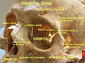
In anatomy, the orbit is the cavity or socket/hole of the skull in which the eye and its appendages are situated. "Orbit" can refer to the bony socket, or it can also be used to imply the contents. In the adult human, the volume of the orbit is 30 millilitres, of which the eye occupies 6.5 ml. The orbital contents comprise the eye, the orbital and retrobulbar fascia, extraocular muscles, cranial nerves II, III, IV, V, and VI, blood vessels, fat, the lacrimal gland with its sac and duct, the eyelids, medial and lateral palpebral ligaments, cheek ligaments, the suspensory ligament, septum, ciliary ganglion and short ciliary nerves.

<span class="mw-page-title-main">Orbicularis oculi muscle</span> Facial muscle that closes the eyelids

The orbicularis oculi is a muscle in the face that closes the eyelids. It arises from the nasal part of the frontal bone, from the frontal process of the maxilla in front of the lacrimal groove, and from the anterior surface and borders of a short fibrous band, the medial palpebral ligament.

<span class="mw-page-title-main">Orbital part of frontal bone</span>

The orbital or horizontal part of the frontal bone consists of two thin triangular plates, the orbital plates, which form the vaults of the orbits, and are separated from one another by a median gap, the ethmoidal notch.

<i>Galesaurus</i> Extinct genus of cynodonts from the Triassic of South Africa

Galesaurus is an extinct genus of carnivorous cynodont therapsid that lived between the Induan and the Olenekian stages of the Early Triassic in what is now South Africa. It was incorrectly classified as a dinosaur by Sir Richard Owen in 1859.

<span class="mw-page-title-main">Greater wing of sphenoid bone</span> Large part of the skull front behind the eye socket

The greater wing of the sphenoid bone, or alisphenoid, is a bony process of the sphenoid bone, positioned in the skull behind each eye. There is one on each side, extending from the side of the body of the sphenoid and curving upward, laterally, and backward.

<span class="mw-page-title-main">Ethmoidal labyrinth</span>

The ethmoidal labyrinth or lateral mass of the ethmoid bone consists of a number of thin-walled cellular cavities, the ethmoid air cells, arranged in three groups, anterior, middle, and posterior, and interposed between two vertical plates of bone; the lateral plate forms part of the orbit, the medial plate forms part of the nasal cavity. In the disarticulated bone many of these cells are opened into, but when the bones are articulated, they are closed in at every part, except where they open into the nasal cavity.

<span class="mw-page-title-main">Squamous part of the frontal bone</span> Upper two-thirds of the forward skull

The squamous part of the frontal bone is the superior portion when viewed in standard anatomical orientation. There are two surfaces of the squamous part of the frontal bone: the external surface, and the internal surface.

<span class="mw-page-title-main">Lacrimal sac</span> Upper, dilated end of the nasolacrimal duct

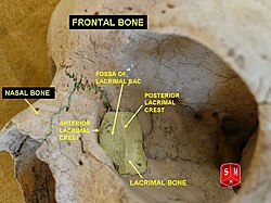
The lacrimal sac or lachrymal sac is the upper dilated end of the nasolacrimal duct, and is lodged in a deep groove formed by the lacrimal bone and frontal process of the maxilla. It connects the lacrimal canaliculi, which drain tears from the eye's surface, and the nasolacrimal duct, which conveys this fluid into the nasal cavity. Lacrimal sac occlusion leads to dacryocystitis.

<span class="mw-page-title-main">Perpendicular plate of palatine bone</span>

The perpendicular plate of palatine bone is the vertical part of the palatine bone, and is thin, of an oblong form, and presents two surfaces and four borders.

<span class="mw-page-title-main">Posterior lacrimal crest</span> Bony ridge on the orbital surface of the lacrimal bone

The posterior lacrimal crest is a vertical bony ridge on the orbital surface of the lacrimal bone. It divides the bone into two parts. It gives origin to the lacrimal part of the orbicularis oculi muscle.

The anterior lacrimal crest is a bony projection on the frontal process of the maxilla. It creates the lateral margin of the lacrimal sac fossa and is continuous with the orbital margin. The medial palpebral ligament is attached to anterior lacrimal crest. It is an important structure to avoid damaging during rhinoplasty.

<span class="mw-page-title-main">Frontal process of maxilla</span> Plate which forms part of the lateral boundary of the nose

The frontal process of the maxilla is a strong plate, which projects upward, medialward, and backward from the maxilla, forming part of the lateral boundary of the nose.

<span class="mw-page-title-main">Body of sphenoid bone</span>

The body of the sphenoid bone, more or less cubical in shape, is hollowed out in its interior to form two large cavities, the sphenoidal sinuses, which are separated from each other by a septum.

<span class="mw-page-title-main">Outline of human anatomy</span> Overview of and topical guide to human anatomy

The following outline is provided as an overview of and topical guide to human anatomy:

References

  1. Maliborski, A; Różycki, R (2014). "Diagnostic imaging of the nasolacrimal drainage system. Part I. Radiological anatomy of lacrimal pathways. Physiology of tear secretion and tear outflow". Med. Sci. Monit. 20: 628–38. doi:10.12659/MSM.890098. PMC   3999077 . PMID   24743297.
  2. Saladin (7 January 2014). Anatomy & Physiology : The Unity of Form and Function (Seventh ed.). New York: McGraw-Hill Education. ISBN   978-0-07-340371-7.[ page needed ]
  3. Romer, Alfred Sherwood; Parsons, Thomas S. (1977). The Vertebrate Body. Philadelphia, PA: Holt-Saunders International. pp. 217–241. ISBN   0-03-910284-X.
  4. Carrano, Matthew T.; Benson, Roger B. J.; Sampson, Scott D. (2012). "The phylogeny of Tetanurae (Dinosauria: Theropoda)". Journal of Systematic Palaeontology . 10 (2): 211–300. Bibcode:2012JSPal..10..211C. doi:10.1080/14772019.2011.630927. S2CID   85354215.