Additional images
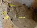 Cranium. Anterior lacrimal crest.Lacrimal bone.
| Anterior lacrimal crest | |
|---|---|
| Details | |
| Part of | maxilla |
| System | skeletal |
| Identifiers | |
| Latin | crista lacrimalis anterior |
| TA98 | A02.1.12.025 |
| TA2 | 782 |
| FMA | 75059 |
| Anatomical terms of bone | |
The anterior lacrimal crest is a bony projection on the frontal process of the maxilla. It creates the lateral margin of the lacrimal sac fossa and is continuous with the orbital margin. The medial palpebral ligament is attached to anterior lacrimal crest. It is an important structure to avoid damaging during rhinoplasty.
The anterior lacrimal crest is a bony projection on the frontal process of the maxilla in the skull. [1] It reaches the junction between the maxilla and the lacrimal bone. [2]
At its junction with the orbital surface is a small tubercle, the lacrimal tubercle, which serves as a guide to the position of the lacrimal sac.
The anterior lacrimal crest is much thicker and stronger than the posterior lacrimal crest. [3] It is one of the thickest parts of the orbit. [4] It is nearly always quite prominent, whilst the posterior lacrimal crest may be less prominent in some people. [5]
The lacrimal sac is directly behind the anterior lacrimal crest, which protects it as part of the fossa for the lacrimal sac. [6]
The anterior lacrimal crest is the site of insertion of the medial palpebral ligament. [1] Some consider this a tendon of the orbicularis oris muscle. [1] The anterior lacrimal crest also protects the lacrimal sac. [6]
The anterior lacrimal crest may be vulnerable to an avulsion fracture due to its connection to the medial palpebral ligament. [4]
The anterior lacrimal crest is vulnerable to damage during osteotomy performed during rhinoplasty, a common plastic surgery. [2] [7] It lies approximately 5 mm from the osteotomy incision line. [7]

Articles related to anatomy include:

The humerus is a long bone in the arm that runs from the shoulder to the elbow. It connects the scapula and the two bones of the lower arm, the radius and ulna, and consists of three sections. The humeral upper extremity consists of a rounded head, a narrow neck, and two short processes. The body is cylindrical in its upper portion, and more prismatic below. The lower extremity consists of 2 epicondyles, 2 processes, and 3 fossae. As well as its true anatomical neck, the constriction below the greater and lesser tubercles of the humerus is referred to as its surgical neck due to its tendency to fracture, thus often becoming the focus of surgeons.

In the human skull, the zygomatic bone, also called cheekbone or malar bone, is a paired irregular bone which articulates with the maxilla, the temporal bone, the sphenoid bone and the frontal bone. It is situated at the upper and lateral part of the face and forms the prominence of the cheek, part of the lateral wall and floor of the orbit, and parts of the temporal fossa and the infratemporal fossa. It presents a malar and a temporal surface; four processes, and four borders.

The lacrimal bone is a small and fragile bone of the facial skeleton; it is roughly the size of the little fingernail. It is situated at the front part of the medial wall of the orbit. It has two surfaces and four borders. Several bony landmarks of the lacrimal bone function in the process of lacrimation or crying. Specifically, the lacrimal bone helps form the nasolacrimal canal necessary for tear translocation. A depression on the anterior inferior portion of the bone, the lacrimal fossa, houses the membranous lacrimal sac. Tears or lacrimal fluid, from the lacrimal glands, collect in this sac during excessive lacrimation. The fluid then flows through the nasolacrimal duct and into the nasopharynx. This drainage results in what is commonly referred to a runny nose during excessive crying or tear production. Injury or fracture of the lacrimal bone can result in posttraumatic obstruction of the lacrimal pathways.

In anatomy, the orbit is the cavity or socket/hole of the skull in which the eye and its appendages are situated. "Orbit" can refer to the bony socket, or it can also be used to imply the contents. In the adult human, the volume of the orbit is 30 millilitres, of which the eye occupies 6.5 ml. The orbital contents comprise the eye, the orbital and retrobulbar fascia, extraocular muscles, cranial nerves II, III, IV, V, and VI, blood vessels, fat, the lacrimal gland with its sac and duct, the eyelids, medial and lateral palpebral ligaments, cheek ligaments, the suspensory ligament, septum, ciliary ganglion and short ciliary nerves.

The pyramid-shaped maxillary sinus is the largest of the paranasal sinuses, located in the maxilla. It drains into the middle meatus of the nose through the semilunar hiatus. It is located to the side of the nasal cavity, and below the orbit.

The orbicularis oculi is a muscle in the face that closes the eyelids. It arises from the nasal part of the frontal bone, from the frontal process of the maxilla in front of the lacrimal groove, and from the anterior surface and borders of a short fibrous band, the medial palpebral ligament.

The orbital or horizontal part of the frontal bone consists of two thin triangular plates, the orbital plates, which form the vaults of the orbits, and are separated from one another by a median gap, the ethmoidal notch.

The greater wing of the sphenoid bone, or alisphenoid, is a bony process of the sphenoid bone, positioned in the skull behind each eye. There is one on each side, extending from the side of the body of the sphenoid and curving upward, laterally, and backward.

The middle cranial fossa is formed by the sphenoid bones, and the temporal bones. It lodges the temporal lobes, and the pituitary gland. It is deeper than the anterior cranial fossa, is narrow medially and widens laterally to the sides of the skull. It is separated from the posterior cranial fossa by the clivus and the petrous crest.

The lacrimal sac or lachrymal sac is the upper dilated end of the nasolacrimal duct, and is lodged in a deep groove formed by the lacrimal bone and frontal process of the maxilla. It connects the lacrimal canaliculi, which drain tears from the eye's surface, and the nasolacrimal duct, which conveys this fluid into the nasal cavity. Lacrimal sac occlusion leads to dacryocystitis.

The posterior lacrimal crest is a vertical bony ridge on the orbital surface of the lacrimal bone. It divides the bone into two parts. It gives origin to the lacrimal part of the orbicularis oculi muscle.

The medial palpebral ligament is a ligament of the face. It attaches to the frontal process of the maxilla, the lacrimal groove, and the tarsus of each eyelid. It has a superficial (anterior) and a deep (posterior) layer, with many surrounding attachments. It connects the medial canthus of each eyelid to the medial part of the orbit. It is a useful point of fixation during eyelid reconstructive surgery.
The zygomatic processes are three processes (protrusions) from other bones of the skull which each articulate with the zygomatic bone. The three processes are:

The frontal process of the maxilla is a strong plate, which projects upward, medialward, and backward from the maxilla, forming part of the lateral boundary of the nose.

In anatomy, the orbital septum is a membranous sheet that acts as the anterior (frontal) boundary of the orbit. It extends from the orbital rims to the eyelids. It forms the fibrous portion of the eyelids.

The lateral margin of the groove of the frontal process of the maxilla is named the anterior lacrimal crest, and is continuous below with the orbital margin; at its junction with the orbital surface is a small tubercle, the lacrimal tubercle, which serves as a guide to the position of the lacrimal sac.

The following outline is provided as an overview of and topical guide to human anatomy:
The lacrimal hamulus is a small, hook-like bony projection of the lacrimal bone. It is a continuation of the posterior lacrimal crest. It articulates with the lacrimal tubercle of the maxilla, and completes the upper orifice of the lacrimal canaliculus. It sometimes exists as a separate piece, and is then called the lesser lacrimal bone.
This glossary explains technical terms commonly employed in the description of dinosaur body fossils. Besides dinosaur-specific terms, it covers terms with wider usage, when these are of central importance in the study of dinosaurs or when their discussion in the context of dinosaurs is beneficial. The glossary does not cover ichnological and bone histological terms, nor does it cover measurements.
![]() This article incorporates text in the public domain from page 161 of the 20th edition of Gray's Anatomy (1918)
This article incorporates text in the public domain from page 161 of the 20th edition of Gray's Anatomy (1918)