The maxilla in vertebrates is the upper fixed bone of the jaw formed from the fusion of two maxillary bones. In humans, the upper jaw includes the hard palate in the front of the mouth. The two maxillary bones are fused at the intermaxillary suture, forming the anterior nasal spine. This is similar to the mandible, which is also a fusion of two mandibular bones at the mandibular symphysis. The mandible is the movable part of the jaw.

The mandibular foramen is an opening on the internal surface of the ramus of the mandible. It allows for divisions of the mandibular nerve and blood vessels to pass through.

The inferior alveolar nerve(IAN) (also the inferior dental nerve) is a sensory branch of the mandibular nerve (CN V3) (which is itself the third branch of the trigeminal nerve (CN V)). The nerve provides sensory innervation to the lower/mandibular teeth and their corresponding gingiva as well as a small area of the face (via its mental nerve).

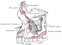
The pyramid-shaped maxillary sinus is the largest of the paranasal sinuses, located in the maxilla. It drains into the middle meatus of the nose through the semilunar hiatus. It is located to the side of the nasal cavity, and below the orbit.

In neuroanatomy, the maxillary nerve (V2) is one of the three branches or divisions of the trigeminal nerve, the fifth (CN V) cranial nerve. It comprises the principal functions of sensation from the maxilla, nasal cavity, sinuses, the palate and subsequently that of the mid-face, and is intermediate, both in position and size, between the ophthalmic nerve and the mandibular nerve.

The inferior alveolar artery is an artery of the head. It is a branch of the maxillary artery. It descends through the infratemporal fossa as part of a neurovascular bundle with the inferior alveolar nerve and vein to the mandibular foramen where it enters and passes anteriorly inside the mandible, suplying the body of mandible and the dental pulp of the lower molar and premolar teeth. Its terminal incisor branch supplies the rest of the lower teeth. Its mental branch exits the mandibula anteriorly through the mental foramen to supply adjacent lip and skin.

In human anatomy, the mandibular canal is a canal within the mandible that contains the inferior alveolar nerve, inferior alveolar artery, and inferior alveolar vein. It runs obliquely downward and forward in the ramus, and then horizontally forward in the body, where it is placed under the alveoli and communicates with them by small openings.

The maxillary artery supplies deep structures of the face. It branches from the external carotid artery just deep to the neck of the mandible.
The descending palatine artery is a branch of the third part of the maxillary artery supplying the hard and soft palate.
The infraorbital artery is a small artery in the head that arises from the maxillary artery and passes through the inferior orbital fissure to enter the orbit, then passes forward along the floor of the orbit, finally exiting the orbit through the infraorbital foramen to reach the face.

The posterior superior alveolar artery is a branch of the maxillary artery. It is one of two or three superior alveolar arteries. It provides arterial suply to the molar and premolar teeth, maxillary sinus and adjacent bone, and the gingiva.
The infratemporal fossa is an irregularly shaped cavity that is a part of the skull. It is situated below and medial to the zygomatic arch. It is not fully enclosed by bone in all directions. It contains superficial muscles, including the lower part of the temporalis muscle, the lateral pterygoid muscle, and the medial pterygoid muscle. It also contains important blood vessels such as the middle meningeal artery, the pterygoid plexus, and the retromandibular vein, and nerves such as the mandibular nerve (CN V3) and its branches.
The anterior superior alveolar nerve (or anterior superior dental nerve) is a branch of the infraorbital nerve (itself a branch of the maxillary nerve (CN V2)). It passes through the canalis sinuosus to reach and innervate upper front teeth. Through its nasal branch, it also innervates parts of the nasal cavity.
The posterior superior alveolar nerves (also posterior superior dental nerves, or posterior superior alveolar branches) are sensory branches of the maxillary nerve (CN V2). They arise within the pterygopalatine fossa as a single trunk. They run on or in the maxilla. They provide sensory innervation to the upper molar teeth and adjacent gum, and the maxillary sinus.

The nerve of the pterygoid canal is formed by the union of the (parasympathetic) greater petrosal nerve and (sympathetic) deep petrosal nerve within the cartilaginous substance filling the foramen lacerum. From the foramen lacerum, the nerve of the pterygoid canal passes through the pterygoid canal to reach the pterygopalatine fossa, ending at the pterygopalatine ganglion.
The infraorbital canal is a canal found at the base of the orbit that opens on to the maxilla. It is continuous with the infraorbital groove and opens onto the maxilla at the infraorbital foramen. The infraorbital nerve and infraorbital artery travel through the canal.

The infraorbital nerve is a branch of the maxillary nerve. It arises in the pterygopalatine fossa. It passes through the inferior orbital fissure to enter the orbit. It travels through the orbit, then enters and traverses the infraorbital canal, exiting the canal at the infraorbital foramen to reach the face. It provides sensory innervation to the skin and mucous membranes around the middle of the face.
In anatomy, a canal is a tubular passage or channel which connects different regions of the body.

The mandibular incisive canal is a bilaterally paired bony canal within the anterior portion of the mandible that extends from the mental foramen (usually) to near the ipsilateral lateral incisor teeth.

In jawed vertebrates, the mandible, lower jaw, or jawbone is a bone that makes up the lower – and typically more mobile – component of the mouth.