Maxillary tuberosity

Last updated
Maxillary
Gray157.png
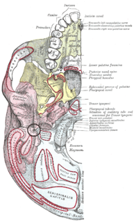
Left maxilla. Outer surface. (Maxillary tuberosity labeled at center right.)
Details
Identifiers
Latin tuber maxillae
TA A02.1.12.016
FMA 57731
Anatomical terms of bone

At the lower part of the infratemporal surface of the maxilla is a rounded eminence, the maxillary tuberosity, especially prominent after the growth of the wisdom tooth; it is rough on its lateral side for articulation with the pyramidal process of the palatine bone and in some cases articulates with the lateral pterygoid plate of the sphenoid.

Maxilla upper jawbone formed from the fusion of two maxillary bones; includes the frontal portion of the palate of the mouth

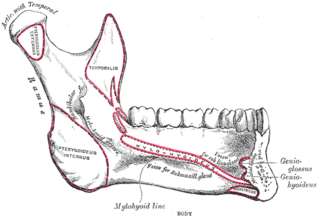
The maxilla in animals is the upper fixed bone of the jaw formed from the fusion of two maxillary bones. The upper jaw includes the hard palate in the front of the mouth. The two maxillary bones are fused at the intermaxillary suture, forming the anterior nasal spine. This is similar to the mandible, which is also a fusion of two mandibular bones at the mandibular symphysis. The mandible is the movable part of the jaw.

Wisdom tooth

A wisdom tooth or third molar is one of the three molars per quadrant of the human dentition. It is the most posterior of the three. The age at which wisdom teeth come through (erupt) is variable, but generally occurs between late teens and early twenties. Most adults have four wisdom teeth, one in each of the four quadrants, but it is possible to have none, fewer, or more, in which case the extras are called supernumerary teeth. Wisdom teeth may get stuck (impacted) against other teeth if there is not enough space for them to come through normally. While this does not cause movement of other teeth, it can cause tooth decay if the impaction makes oral hygiene difficult. Wisdom teeth which are partially erupted through the gum may also cause inflammation and infection in the surrounding gum tissues, termed pericoronitis. Wisdom teeth are often extracted when or even before these problems occur. However some recommend against the prophylactic extraction of disease-free impacted wisdom teeth.

Sphenoid bone unpaired bone situated at the front middle of the skull in front of the temporal bone and basilar part of the occipital bone

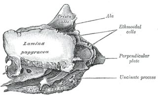
The sphenoid bone is an unpaired bone of the neurocranium. It is situated in the middle of the skull towards the front, in front of the temporal bone and the basilar part of the occipital bone. The sphenoid bone is one of the seven bones that articulate to form the orbit. Its shape somewhat resembles that of a butterfly or bat with its wings extended.

It gives origin to a few fibers of the Medial pterygoid muscle.

Medial pterygoid muscle

The medial pterygoid, is a thick, quadrilateral muscle of mastication.

Related Research Articles

Nasal bone facial bone

The nasal bones are two small oblong bones, varying in size and form in different individuals; they are placed side by side at the middle and upper part of the face and by their junction, form the bridge of the nose.

Inferior nasal concha facial bone

The inferior nasal concha is one of the three paired nasal conchae in the nose. It extends horizontally along the lateral wall of the nasal cavity and consists of a lamina of spongy bone, curled upon itself like a scroll,. The inferior nasal conchae are considered a pair of facial bones. As the air passes through the turbinates, the air is churned against these mucosa-lined bones in order to receive warmth, moisture and cleansing. Superior to inferior nasal concha are the middle nasal concha and superior nasal concha which arise from the cranial portion of the skull. Hence, these two are considered as a part of the cranial bones.

Inguinal ligament This is formed due to inward folding of externus abdominus muscle

The inguinal ligament is a band running from the pubic tubercle to the anterior superior iliac spine. It forms the base of the inguinal canal through which an indirect inguinal hernia may develop.

Anterior superior iliac spine

The anterior superior iliac spine is a bony projection of the iliac bone and an important landmark of surface anatomy. It refers to the anterior extremity of the iliac crest of the pelvis, which provides attachment for the inguinal ligament, and the sartorius muscle. The tensor fasciae latae muscle attaches about 5 cm away at the iliac tubercle.

Stylomastoid foramen

Between the styloid and mastoid processes of the temporal bone is the stylomastoid foramen

Supraorbital foramen

The supraorbital foramen, is a bony elongated opening located above the orbit and under the forehead. The supraorbital foramen lies directly under the eyebrow. Sometimes this foramen is incomplete and is then known as the suprorbital notch.

Condyloid process

The condyloid process or condylar process is the process on the human mandible and some other species' mandibles that ends in a condyle, the mandibular condyle. It is thicker than the coronoid process of the mandible and consists of two portions: the condyle and the constricted portion which supports it, the neck.

Pterygomaxillary fissure

The pterygomaxillary fissure is a fissure of the human skull. It is vertical, and descends at right angles from the medial end of the inferior orbital fissure. It is a triangular interval, formed by the divergence of the maxilla from the pterygoid process of the sphenoid.

Sphenopalatine foramen

The sphenopalatine foramen is a foramen in the skull that connects the nasal cavity with the pterygopalatine fossa.

Ascending palatine artery

The ascending palatine artery is an artery in the head that branches off the facial artery and runs up the superior pharyngeal constrictor muscle.

Anterior nasal spine

The anterior nasal spine, or anterior nasal spine of maxilla, is a bony projection in the skull that serves as a cephalometric landmark. The anterior nasal spine is the projection formed by the fusion of the two maxillary bones at the intermaxillary suture. It is placed at the level of the nostrils, at the uppermost part of the philtrum and rarely fractures.

Infraorbital groove

Not to be confused with the inferior orbital fissure, which is just lateral to the infraorbital groove.

Zygomatic process

Each Zygomatic process is the part of a bone which articulates with the zygomatic bone. The three processes are:

Frontal process of maxilla

The frontal process of maxilla is a strong plate, which projects upward, medialward, and backward from the maxilla, forming part of the lateral boundary of the nose.

Palatine process of maxilla

In human anatomy of the mouth, the palatine process of maxilla, is a thick, horizontal process of the maxilla. It forms the anterior three quarters of the hard palate, the horizontal plate of the palatine bone making up the rest.

Median umbilical ligament

The median umbilical ligament is a structure in human anatomy. It is a shrivelled piece of tissue that represents the remnant of the embryonic urachus.

Orbital lamina of ethmoid bone

The orbital lamina of ethmoid bone, is a smooth, oblong bone plate which forms the lateral surface of the labyrinth of the ethmoid bone in the skull. The plate covers in the middle and posterior ethmoidal cells and forms a large part of the medial wall of the orbit.

Pterygoid fovea

The pterygoid fovea is a concave surface on the uppermost medial side of the ramus of the mandible.

References

This article incorporates text in the public domain from page 158 of the 20th edition of Gray's Anatomy (1918)

The public domain consists of all the creative works to which no exclusive intellectual property rights apply. Those rights may have expired, been forfeited, expressly waived, or may be inapplicable.

<i>Grays Anatomy</i> English-language textbook of human anatomy

Gray's Anatomy is an English language textbook of human anatomy originally written by Henry Gray and illustrated by Henry Vandyke Carter. Earlier editions were called Anatomy: Descriptive and Surgical, Anatomy of the Human Body and Gray's Anatomy: Descriptive and Applied, but the book's name is commonly shortened to, and later editions are titled, Gray's Anatomy. The book is widely regarded as an extremely influential work on the subject, and has continued to be revised and republished from its initial publication in 1858 to the present day. The latest edition of the book, the 41st, was published in September 2015.