A body orifice is any opening in the body of an animal.

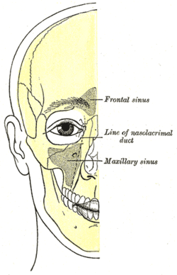
The lacrimal bone is a small and fragile bone of the facial skeleton; it is roughly the size of the little fingernail. It is situated at the front part of the medial wall of the orbit. It has two surfaces and four borders. Several bony landmarks of the lacrimal bone function in the process of lacrimation or crying. Specifically, the lacrimal bone helps form the nasolacrimal canal necessary for tear translocation. A depression on the anterior inferior portion of the bone, the lacrimal fossa, houses the membranous lacrimal sac. Tears or lacrimal fluid, from the lacrimal glands, collect in this sac during excessive lacrimation. The fluid then flows through the nasolacrimal duct and into the nasopharynx. This drainage results in what is commonly referred to a runny nose during excessive crying or tear production. Injury or fracture of the lacrimal bone can result in posttraumatic obstruction of the lacrimal pathways.

The inferior nasal concha is one of the three paired nasal conchae in the nose. It extends horizontally along the lateral wall of the nasal cavity and consists of a lamina of spongy bone, curled upon itself like a scroll,. The inferior nasal conchae are considered a pair of facial bones. As the air passes through the turbinates, the air is churned against these mucosa-lined bones in order to receive warmth, moisture and cleansing. Superior to inferior nasal concha are the middle nasal concha and superior nasal concha which both arise from the ethmoid bone, of the cranial portion of the skull. Hence, these two are considered as a part of the cranial bones.

In anatomy, the orbit is the cavity or socket/hole of the skull in which the eye and its appendages are situated. "Orbit" can refer to the bony socket, or it can also be used to imply the contents. In the adult human, the volume of the orbit is 30 millilitres, of which the eye occupies 6.5 ml. The orbital contents comprise the eye, the orbital and retrobulbar fascia, extraocular muscles, cranial nerves II, III, IV, V, and VI, blood vessels, fat, the lacrimal gland with its sac and duct, the eyelids, medial and lateral palpebral ligaments, cheek ligaments, the suspensory ligament, septum, ciliary ganglion and short ciliary nerves.

The canal containing the nasolacrimal duct is called the nasolacrimal canal.
The lacrimal apparatus is the physiological system containing the orbital structures for tear production and drainage.
It consists of:
The lacrimal canaliculi are the small channels in each eyelid that drain lacrimal fluid, from the lacrimal puncta to the lacrimal sac. This forms part of the lacrimal apparatus that drains lacrimal fluid from the surface of the eye to the nasal cavity.

The lacrimal sac or lachrymal sac is the upper dilated end of the nasolacrimal duct, and is lodged in a deep groove formed by the lacrimal bone and frontal process of the maxilla. It connects the lacrimal canaliculi, which drain tears from the eye's surface, and the nasolacrimal duct, which conveys this fluid into the nasal cavity. Lacrimal sac occlusion leads to dacryocystitis.

On the nasal surface of the body of the maxilla, in front of the opening of the sinus is a deep groove, the lacrimal groove, which is converted into the nasolacrimal canal, by the lacrimal bone and inferior nasal concha; this canal opens into the inferior meatus of the nose and transmits the nasolacrimal duct.

The lacrimal punctum or lacrimal point is a minute opening on the summits of the lacrimal papillae, seen on the margins of the eyelids at the lateral extremity of the lacrimal lake. There are two lacrimal puncta in the medial (inside) portion of each eyelid. Normally, the puncta dip into the lacrimal lake.
Oculoplastics, or oculoplastic surgery, includes a wide variety of surgical procedures that deal with the orbit, eyelids, tear ducts, and the face. It also deals with the reconstruction of the eye and associated structures.

Dacryocystorhinostomy (DCR) is a surgical procedure to restore the flow of tears into the nose from the lacrimal sac when the nasolacrimal duct does not function.

Dacryocystitis is an infection of the lacrimal sac, secondary to obstruction of the nasolacrimal duct at the junction of lacrimal sac. The term derives from the Greek dákryon (tear), cysta (sac), and -itis (inflammation). It causes pain, redness, and swelling over the inner aspect of the lower eyelid and epiphora. When nasolacrimal duct obstruction is secondary to a congenital barrier it is referred to as dacryocystocele. It is most commonly caused by Staphylococcus aureus and Streptococcus pneumoniae. The most common complication is corneal ulceration, frequently in association with S. pneumoniae. The mainstays of treatment are oral antibiotics, warm compresses, and relief of nasolacrimal duct obstruction by dacryocystorhinostomy.

Epiphora is an overflow of tears onto the face, other than caused by normal crying. It is a clinical sign or condition that constitutes insufficient tear film drainage from the eyes, in that tears will drain down the face rather than through the nasolacrimal system.

Marsupialization is the surgical technique of cutting a slit into an abscess or cyst and suturing the edges of the slit to form a continuous surface from the exterior surface to the interior surface of the cyst or abscess. Sutured in this fashion, the site remains open and can drain freely. This technique is used to treat a cyst or abscess when a single draining would not be effective and complete removal of the surrounding structure would not be desirable. The technique is often applied to Gartner's duct cysts, pancreatic cysts, pilonidal cysts, and Bartholin's cysts.
Dacryocystocele (Dacryocystitis) or timo cyst is a benign, bluish-gray mass in the inferomedial canthus that develops within a few days or weeks after birth. The uncommon condition forms as a result as a consequence of narrowing or obstruction of the nasolacrimal duct, usually during prenatal development. Nasolacrimal duct obstruction disrupts the lacrimal drainage system, eventually creating a swelling cyst in the lacrimal sac area by the nasal cavity. The location of the cyst can cause respiratory dysfunction, compromising the airway. The obstruction ultimately leads to epiphora, an abundance of tear production.

The accessory visual structures are the protecting and supporting structures (adnexa) of the eye, including the eyebrow, eyelids, and lacrimal apparatus. The eyebrows, eyelids, eyelashes, lacrimal gland and drainage apparatus all play a crucial role with regards to globe protection, lubrication, and minimizing the risk of ocular infection. The adnexal structures also help to keep the cornea moist and clean.

Nasolacrimal duct obstruction is the obstruction of the nasolacrimal duct and may be either congenital or acquired. Obstruction of the nasolacrimal duct leads to the excess overflow of tears called epiphora.

In anatomy, the term nasal meatus can refer to any of the three meatuses (passages) through the skull's nasal cavity: the superior meatus, middle meatus, and inferior meatus.

Dacryoscintigraphy (DSG), also known as lacrimal scintigraphy, is a nuclear medicine technique for imaging the lacrimal apparatus. It is used to identify obstructions, for example in the lacrimal duct, nasal cavity or nasolacrimal duct.