Coxiella burnetii

Last updated

Coxiella burnetii
Coxiella burnetii, the bacteria that causes Q Fever.jpg
A dry fracture of a Vero cell exposing the contents of a vacuole where Coxiella burnetii is growing
Scientific classification OOjs UI icon edit-ltr.svg
Domain: Bacteria
Phylum: Pseudomonadota
Class: Gammaproteobacteria
Order: Legionellales
Family: Coxiellaceae
Genus: Coxiella
Species:
C. burnetii
Binomial name
Coxiella burnetii
(Derrick 1939)
Philip 1948

Coxiella burnetii is an obligate intracellular bacterial pathogen, and is the causative agent of Q fever. [1] The genus Coxiella is morphologically similar to Rickettsia , but with a variety of genetic and physiological differences. C. burnetii is a small Gram-negative, coccobacillary bacterium that is highly resistant to environmental stresses such as high temperature, osmotic pressure, and ultraviolet light. These characteristics are attributed to a small cell variant form of the organism that is part of a biphasic developmental cycle, including a more metabolically and replicatively active large cell variant form. [2] It can survive standard disinfectants, and is resistant to many other environmental changes like those presented in the phagolysosome. [3]

Contents

History and naming

Research in the 1920s and 1930s identified what appeared to be a new type of Rickettsia, isolated from ticks, that was able to pass through filters. The first description of what may have been Coxiella burnetii was published in 1930 by Hideyo Noguchi, but since his samples did not survive, it remains unclear as to whether it was the same organism. The definitive descriptions were published in the late 1930s as part of research into the cause of Q fever, by Edward Holbrook Derrick and Macfarlane Burnet in Australia, and Herald Rea Cox and Gordon Davis at the Rocky Mountain Laboratory (RML) in the United States. [4]

The RML team proposed the name Rickettsia diaporica, derived from the Greek word for having the ability to pass through filter pores, to avoid naming it after either Cox or Davis if indeed Noguchi's description had priority. Around the same time, Derrick proposed the name Rickettsia burnetii, in recognition of Burnet's contribution in identifying the organism as a Rickettsia. As it became clear that the species differed significantly from other Rickettsia, it was first elevated to a subgenus named after Cox, Coxiella, and then in 1948 to its own genus of that name, proposed by Cornelius B. Philip, another RML researcher. [4] Research in the 1960s1970s by French Canadian-American microbiologist and virologist Paul Fiset was instrumental in the development of the first successful Q fever vaccine. [5]

Coxiella was difficult to study because it could not be reproduced outside a host. However, in 2009, scientists reported a technique allowing the bacteria to grow in an axenic culture and suggested the technique may be useful for study of other pathogens. [6]

Pathogenesis

Immunohistochemical detection of C. burnetii in resected cardiac valve of a 60-year-old man with Q fever endocarditis, Cayenne, French Guiana, monoclonal antibody against C. burnetii and hematoxylin were used for staining: Original magnification x50 Immunohistochemical detection of Coxiella burnetii in resected cardiac valve of a 60-year-old man with Q fever endocarditis.jpg
Immunohistochemical detection of C. burnetii in resected cardiac valve of a 60-year-old man with Q fever endocarditis, Cayenne, French Guiana, monoclonal antibody against C. burnetii and hematoxylin were used for staining: Original magnification ×50

Of the many C. burnetii strains, two of the most studied are the Nine Mile phase I and Priscilla phase I strain. In recent years, more strains have been studied. Nonetheless, it has been demonstrated that the Nine Mile strain is one of the most virulent strains of C. burnetti with as few as four organisms needed to cause infection. This is particularly relevant as murine rodents are poorly susceptible to C. burnetii, necessitating a higher dose and a more virulent dose to inoculate murine rodents for disease study. [7]

The ID50 (the dose needed to infect 50% of experimental subjects) is one via inhalation; i.e., inhalation of one organism will yield disease in 50% of the population. This is an extremely low infectious dose (only 1-10 organisms required), making C. burnetii one of the most infectious known organisms. [8] [9] Disease occurs in two stages: an acute stage that presents with headaches, chills, and respiratory symptoms, and an insidious chronic stage.

C. burnettii infections begins within the alveoli. Upon inhalation, it targets alveolar macrophages and passively enters them via actin-dependent phagocytosis. After initial binding, it is suggested that C. burnetii enters phagocytotic cells via passive actin-dependent phagocytosis and enters non-professional phagocytes via an active zipper mechanism. C. burnetii exploits the αVβ3 integrin to enter using RAC1-dependent phagocytosis, which is believed to have evolved as a mechanism to avoid the induction of an inflammatory response. [10]

Following infection, C. burnetii has a biphasic developmental cycle, which consists of small cell variant (SCV) and large cell variant (LCV) morphological forms, which are both infectious. As the SCV is metabolically repressed and resistant to many environmental stressors, it is likely the form that initiates natural infections. Having entered a host cell, C. burnetii SCVs transit through the phagolysosomal maturation pathway. In the first six hours post-infection, endosomes, autophagosomes, and lysosomes containing acid phosphatase fuse with the nascent phagosome to form early PV, which fosters the transition from SCV to LCV. Resultantly, C. burnetii is metabolically activated and produces the T4SS to translocate effector proteins into the host cytoplasm. After 6 days, C. burnetii transitions back to SCV. [7] [11]

While most infections clear up spontaneously, treatment with tetracycline or doxycycline appears to reduce the symptomatic duration and reduce the likelihood of chronic infection. A combination of erythromycin and rifampin is highly effective in curing the disease, and vaccination with Q-VAX vaccine (CSL) is effective for prevention of it.[ citation needed ]

The bacteria use a type IVB secretion system known as Icm/Dot (intracellular multiplication / defect in organelle trafficking genes) to inject over 100 effector proteins into the host. These effectors increase the bacteria's ability to survive and grow inside the host cell by modulating many host cell pathways, including blocking cell death, inhibiting immune reactions, and altering vesicle trafficking. [12] [13] [14] In Legionella pneumophila , which uses the same secretion system and also injects effectors, survival is enhanced because these proteins interfere with fusion of the bacteria-containing vacuole with the host's degradation endosomes. [15]

Use as a biological weapon

The United States ended its biological warfare program in 1969. When it did, C. burnetii was one of seven agents it had standardized as biological weapons. [16]

Genomics

At least 75 [17] completely sequenced genomes of Coxiella burnetii strains exist, [18] which contain about 2.1 Mbp of DNA each and encode around 2,100 open reading frames; 746 (or about 35%) of these genes have no known function.

In bacteria small regulatory RNAs are activated during stress and virulence conditions. Coxiella burnetii small RNAs (CbSRs 1, 11, 12, and 14) are encoded within intergenic region (IGR). CbSRs 2, 3, 4 and 9 are located antisense to identified ORFs. The CbSRs are up-regulated during intracellular growth in host cells. [19]

All C. burnetii isolates either carry one of four conserved independently-replicating large plasmids (QpH1, QpDG, QpRS, or QpDV) or a chromosomal element derived from QpRS. QpH1 carries virluence factors important for the bacterium's survival inside mouse macrophages [20] and Vero cells; growth on axenic media is unaffected. QpH1 also contains a toxin-antitoxin system. [21] Among all plasmids, 8 conserved genes code for proteins that are inserted into the host cell via the secretion system. [21]

Additional images

Related Research Articles

<i>Salmonella</i> Genus of prokaryotes

Salmonella is a genus of rod-shaped (bacillus) gram-negative bacteria of the family Enterobacteriaceae. The two known species of Salmonella are Salmonella enterica and Salmonella bongori. S. enterica is the type species and is further divided into six subspecies that include over 2,600 serotypes. Salmonella was named after Daniel Elmer Salmon (1850–1914), an American veterinary surgeon.

<span class="mw-page-title-main">Q fever</span> Coxiella burnetii infection

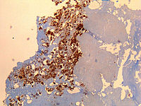
Q fever or query fever is a disease caused by infection with Coxiella burnetii, a bacterium that affects humans and other animals. This organism is uncommon, but may be found in cattle, sheep, goats, and other domestic mammals, including cats and dogs. The infection results from inhalation of a spore-like small-cell variant, and from contact with the milk, urine, feces, vaginal mucus, or semen of infected animals. Rarely, the disease is tick-borne. The incubation period can range from 9 to 40 days. Humans are vulnerable to Q fever, and infection can result from even a few organisms. The bacterium is an obligate intracellular pathogenic parasite.

<i>Rickettsia</i> Genus of bacteria

Rickettsia is a genus of nonmotile, gram-negative, nonspore-forming, highly pleomorphic bacteria that may occur in the forms of cocci, bacilli, or threads. The genus was named after Howard Taylor Ricketts in honor of his pioneering work on tick-borne spotted fever.

<i>Candida albicans</i> Species of fungus

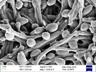
Candida albicans is an opportunistic pathogenic yeast that is a common member of the human gut flora. It can also survive outside the human body. It is detected in the gastrointestinal tract and mouth in 40–60% of healthy adults. It is usually a commensal organism, but it can become pathogenic in immunocompromised individuals under a variety of conditions. It is one of the few species of the genus Candida that cause the human infection candidiasis, which results from an overgrowth of the fungus. Candidiasis is, for example, often observed in HIV-infected patients. C. albicans is the most common fungal species isolated from biofilms either formed on (permanent) implanted medical devices or on human tissue. C. albicans, C. tropicalis, C. parapsilosis, and C. glabrata are together responsible for 50–90% of all cases of candidiasis in humans. A mortality rate of 40% has been reported for patients with systemic candidiasis due to C. albicans. By one estimate, invasive candidiasis contracted in a hospital causes 2,800 to 11,200 deaths yearly in the US. Nevertheless, these numbers may not truly reflect the true extent of damage this organism causes, given new studies indicating that C. albicans can cross the blood–brain barrier in mice.

Rickettsia prowazekii is a species of gram-negative, alphaproteobacteria, obligate intracellular parasitic, aerobic bacillus bacteria that is the etiologic agent of epidemic typhus, transmitted in the feces of lice. In North America, the main reservoir for R. prowazekii is the flying squirrel. R. prowazekii is often surrounded by a protein microcapsular layer and slime layer; the natural life cycle of the bacterium generally involves a vertebrate and an invertebrate host, usually an arthropod, typically the human body louse. A form of R. prowazekii that exists in the feces of arthropods remains stably infective for months. R. prowazekii also appears to be the closest free-living relative of mitochondria, based on genome sequencing.

<i>Rickettsia rickettsii</i> Species of bacterium

Rickettsia rickettsii is a Gram-negative, intracellular, coccobacillus bacterium that was first discovered in 1902. R. rickettsii is the causative agent of Rocky Mountain Spotted Fever and is transferred to its host via a tick bite. It is one of the most pathogenic Rickettsia species and affects a large majority of the Western Hemisphere, most commonly the Americas. The pathogenic agent has been found on every continent, except Antarctica; however, Rocky Mountain Spotted Fever occurs mostly in North, Central, and South America. This prevalence is due to R. rickettsi thriving in warm, damp environments.

<i>Legionella pneumophila</i> Species of bacterium

Legionella pneumophila is an aerobic, pleomorphic, flagellated, non-spore-forming, Gram-negative bacterium of the genus Legionella. L. pneumophila is the primary human pathogenic bacterium in this group. In nature, L. pneumophila infects freshwater and soil amoebae of the genera Acanthamoeba and Naegleria. This pathogen is found commonly near freshwater environments and will then invade the amoebae found in these environments, using them to carry out metabolic functions.

<i>Francisella tularensis</i> Species of bacterium

Francisella tularensis is a pathogenic species of Gram-negative coccobacillus, an aerobic bacterium. It is nonspore-forming, nonmotile, and the causative agent of tularemia, the pneumonic form of which is often lethal without treatment. It is a fastidious, facultative intracellular bacterium, which requires cysteine for growth. Due to its low infectious dose, ease of spread by aerosol, and high virulence, F. tularensis is classified as a Tier 1 Select Agent by the U.S. government, along with other potential agents of bioterrorism such as Yersinia pestis, Bacillus anthracis, and Ebola virus. When found in nature, Francisella tularensis can survive for several weeks at low temperatures in animal carcasses, soil, and water. In the laboratory, F. tularensis appears as small rods, and is grown best at 35–37 °C.

<span class="mw-page-title-main">Phagosome</span>

In cell biology, a phagosome is a vesicle formed around a particle engulfed by a phagocyte via phagocytosis. Professional phagocytes include macrophages, neutrophils, and dendritic cells (DCs).

Intracellular parasites are microparasites that are capable of growing and reproducing inside the cells of a host. They are also called intracellular pathogens.

<i>Shigella flexneri</i> Species of bacterium

Shigella flexneri is a species of Gram-negative bacteria in the genus Shigella that can cause diarrhea in humans. Several different serogroups of Shigella are described; S. flexneri belongs to group B. S. flexneri infections can usually be treated with antibiotics, although some strains have become resistant. Less severe cases are not usually treated because they become more resistant in the future. Shigella are closely related to Escherichia coli, but can be differentiated from E.coli based on pathogenicity, physiology and serology.

<span class="mw-page-title-main">Phagolysosome</span> Cytoplasmic body

In biology, a phagolysosome, or endolysosome, is a cytoplasmic body formed by the fusion of a phagosome with a lysosome in a process that occurs during phagocytosis. Formation of phagolysosomes is essential for the intracellular destruction of microorganisms and pathogens. It takes place when the phagosome's and lysosome's membranes 'collide', at which point the lysosomal contents—including hydrolytic enzymes—are discharged into the phagosome in an explosive manner and digest the particles that the phagosome had ingested. Some products of the digestion are useful materials and are moved into the cytoplasm; others are exported by exocytosis.

In biology, axenic describes the state of a culture in which only a single species, variety, or strain of organism is present and entirely free of all other contaminating organisms. The earliest axenic cultures were of bacteria or unicellular eukaryotes, but axenic cultures of many multicellular organisms are also possible. Axenic culture is an important tool for the study of symbiotic and parasitic organisms in a controlled environment.

<i>Ehrlichia</i> Genus of bacteria

Ehrlichia is a genus of Rickettsiales bacteria that are transmitted to vertebrates by ticks. These bacteria cause the disease ehrlichiosis, which is considered zoonotic, because the main reservoirs for the disease are animals.

<i>Orientia tsutsugamushi</i> Species of bacterium

Orientia tsutsugamushi is a mite-borne bacterium belonging to the family Rickettsiaceae and is responsible for a disease called scrub typhus in humans. It is a natural and an obligate intracellular parasite of mites belonging to the family Trombiculidae. With a genome of only 2.0–2.7 Mb, it has the most repeated DNA sequences among bacterial genomes sequenced so far. The disease, scrub typhus, occurs when infected mite larvae accidentally bite humans. Primarily indicated by undifferentiated febrile illnesses, the infection can be complicated and often fatal.

<i>Rhodococcus equi</i> Species of bacterium

Rhodococcus equi is a Gram-positive coccobacillus bacterium. The organism is commonly found in dry and dusty soil and can be important for diseases of domesticated animals. The frequency of infection can reach near 60%. R. equi is an important pathogen causing pneumonia in foals. Since 2008, R. equi has been known to infect wild boar and domestic pigs. R. equi can infect humans. At-risk groups are immunocompromised people, such as HIV-AIDS patients or transplant recipients. Rhodococcus infection in these patients resemble clinical and pathological signs of pulmonary tuberculosis. It is facultative intracellular.

<i>Bacillus anthracis</i> Species of bacterium

Bacillus anthracis is a gram-positive and rod-shaped bacterium that causes anthrax, a deadly disease to livestock and, occasionally, to humans. It is the only permanent (obligate) pathogen within the genus Bacillus. Its infection is a type of zoonosis, as it is transmitted from animals to humans. It was discovered by a German physician Robert Koch in 1876, and became the first bacterium to be experimentally shown as a pathogen. The discovery was also the first scientific evidence for the germ theory of diseases.

Rickettsia typhi is a small, aerobic, obligate intracellular, rod shaped gram negative bacterium. It belongs to the typhus group of the Rickettsia genus, along with R. prowazekii. R. typhi has an uncertain history, as it may have long gone shadowed by epidemic typhus. This bacterium is recognized as a biocontainment level 2/3 organism. R. typhi is a flea-borne disease that is best known to be the causative agent for the disease murine typhus, which is an endemic typhus in humans that is distributed worldwide. As with all rickettsial organisms, R. typhi is a zoonotic agent that causes the disease murine typhus, displaying non-specific mild symptoms of fevers, headaches, pains and rashes. There are two cycles of R. typhi transmission from animal reservoirs containing R. typhi to humans: a classic rat-flea-rat cycle that is most well studied and common, and a secondary periodomestic cycle that could involve cats, dogs, opossums, sheep, and their fleas.

Rickettsia australis is a bacterium that causes a medical condition called Queensland tick typhus. The probable vectors are the tick species, Ixodes holocyclus and Ixodes tasmani. Small marsupials are suspected reservoirs of this bacterium.

<span class="mw-page-title-main">Paul Fiset</span> Canadian-American microbiologist (1922–2001)

Paul Fiset was a Canadian-American microbiologist and virologist. His research helped to develop one of the first successful Q fever vaccines, noted by The New York Times. Fiset was born in Quebec, Canada, and attended Laval University, where he earned a Doctor of Medicine degree in 1949. He subsequently attended Cambridge University, where he received a PhD degree in 1956. As a professor at the University of Maryland School of Medicine, he also researched other bacterial diseases such as typhus and Rocky Mountain spotted fever, in addition to Q fever.

References

  1. Shaw EI, Voth DE (January 2019). "Coxiella burnetii: A Pathogenic Intracellular Acidophile". Microbiology. 165 (1): 1–3. doi: 10.1099/mic.0.000707 . PMC   6600347 . PMID   30422108.
  2. Voth DE, Heinzen RA (April 2007). "Lounging in a lysosome: the intracellular lifestyle of Coxiella burnetii". Cellular Microbiology. 9 (4): 829–40. doi: 10.1111/j.1462-5822.2007.00901.x . PMID   17381428.
  3. Sankaran N (2000). "Coxiella burnetii" . Microbes and people : an A-Z of microorganisms in our lives. Phoenix, Arizona: The Oryx Press. pp.  72. ISBN   1-57356-217-3. "In contrast to other rickettsiae, which are highly sensitive and easily killed by chemical disinfectants and changes in their surroundings, C. burnetii is highly resistant" & "Q fever". Centers for Disease Control and Prevention; National Center for Infectious Diseases; Division of Viral and Rickettsial Diseases; Viral and Rickettsial Zoonoses Branch. 2003-02-13. Retrieved 2006-05-24. "The organisms are resistant to heat, drying, and many common disinfectants."
  4. 1 2 McDade JE (1990). "Historical Aspects of Q Fever". In Marrie TJ (ed.). Q Fever, Volume I: The Disease. CRC Press. pp. 5–22. ISBN   0-8493-5984-8.
  5. Saxon, Wolfgang (March 8, 2001). "Dr. Paul Fiset, 78, Microbiologist And Developer of Q Fever Vaccine". New York Times . p. C-17.
  6. Omsland A, Cockrell DC, Howe D, Fischer ER, Virtaneva K, Sturdevant DE, et al. (March 2009). "Host cell-free growth of the Q fever bacterium Coxiella burnetii". Proceedings of the National Academy of Sciences of the United States of America. 106 (11): 4430–4. Bibcode:2009PNAS..106.4430O. doi: 10.1073/pnas.0812074106 . PMC   2657411 . PMID   19246385.
  7. 1 2 Eldin, Carole; Mélenotte, Cléa; Mediannikov, Oleg; Ghigo, Eric; Million, Matthieu; Edouard, Sophie; Mege, Jean-Louis; Maurin, Max; Raoult, Didier (January 2017). "From Q Fever to Coxiella burnetii Infection: a Paradigm Change". Clinical Microbiology Reviews. 30 (1): 115–190. doi:10.1128/CMR.00045-16. ISSN   0893-8512. PMC   5217791 . PMID   27856520.
  8. Tigertt WD, Benenson AS, Gochenour WS (September 1961). "Airborne Q fever". Bacteriological Reviews. 25 (3): 285–93. doi:10.1128/br.25.3.285-293.1961. PMC   441106 . PMID   13921201.
  9. "Q fever caused by Coxiella burnetii". Centers for Disease Control. 15 January 2019.
  10. Dragan, Amanda L.; Voth, Daniel E. (April 2020). "Coxiella burnetii: international pathogen of mystery". Microbes and Infection. 22 (3): 100–110. doi:10.1016/j.micinf.2019.09.001. ISSN   1286-4579. PMC   7101257 . PMID   31574310.
  11. Dragan, Amanda L.; Voth, Daniel E. (2020-04-01). "Coxiella burnetii: international pathogen of mystery". Microbes and Infection. 22 (3): 100–110. doi:10.1016/j.micinf.2019.09.001. ISSN   1286-4579. PMC   7101257 . PMID   31574310.
  12. Lührmann A, Nogueira CV, Carey KL, Roy CR (November 2010). "Inhibition of pathogen-induced apoptosis by a Coxiella burnetii type IV effector protein". Proceedings of the National Academy of Sciences of the United States of America. 107 (44): 18997–9001. Bibcode:2010PNAS..10718997L. doi: 10.1073/pnas.1004380107 . PMC   2973885 . PMID   20944063.
  13. Clemente TM, Mulye M, Justis AV, Nallandhighal S, Tran TM, Gilk SD (October 2018). Freitag NE (ed.). "Coxiella burnetii Blocks Intracellular Interleukin-17 Signaling in Macrophages". Infection and Immunity. 86 (10). doi:10.1128/IAI.00532-18. PMC   6204741 . PMID   30061378.
  14. Newton HJ, Kohler LJ, McDonough JA, Temoche-Diaz M, Crabill E, Hartland EL, Roy CR (July 2014). Valdivia RH (ed.). "A screen of Coxiella burnetii mutants reveals important roles for Dot/Icm effectors and host autophagy in vacuole biogenesis". PLOS Pathogens. 10 (7): e1004286. doi: 10.1371/journal.ppat.1004286 . PMC   4117601 . PMID   25080348.
  15. Pan X, Lührmann A, Satoh A, Laskowski-Arce MA, Roy CR (June 2008). "Ankyrin repeat proteins comprise a diverse family of bacterial type IV effectors". Science. 320 (5883): 1651–4. Bibcode:2008Sci...320.1651P. doi:10.1126/science.1158160. PMC   2514061 . PMID   18566289.
  16. Croddy, Eric C.; Hart, C. Perez-Armendariz J. (2002). Chemical and Biological Warfare. Springer. pp. 30–31. ISBN   0-387-95076-1.
  17. Abou Abdallah, Rita; Million, Matthieu; Delerce, Jeremy; Anani, Hussein; Diop, Awa; Caputo, Aurelia; Zgheib, Rita; Rousset, Elodie; Sidi Boumedine, Karim; Raoult, Didier; Fournier, Pierre-Edouard (21 November 2022). "Pangenomic analysis of Coxiella burnetii unveils new traits in genome architecture". Frontiers in Microbiology. 13. doi: 10.3389/fmicb.2022.1022356 . PMC   9721466 . PMID   36478861.
  18. "Genome - NCBI". National Center for Biotechnology Information, U.S. National Library of Medicine. Archived from the original on 2011-11-28. Retrieved 1 January 2022.
  19. Warrier I, Hicks LD, Battisti JM, Raghavan R, Minnick MF (2014). "Identification of novel small RNAs and characterization of the 6S RNA of Coxiella burnetii". PLOS ONE. 9 (6): e100147. Bibcode:2014PLoSO...9j0147W. doi: 10.1371/journal.pone.0100147 . PMC   4064990 . PMID   24949863.
  20. Luo, Shengdong; Lu, Shanshan; Fan, Huahao; Chen, Zeliang; Sun, Zhihui; Hu, Yan; Li, Ruisheng; An, Xiaoping; Uversky, Vladimir N.; Tong, Yigang; Song, Lihua (8 April 2021). "The Coxiella burnetii QpH1 Plasmid Is a Virulence Factor for Colonizing Bone Marrow-Derived Murine Macrophages". Journal of Bacteriology. 203 (9). doi:10.1128/jb.00588-20. PMC   8092169 . PMID   33558394.
  21. 1 2 Wachter, S; Cockrell, DC; Miller, HE; Virtaneva, K; Kanakabandi, K; Darwitz, B; Heinzen, RA; Beare, PA (December 2022). "The endogenous Coxiella burnetii plasmid encodes a functional toxin-antitoxin system". Molecular Microbiology. 118 (6): 744–764. doi:10.1111/mmi.15001. PMC   10098735 . PMID   36385554.