Burkholderia pseudomallei

Last updated

Burkholderia pseudomallei
Bps close.JPG
Burkholderia pseudomallei colonies on Ashdown's agar showing the characteristic cornflower head morphology
Scientific classification OOjs UI icon edit-ltr.svg
Domain: Bacteria
Phylum: Pseudomonadota
Class: Betaproteobacteria
Order: Burkholderiales
Family: Burkholderiaceae
Genus: Burkholderia
Species:
B. pseudomallei
Binomial name
Burkholderia pseudomallei
(Whitmore 1913)
Yabuuchi et al. 1993 [1]
Synonyms

Bacillus pseudomallei Whitmore 1913
Bacterium whitmoriStanton and Fletcher 1921
Malleomyces pseudomalleiBreed 1939
Loefflerella pseudomalleiBrindle and Cowan 1951
Pfeiferella pseudomallei
Pseudomonas pseudomallei(Whitmore 1913) Haynes 1957

Contents

Burkholderia pseudomallei (also known as Pseudomonas pseudomallei) is a Gram-negative, bipolar, aerobic, motile rod-shaped bacterium. [2] It is a soil-dwelling bacterium endemic in tropical and subtropical regions worldwide, particularly in Thailand and northern Australia. [3] It was reported in 2008 that there had been an expansion of the affected regions due to significant natural disasters, and it could be found in Southern China, Hong Kong, and countries in America. [4] B. pseudomallei, amongst other pathogens, has been found in monkeys imported into the United States from Asia for laboratory use, posing a risk that the pathogen could be introduced into the country. [5]

Although it is mainly a soil-dwelling bacteria, a study performed by Apinya Pumpuang and others showed that Burkholderia pseudomallei survived in distilled water for 16 years, demonstrating that it is capable of living in water if a specific environment is provided. [6] It is resistant to a variety of harsh conditions including nutrient deficiency, extreme temperature or pH. [7] It infects humans, causing the disease melioidosis; [8] mortality is 20–50% even with treatment. The CDC classifies it as a "Tier 1 select agent" with potential as a bioterrorism agent. [5] It infects other animals, most commonly livestock such as goats, pigs, and sheep, less frequently. [9] It is also capable of infecting plants in a laboratory setting. [10]

Burkholderia pseudomallei measures 2–5 μm in length and 0.4–0.8 μm in diameter and is capable of self-propulsion using flagella. The bacteria can grow in a number of artificial nutrient environments, especially betaine- and arginine-containing ones.

In vitro , optimal proliferation temperature is reported around 40 °C in neutral or slightly acidic environments (pH 6.8–7.0). The majority of strains are capable of oxidation, not fermentation, of sugars without gas formation (most importantly, glucose and galactose; older cultures are reported to also metabolize maltose and starch). Bacteria produce both exo- and endotoxins. The role of the toxins identified in the process of melioidosis symptom development has not been fully elucidated. [11]

Identification

Burkholderia pseudomallei is not fastidious and grows on a large variety of culture media (blood agar, MacConkey agar, EMB, etc.). Ashdown's medium (or Burkholderia cepacia medium) may be used for selective isolation. [12] Cultures typically become positive in 24 to 48 hours (this rapid growth rate differentiates the organism from B. mallei , which typically takes a minimum of 72 hours to grow). Colonies are wrinkled, have a metallic appearance, and possess an earthy odor. On Gram staining, the organism is a Gram-negative rod with a characteristic "safety pin" appearance (bipolar staining). On sensitivity testing, the organism appears highly resistant (it is innately resistant to many antibiotics including colistin and gentamicin) and that again differentiates it from B. mallei, which is in contrast, exquisitely sensitive to many antibiotics. For environmental specimens only, differentiation from the nonpathogenic B. thailandensis using an arabinose test is necessary (B. thailandensis is never isolated from clinical specimens). [13] The laboratory identification of B. pseudomallei has been described in the literature. [14]

The classic textbook description of B. pseudomallei in clinical samples is of an intracellular, bipolar-staining, Gram-negative rod, but this is of little value in identifying the organism from clinical samples. [14] Some [15] suggest the Wayson stain is useful for this purpose, but this has been shown not to be the case. [16]

Laboratory identification of B. pseudomallei can be difficult, especially in Western countries where it is rarely seen. The large, wrinkled colonies look like environmental contaminants, so are often discarded as being of no clinical significance. Colony morphology is very variable and a single strain may display multiple colony types, [17] [18] so inexperienced laboratory staff may mistakenly believe the growth is not pure. The organism grows more slowly than other bacteria that may be present in clinical specimens, and in specimens from nonsterile sites, is easily overgrown. Nonsterile specimens should, therefore, be cultured in selective media (e.g., Ashdown's [19] [20] or B. cepacia medium). [12] For heavily contaminated samples, such as feces, a modified version of Ashdown's that includes norfloxacin, amoxicillin, and polymyxin B has been proposed. [21] In blood culture, the BacT/ALERT MB system (normally used for culturing mycobacteria) by bioMérieux has been shown to have superior yields compared to conventional blood culture media. [22]

Even when the isolate is recognized to be significant, commonly used identification systems may misidentify the organism as Chromobacterium violaceum or other nonfermenting, Gram-negative bacilli such as Burkholderia cepacia or Pseudomonas aeruginosa . [23] [24] Again, because the disease is rarely seen in Western countries, identification of B. pseudomallei in cultures may not actually trigger alarms in physicians unfamiliar with the disease. [25] Routine biochemical methods for identification of bacteria vary widely in their identification of this organism: the API 20NE system accurately identifies B. pseudomallei in 99% of cases, [26] as does the automated Vitek 1 system, but the automated Vitek 2 system only identifies 19% of isolates. [24]

The pattern of resistance to antimicrobials is distinctive, and helps to differentiate the organism from P. aeruginosa. The majority of B. pseudomallei isolates are intrinsically resistant to all aminoglycosides (via an efflux pump mechanism), [27] but sensitive to co-amoxiclav: [28] this pattern of resistance almost never occurs in P. aeruginosa and is helpful in identification. [29] Unfortunately, the majority of strains in Sarawak, Borneo, are susceptible to aminoglycosides and macrolides, which means the conventional recommendations for isolation and identification do not apply there. [30]

Molecular methods (PCR) of diagnosis are possible, but not routinely available for clinical diagnosis. [31] [32] Fluorescence in situ hybridisation has also been described, but has not been clinically validated, and it is not commercially available. [33] In Thailand, a latex agglutination assay is widely used, [26] while a rapid immunofluorescence technique is also available in a small number of centres. [34]

Characteristics

Morphological, physiological, and biochemical characteristics of Burkholderia pseudomallei are shown in the Table below.

Test typeTestCharacteristics
Colony charactersSize2–5 μm in length and 0.4–0.8 μm in diameter
TypeRound
ColorWhitish
ShapeMultiple
Morphological charactersShapeRod (Variable)
Physiological charactersMotility+
Growth at 6.5% NaCl+
Biochemical charactersGram staining-
Oxidase+
Catalase+ [35]
Oxidative-Fermentative
Motility+
Methyl Red
Voges-Proskauer
Indole- [36]
H2S Production-
Urease
Nitrate reductase+
β-Galactosidase
Hydrolysis ofGelatin+
Casein
Utilization ofGlycerol+
Galactose+
D-Glucose+
D-Fructose+
D-Mannose+
MannitolVariable

Note: + = Positive, – =Negative

Disinfection

Burkholderia pseudomallei is susceptible to numerous disinfectants, including benzalkonium chloride, iodine, mercuric chloride, potassium permanganate, 1% sodium hypochlorite, 70% ethanol, 2% glutaraldehyde, and to a lesser extent, phenolic preparations. [37] B. pseudomallei is effectively killed by the commercial disinfectants, Perasafe and Virkon. [38] The microorganism can also be destroyed by heating to above 74 °C for 10 min or by ultraviolet irradiation. [39]

Medical importance

Burkholderia pseudomallei infection in humans is called melioidosis or Whitmore's disease. It is spread though direct contact with water or soil that holds the bacteria. There have been few cases of transmission of the bacteria perinatally. [40] Its mortality is 20 to 50% even with treatment. [28]

Antibiotic treatment and sensitivity testing

The antibiotic of choice is ceftazidime. [28] While various antibiotics are active in vitro (e.g., chloramphenicol, doxycycline, co-trimoxazole), they have been proven to be inferior in vivo for the treatment of acute melioidosis. [41] Disc diffusion tests are unreliable when looking for co-trimoxazole resistance in B. pseudomallei (they greatly overestimate resistance) and Etests or agar dilution tests should be used in preference. [42] [43] The actions of co-trimoxazole and doxycycline are antagonistic, which suggests these two drugs ought not to be used together. [44]

The organism is intrinsically resistant to gentamicin [45] and colistin, and this fact is helpful in the identification of the organism. [46] Kanamycin is used to kill B. pseudomallei in the laboratory, but the concentrations used are much higher than those achievable in humans. [47]

Pathogenicity mechanisms and virulence factors

Burkholderia pseudomallei is an opportunistic pathogen. An environmental organism, it has no requirement to pass through an animal host to replicate. From the point of view of the bacterium, human infection is a developmental "dead end". [48]

Strains which cause disease in humans differ from those causing disease in other animals, by possessing certain genomic islands. [49] It may have the ability to cause disease in humans because of DNA it has acquired from other microorganisms. [49] Its mutation rate is also high, and the organism continues to evolve even after infecting a host. [50]

Burkholderia pseudomallei is able to invade cells (it is an intracellular pathogen). [51] It is able to polymerise actin, and to spread from cell to cell, causing cell fusion and the formation of multinucleated giant cells. [52] It possesses a uniquely fusogenic type VI secretion system that is required for cell-cell spread and virulence in mammalian hosts. [53] The bacterium also expresses a toxin called lethal factor 1. [54] B. pseudomallei is one of the first Proteobacteria to be identified as containing an active type VI secretion system. It is also the only organism identified that contains up to six different type VI secretion systems. [55]

B. pseudomallei is intrinsically resistant to many antimicrobial agents by virtue of its efflux pump mechanism. This mediates resistance to aminoglycosides (AmrAB-OprA), tetracyclines, fluoroquinolones, and macrolides (BpeAB-OprB). [56]

Vaccine candidates

As of 2023 no vaccine had been licensed, although many had been evaluated in pre-clinical studies. [57] [58]

Vaccine candidates have been suggested. Aspartate-β-semialdehyde dehydrogenase (asd) gene deletion mutants are auxotrophic for diaminopimelate (DAP) in rich media and auxotrophic for DAP, lysine, methionine and threonine in minimal media. [59] The Δasd bacterium (bacterium with the asd gene removed) protects against inhalational melioidosis in mice. [60]

Transformation

Burkholderia pseudomoallei can go through transformation. The bacteria is able to uptake a free plasmid using electroporation and the plasmid material will integrate into the host DNA when they are electrocompetent. An example is given by an experiment performed by Mack K. and Titball R. W., which showed that a Burkholderia pseudomallei 4845 was capable of transformation by electroporation, utilizing the incQ plasmid pKT230. [61]

Related Research Articles

<span class="mw-page-title-main">Melioidosis</span> Human disease

Melioidosis is an infectious disease caused by a gram-negative bacterium called Burkholderia pseudomallei. Most people exposed to B. pseudomallei experience no symptoms; however, those who do experience symptoms have signs and symptoms that range from mild, such as fever and skin changes, to severe with pneumonia, abscesses, and septic shock that could cause death. Approximately 10% of people with melioidosis develop symptoms that last longer than two months, termed "chronic melioidosis".

<span class="mw-page-title-main">Glanders</span> Horse disease that can be transmitted to humans

Glanders is a contagious zoonotic infectious disease that occurs primarily in horses, mules, and donkeys. It can be contracted by other animals, such as dogs, cats, pigs, goats, and humans. It is caused by infection with the bacterium Burkholderia mallei.

<i>Burkholderia</i> Genus of bacteria

Burkholderia is a genus of Pseudomonadota whose pathogenic members include the Burkholderia cepacia complex, which attacks humans and Burkholderia mallei, responsible for glanders, a disease that occurs mostly in horses and related animals; Burkholderia pseudomallei, causative agent of melioidosis; and Burkholderia cepacia, an important pathogen of pulmonary infections in people with cystic fibrosis (CF). Burkholderia species is also found in marine environments. S.I. Paul et al. (2021) isolated and characterized Burkholderia cepacia from marine sponges of the Saint Martin's Island of the Bay of Bengal, Bangladesh.

<span class="mw-page-title-main">Burkholderiaceae</span> Family of bacteria

The Burkholderiaceae are a family of bacteria included in the order Burkholderiales. It includes some pathogenic species, such as Burkholderia mallei (glanders) and Burkholderia pseudomallei (melioidosis). This family was found to be enriched in scale-eating pupfish guts, even after being fed a common laboratory diet, suggesting it may aid in scale-digestion.

<i>Staphylococcus haemolyticus</i> Species of bacterium

Staphylococcus haemolyticus is a member of the coagulase-negative staphylococci (CoNS). It is part of the skin flora of humans, and its largest populations are usually found at the axillae, perineum, and inguinal areas. S. haemolyticus also colonizes primates and domestic animals. It is a well-known opportunistic pathogen, and is the second-most frequently isolated CoNS. Infections can be localized or systemic, and are often associated with the insertion of medical devices. The highly antibiotic-resistant phenotype and ability to form biofilms make S. haemolyticus a difficult pathogen to treat. Its most closely related species is Staphylococcus borealis.

<i>Pseudomonas aeruginosa</i> Species of bacterium

Pseudomonas aeruginosa is a common encapsulated, Gram-negative, aerobic–facultatively anaerobic, rod-shaped bacterium that can cause disease in plants and animals, including humans. A species of considerable medical importance, P. aeruginosa is a multidrug resistant pathogen recognized for its ubiquity, its intrinsically advanced antibiotic resistance mechanisms, and its association with serious illnesses – hospital-acquired infections such as ventilator-associated pneumonia and various sepsis syndromes. P. aeruginosa is able to selectively inhibit various antibiotics from penetrating its outer membrane - and has high resistance to several antibiotics, according to the World Health Organization P. aeruginosa poses one of the greatest threats to humans in terms of antibiotic resistance.

Bartonellosis is an infectious disease produced by bacteria of the genus Bartonella. Bartonella species cause diseases such as Carrión's disease, trench fever, cat-scratch disease, bacillary angiomatosis, peliosis hepatis, chronic bacteremia, endocarditis, chronic lymphadenopathy, and neurological disorders.

<i>Burkholderia cepacia</i> complex Species of bacterium

Burkholderia cepacia complex (BCC) is a species complex consisting of Burkholderia cepacia and at least 20 different biochemically similar species of Gram-negative bacteria. They are catalase-producing and lactose-nonfermenting. Members of BCC are opportunistic human pathogens that most often cause pneumonia in immunocompromised individuals with underlying lung disease. Patients with sickle-cell haemoglobinopathies are also at risk. The species complex also attacks young onion and tobacco plants, and displays a remarkable ability to digest oil.

<i>Burkholderia mallei</i> Species of bacterium

Burkholderia mallei is a Gram-negative, bipolar, aerobic bacterium, a human and animal pathogen of genus Burkholderia causing glanders; the Latin name of this disease (malleus) gave its name to the species causing it. It is closely related to B. pseudomallei, and by multilocus sequence typing it is a subspecies of B. pseudomallei.B. mallei evolved from B. pseudomallei by selective reduction and deletions from the B. pseudomallei genome. Unlike B. pseudomallei and other genus members, B. mallei is nonmotile; its shape is coccobacillary measuring some 1.5–3.0 μm in length and 0.5–1.0 μm in diameter with rounded ends.

Dientamoebiasis is a medical condition caused by infection with Dientamoeba fragilis, a single-cell parasite that infects the lower gastrointestinal tract of humans. It is an important cause of traveler's diarrhea, chronic abdominal pain, chronic fatigue, and failure to thrive in children.

<i>Chromobacterium violaceum</i> Species of bacterium

Chromobacterium violaceum is a Gram-negative, facultative anaerobic, non-sporing coccobacillus. It is motile with the help of a single flagellum which is located at the pole of the coccobacillus. Usually, there are one or two more lateral flagella as well. It is part of the normal flora of water and soil of tropical and sub-tropical regions of the world. It produces a natural antibiotic called violacein, which may be useful for the treatment of colon and other cancers. It grows readily on nutrient agar, producing distinctive smooth low convex colonies with a characteristic striking dark violet metallic sheen. Some strains of the bacteria which do not produce this pigment have also been reported. It has the ability to break down tarballs.

<i>Aeromonas hydrophila</i> Species of heterotrophic, Gram-negative, bacterium

Aeromonas hydrophila is a heterotrophic, Gram-negative, rod-shaped bacterium mainly found in areas with a warm climate. This bacterium can be found in fresh or brackish water. It can survive in aerobic and anaerobic environments, and can digest materials such as gelatin and hemoglobin. A. hydrophila was isolated from humans and animals in the 1950s. It is the best known of the species of Aeromonas. It is resistant to most common antibiotics and cold temperatures and is oxidase- and indole-positive. Aeromonas hydrophila also has a symbiotic relationship as gut flora inside of certain leeches, such as Hirudo medicinalis.

<i>Burkholderia cenocepacia</i> Species of bacterium

Burkholderia cenocepacia is a Gram-negative, rod-shaped bacterium that is commonly found in soil and water environments and may also be associated with plants and animals, particularly as a human pathogen. It is one of over 20 species in the Burkholderia cepacia complex (Bcc) and is notable due to its virulence factors and inherent antibiotic resistance that render it a prominent opportunistic pathogen responsible for life-threatening, nosocomial infections in immunocompromised patients, such as those with cystic fibrosis or chronic granulomatous disease. The quorum sensing systems CepIR and CciIR regulate the formation of biofilms and the expression of virulence factors such as siderophores and proteases. Burkholderia cenocepacia may also cause disease in plants, such as in onions and bananas. Additionally, some strains serve as plant growth-promoting rhizobacteria.

Burkholderia gladioli is a species of aerobic gram-negative rod-shaped bacteria that causes disease in both humans and plants. It can also live in symbiosis with plants and fungi and is found in soil, water, the rhizosphere, and in many animals. It was formerly known as Pseudomonas marginata.

<i>Burkholderia thailandensis</i> Species of bacterium

Burkholderia thailandensis is a nonfermenting motile, Gram-negative bacillus that occurs naturally in soil. It is closely related to Burkholderia pseudomallei, but unlike B. pseudomallei, it only rarely causes disease in humans or animals. The lethal inoculum is approximately 1000 times higher than for B. pseudomallei. It is usually distinguished from B. pseudomallei by its ability to assimilate arabinose. Other differences between these species include lipopolysaccharide composition, colony morphology, and differences in metabolism.

<span class="mw-page-title-main">Ashdown's medium</span>

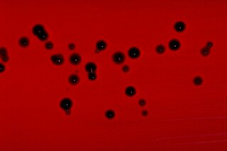
Ashdown's medium is a selective culture medium for the isolation and characterisation of Burkholderia pseudomallei.

<span class="mw-page-title-main">Pathogenic bacteria</span> Disease-causing bacteria

Pathogenic bacteria are bacteria that can cause disease. This article focuses on the bacteria that are pathogenic to humans. Most species of bacteria are harmless and are often beneficial but others can cause infectious diseases. The number of these pathogenic species in humans is estimated to be fewer than a hundred. By contrast, several thousand species are part of the gut flora present in the digestive tract.

<i>Actinobacillus</i> Genus of bacteria

Actinobacillus is a genus of Gram-negative, nonmotile and non-spore-forming, oval to rod-shaped bacteria occurring as parasites or pathogens in mammals, birds, and reptiles. It is a member of the family Pasteurellaceae. The bacteria are facultatively anaerobic or aerobic, capable of fermenting carbohydrates, and of reducing nitrates. The genomic DNA contains between 40 and 47 mol % guanine plus cytosine.

<i>Proteus penneri</i> Species of bacterium

Proteus penneri is a Gram-negative, facultatively anaerobic, rod-shaped bacterium. It is an invasive pathogen and a cause of nosocomial infections of the urinary tract or open wounds. Pathogens have been isolated mainly from the urine of patients with abnormalities in the urinary tract, and from stool. P. penneri strains are naturally resistant to numerous antibiotics, including penicillin G, amoxicillin, cephalosporins, oxacillin, and most macrolides, but are naturally sensitive to aminoglycosides, carbapenems, aztreonam, quinolones, sulphamethoxazole, and co-trimoxazole. Isolates of P. penneri have been found to be multiple drug-resistant (MDR) with resistance to six to eight drugs. β-lactamase production has also been identified in some isolates.

Ornibactin is a siderophore, or small iron-binding compound secreted by bacteria to transport iron into the cell. Ornibactin is produced by Burkholderia cenocepacia under iron-deficient conditions. B. cenocepacia is known to opportunistically infect humans, specifically ones suffering from cystic fibrosis.

References

  1. Yabuuchi, E; Kosako, Y; Oyaizu, H; Yano, I; Hotta, H; Hashimoto, Y; Ezaki, T; Arakawa, M (1992). "Proposal of Burkholderia gen. nov. and transfer of seven species of the genus Pseudomonas homology group II to the new genus, with the type species Burkholderia cepacia (Palleroni and Holmes 1981) comb. nov". Microbiol Immunol. 36 (12): 1251–75. doi: 10.1111/j.1348-0421.1992.tb02129.x . PMID   1283774.
  2. "Burkholderia pseudomallei". VirginiaTech Pathogen Database. Archived from the original on 2006-09-01. Retrieved 2006-03-26.
  3. Limmathurotsakul, Direk; Golding, Nick; Dance, David A. B.; Messina, Jane P.; Pigott, David M.; Moyes, Catherine L.; Rolim, Dionne B.; Bertherat, Eric; Day, Nicholas P. J. (2016-01-11). "Predicted global distribution of Burkholderia pseudomallei and burden of melioidosis". Nature Microbiology. 1 (1): 15008. doi:10.1038/nmicrobiol.2015.8. ISSN   2058-5276. PMC   4746747 . PMID   26877885.
  4. Currie, Bart J.; Dance, David A.B.; Cheng, Allen C. (December 2008). "The global distribution of Burkholderia pseudomallei and melioidosis: an update". Transactions of the Royal Society of Tropical Medicine and Hygiene. 102: S1–S4. doi:10.1016/S0035-9203(08)70002-6. PMID   19121666.
  5. 1 2 Colley, Claire (18 December 2022). "US public not warned that monkeys imported from Cambodia carried deadly pathogens". The Guardian.
  6. Pumpuang, Apinya; Chantratita, Narisara; Wikraiphat, Chanthiwa; Saiprom, Natnaree; Day, Nicholas P.J.; Peacock, Sharon J.; Wuthiekanun, Vanaporn (October 2011). "Survival of Burkholderia pseudomallei in distilled water for 16 years". Transactions of the Royal Society of Tropical Medicine and Hygiene. 105 (10): 598–600. doi:10.1016/j.trstmh.2011.06.004. ISSN   0035-9203. PMC   3183224 . PMID   21764093.
  7. Cheng, Allen C.; Currie, Bart J. (April 2005). "Melioidosis: Epidemiology, Pathophysiology, and Management". Clinical Microbiology Reviews. 18 (2): 383–416. doi:10.1128/CMR.18.2.383-416.2005. ISSN   0893-8512. PMC   1082802 . PMID   15831829.
  8. "Melioidosis". CDC. 2022-07-27.
  9. "Melioidosis". New Jersey Department of Agriculture. 2003.
  10. Lee YH, Chen Y, Ouyang X, Gan YH (2010). "Identification of tomato plant as a novel host model for Burkholderia pseudomallei". BMC Microbiol. 10: 28. doi: 10.1186/1471-2180-10-28 . PMC   2823722 . PMID   20109238.
  11. Haase A, Janzen J, Barrett S, Currie B (July 1997). "Toxin production by Burkholderia pseudomallei strains and correlation with severity of melioidosis". Journal of Medical Microbiology. 46 (7): 557–63. doi: 10.1099/00222615-46-7-557 . PMID   9236739.
  12. 1 2 Peacock SJ, Chieng G, Cheng AC, et al. (October 2005). "Comparison of Ashdown's medium, Burkholderia cepacia medium, and Burkholderia pseudomallei selective agar for clinical isolation of Burkholderia pseudomallei". Journal of Clinical Microbiology. 43 (10): 5359–61. doi:10.1128/JCM.43.10.5359-5361.2005. PMC   1248505 . PMID   16208018.
  13. Chaiyaroj SC, Kotrnon K, Koonpaew S, Anantagool N, White NJ, Sirisinha S (1999). "Differences in genomic macrorestriction patterns of arabinose-positive (Burkholderia thailandensis) and arabinose-negative Burkholderia pseudomallei". Microbiology and Immunology. 43 (7): 625–30. doi: 10.1111/j.1348-0421.1999.tb02449.x . PMID   10529102.
  14. 1 2 Walsh AL, Wuthiekanun V (1996). "The laboratory diagnosis of melioidosis". Br J Biomed Sci. 53 (4): 249–53. PMID   9069100.
  15. Brundage WG, Thuss CJ, Walden DC (March 1968). "Four fatal cases of melioidosis in U. S. soldiers in Vietnam. Bacteriologic and pathologic characteristics". The American Journal of Tropical Medicine and Hygiene. 17 (2): 183–91. doi:10.4269/ajtmh.1968.17.183. PMID   4869109.
  16. Sheridan EA, Ramsay AR, Short JM, Stepniewska K, Wuthiekanun V, Simpson AJ (May 2007). "Evaluation of the Wayson stain for the rapid diagnosis of melioidosis". Journal of Clinical Microbiology. 45 (5): 1669–70. doi:10.1128/JCM.00396-07. PMC   1865910 . PMID   17360835.
  17. Chantratita N, Wuthiekanun V, Boonbumrung K, et al. (2007). "Biological relevance of colony morphology and phenotypic switching by Burkholderia pseudomallei.". J Bacteriol. 189 (3): 807–17. doi:10.1128/JB.01258-06. PMC   1797308 . PMID   17114252.
  18. Pumpuang A, Chantratita N, Wikraiphat C, et al. (2011). "Survival of Burkholderia pseudomallei in distilled water for 16 years". Trans R Soc Trop Med Hyg. 105 (10–2): 598–600. doi:10.1016/j.trstmh.2011.06.004. PMC   3183224 . PMID   21764093.
  19. Ashdown LR (1979). "An improved screening technique for isolation of Pseudomonas pseudomallei from clinical specimens". Pathology. 11 (2): 293–7. doi:10.3109/00313027909061954. PMID   460953.
  20. Roesnita B; Tay ST; Puthucheary SD; Sam IC. (2012). "Diagnostic use of Burkholderia pseudomallei selective media in a low prevalence setting". Trans R Soc Trop Med Hyg. 106 (2): 131–3. doi:10.1016/j.trstmh.2011.10.007. PMID   22112687.
  21. Goodyear A, Strange L, Rholl DA, et al. (2013). "An improved selective culture medium enhances the isolation of Burkholderia pseudomallei from contaminated specimens". Am J Trop Med Hyg. 89 (5): 973–82. doi:10.4269/ajtmh.13-0119. PMC   3820346 . PMID   24062483.
  22. Jorakate P, Higdon M, Kaewpan A, et al. (2015). "Contribution of the BacT/ALERT MB Mycobacteria Bottle to bloodstream infection surveillance in Thailand: added yield for Burkholderia pseudomallei.". J Clin Microbiol. 53 (3): 910–4. doi:10.1128/JCM.02008-14. PMC   4390673 . PMID   25588650.
  23. Inglis TJ, Chiang D, Lee GS, Chor-Kiang L (February 1998). "Potential misidentification of Burkholderia pseudomallei by API 20NE". Pathology. 30 (1): 62–4. doi:10.1080/00313029800169685. PMID   9534210. S2CID   31987728.
  24. 1 2 Lowe P, Engler C, Norton R (December 2002). "Comparison of automated and nonautomated systems for identification of Burkholderia pseudomallei". Journal of Clinical Microbiology. 40 (12): 4625–7. doi:10.1128/JCM.40.12.4625-4627.2002. PMC   154629 . PMID   12454163.
  25. Kite-Powell A, Livengood JR, Suarez J, et al. (2006). "Imported MelioidosisSouth Florida, 2005". Morb Mortal Wkly Rep. 55 (32): 873–6. PMID   16915220.
  26. 1 2 Amornchai P, Chierakul W, Wuthiekanun V, et al. (November 2007). "Accuracy of Burkholderia pseudomallei identification using the API 20NE system and a latex agglutination test". Journal of Clinical Microbiology. 45 (11): 3774–6. doi:10.1128/JCM.00935-07. PMC   2168515 . PMID   17804660.
  27. Moore RA, DeShazer D, Reckseidler S, Weissman A, Woods DE (March 1999). "Efflux-mediated aminoglycoside and macrolide resistance in Burkholderia pseudomallei". Antimicrobial Agents and Chemotherapy. 43 (3): 465–70. doi:10.1128/AAC.43.3.465. PMC   89145 . PMID   10049252.
  28. 1 2 3 Wuthiekanun V, Peacock SJ (June 2006). "Management of melioidosis". Expert Review of Anti-infective Therapy. 4 (3): 445–55. doi:10.1586/14787210.4.3.445. PMID   16771621. S2CID   35619761.
  29. Hodgson K, Engler C, Govan B, et al. (2009). "A comparison of routine bench and molecular diagnostic methods in the identification of Burkholderia pseudomallei". J Clin Microbiol. 47 (5): 1578–80. doi:10.1128/JCM.02507-08. PMC   2681847 . PMID   19279182.
  30. Podin Y, Sarovich DS, Price EP, Kaestli M, Mayo M, Hii K, et al. (2013). "Burkholderia pseudomallei from Sarawak, Malaysian Borneo are predominantly susceptible to aminoglycosides and macrolides". Antimicrob Agents Chemother. 58 (1): 162–6. doi:10.1128/AAC.01842-13. PMC   3910780 . PMID   24145517.
  31. Ruppitsch W, Stöger A, Indra A, et al. (March 2007). "Suitability of partial 16S ribosomal RNA gene sequence analysis for the identification of dangerous bacterial pathogens". Journal of Applied Microbiology. 102 (3): 852–9. doi:10.1111/j.1365-2672.2006.03107.x. PMID   17309636. S2CID   24843231.
  32. Wattiau P, Van Hessche M, Neubauer H, Zachariah R, Wernery U, Imberechts H (March 2007). "Identification of Burkholderia pseudomallei and related bacteria by multiple-locus sequence typing-derived PCR and real-time PCR". Journal of Clinical Microbiology. 45 (3): 1045–8. doi:10.1128/JCM.02350-06. PMC   1829090 . PMID   17251403.
  33. Hagen RM, Frickmann H, Elschner M, et al. (2011). "Rapid identification of Burkholderia pseudomallei and Burkholderia mallei by fluorescence in situ hybridization (FISH) from culture and paraffin-embedded tissue samples". International Journal of Medical Microbiology . 301 (7): 585–90. doi:10.1016/j.ijmm.2011.04.017. PMID   21658996.
  34. Wuthiekanun V, Desakorn V, Wongsuvan G, et al. (April 2005). "Rapid immunofluorescence microscopy for diagnosis of melioidosis". Clinical and Diagnostic Laboratory Immunology. 12 (4): 555–6. doi:10.1128/CDLI.12.4.555-556.2005. PMC   1074392 . PMID   15817767.
  35. "Burkholderia Pseudomallei". www.antimicrobe.org. Retrieved 2022-04-21.
  36. Hemarajata, Peera; Baghdadi, Jonathan D.; Hoffman, Risa; Humphries, Romney M. (December 2016). "Burkholderia pseudomallei: Challenges for the Clinical Microbiology Laboratory". Journal of Clinical Microbiology. 54 (12): 2866–2873. doi:10.1128/JCM.01636-16. ISSN   0095-1137. PMC   5121373 . PMID   27654336.
  37. Miller, WR; Pannell, L; Cravitz, L; Tanner, WA; Ingalls, MS (1948). "Studies on certain biological characteristics of Malleomyces mallei and Malleomyces pseudomallei: I. Morphology, cultivation, viability, and isolation from contaminated specimens". J Bacteriol. 55 (1): 115–126. doi:10.1128/JB.55.1.115-126.1948. PMC   518415 . PMID   16561426.
  38. Wuthiekanun V, Wongsuwan G, Pangmee S, Teerawattanasook N, Day NP, Peacock SJ (2010). "Perasafe, Virkon and bleach are bactericidal for Burkholderia pseudomallei, a select agent and the cause of melioidosis". J Hosp Infect. 77 (2): 183–4. doi:10.1016/j.jhin.2010.06.026. PMC   3036794 . PMID   20832143.
  39. Rose, L. J.; O'Connell, H. (2009-05-01). "UV Light Inactivation of Bacterial Biothreat Agents". Applied and Environmental Microbiology. 75 (9): 2987–2990. Bibcode:2009ApEnM..75.2987R. doi:10.1128/AEM.02180-08. ISSN   0099-2240. PMC   2681683 . PMID   19270145.
  40. Rodríguez, José Y.; Huertas, Mónica G.; Rodríguez, Gerson J.; Vargas-Otalora, Sandra; Benıtez-Peñuela, Miguel A.; Esquea, Kelin; Rios, Rafael; Mendoza, Laura R.; Diaz, Lorena; Reyes, Jinnethe; Álvarez-Moreno, Carlos A. (2020-11-04). "Case Report: Gestational Melioidosis through Perinatal Transmission". The American Journal of Tropical Medicine and Hygiene. 103 (5): 1838–1840. doi:10.4269/ajtmh.20-0223. ISSN   0002-9637. PMC   7646751 . PMID   32748772.
  41. White NJ, Dance DA, Chaowagul W, Wattanagoon Y, Wuthiekanun V, Pitakwatchara N (September 1989). "Halving of mortality of severe melioidosis by ceftazidime". Lancet. 2 (8665): 697–701. doi:10.1016/S0140-6736(89)90768-X. PMID   2570956. S2CID   28919574.
  42. Lumbiganon P, Tattawasatra U, Chetchotisakd P, et al. (2000). "Comparison between the antimicrobial susceptibility of Burkholderia pseudomallei to trimethoprim-sulfamethoxazole by standard disk diffusion method and by minimal inhibitory concentration determination". J Med Assoc Thai. 83 (8): 856–60. PMID   10998837.
  43. Wuthiekanun V, Cheng AC, Chierakul W, et al. (2005). "Trimethoprim/sulfamethoxazole resistance in clinical isolates of Burkholderia pseudomallei". J Antimicrob Chemother. 55 (6): 1029–31. doi: 10.1093/jac/dki151 . PMID   15886263.
  44. Saraya S, Soontornpas C, Chindavijak B, Mootsikapun P (2009). "In vitro interactions between cotrimoxazole and doxycycline in Burkholderia pseudomallei: how important is this combination in maintenance therapy of melioidosis?". Indian J Med Microbiol. 27 (1): 88–9. doi: 10.1016/S0255-0857(21)01773-4 . hdl: 1807/53689 . PMID   19172079.
  45. Trunck LA; Propst, KL; Wuthiekanun, V; Tuanyok, A; Beckstrom-Sternberg, SM; Beckstrom-Sternberg, JS; Peacock, SJ; Keim, P; et al. (2009). Picardeau, Mathieu (ed.). "Molecular basis of rare aminoglycoside susceptibility and pathogenesis of Burkholderia pseudomallei clinical isolates from Thailand". PLOS Negl Trop Dis. 3 (9): e519. doi: 10.1371/journal.pntd.0000519 . PMC   2737630 . PMID   19771149.
  46. Ashdown, LR (1979). "Identification of Pseudomonas pseudomallei in the clinical laboratory". J Clin Pathol. 32 (5): 500–4. doi:10.1136/jcp.32.5.500. PMC   1145715 . PMID   381328.
  47. Kespichayawattana W, Intachote P, Utaisincharoen P, Stitaya Sirisinha S (2004). "Virulent Burkholderia pseudomallei is more efficient than avirulent Burkholderia thailandensis in invasion of and adherence to cultured human epithelial cells". Microbial Pathogenesis. 36 (5): 287–9. doi:10.1016/j.micpath.2004.01.001. PMID   15043863.
  48. Nandi T, Ong C, Singh AP, Boddey J, Atkins T, Sarkar-Tyson M, Essex-Lopresti AE, Chua HH, Pearson T, Kreisberg JF, Nilsson C, Ariyaratne P, Ronning C, Losada L, Ruan Y, Sung WK, Woods D, Titball RW, Beacham I, Peak I, Keim P, Nierman WC, Tan P (2010). Guttman DS (ed.). "A genomic survey of positive selection in Burkholderia pseudomallei provides insights into the evolution of accidental virulence". PLOS Pathog. 6 (4): e1000845. doi: 10.1371/journal.ppat.1000845 . PMC   2848565 . PMID   20368977.
  49. 1 2 Sim SH, Yu Y, Lin CH, et al. (October 2008). Achtman M (ed.). "The core and accessory genomes of Burkholderia pseudomallei: implications for human melioidosis". PLOS Pathog. 4 (10): e1000178. doi: 10.1371/journal.ppat.1000178 . PMC   2564834 . PMID   18927621.
  50. Price EP, Hornstra HM, Limmathurotsakul D, et al. (2010). Guttman DS (ed.). "Within-host evolution of Burkholderia pseudomallei in four cases of acute melioidosis". PLOS Pathog. 6 (1): e1000725. doi: 10.1371/journal.ppat.1000725 . PMC   2799673 . PMID   20090837.
  51. Wiersinga WJ, van der Poll T, White NJ, Day NP, Peacock SJ (2006). "Melioidosis: insights into the pathogenicity of Burkholderia pseudomallei". Nature Reviews Microbiology. 4 (4): 272–82. doi: 10.1038/nrmicro1385 . PMID   16541135. S2CID   23909148.
  52. Kespichayawattana W, Rattanachetkul S, Wanun T, et al. (2000). "Burkholderia pseudomallei induces cell fusion and actin-associated membrane protrusion: a possible mechanism for cell-to-cell spreading". Infect. Immun. 68 (9): 5377–84. doi:10.1128/IAI.68.9.5377-5384.2000. PMC   101801 . PMID   10948167.
  53. Toesca, Isabelle J.; French, Christopher T.; Miller, Jeff F. (2014-04-01). "The Type VI secretion system spike protein VgrG5 mediates membrane fusion during intercellular spread by pseudomallei group Burkholderia species". Infection and Immunity. 82 (4): 1436–1444. doi:10.1128/IAI.01367-13. ISSN   1098-5522. PMC   3993413 . PMID   24421040.
  54. Cruz-Migoni A, Hautbergue GM, Artymiuk PJ, et al. (2011). "A Burkholderia pseudomallei toxin inhibits helicase activity of translation factor eIF4A". Science. 334 (6057): 821–4. Bibcode:2011Sci...334..821C. doi:10.1126/science.1211915. PMC   3364511 . PMID   22076380.
  55. Shalom G, Shaw JG, Thomas MS (August 2007). "In vivo expression technology identifies a type VI secretion system locus in Burkholderia pseudomallei that is induced upon invasion of macrophages". Microbiology. 153 (Pt 8): 2689–99. doi: 10.1099/mic.0.2007/006585-0 . PMID   17660433.
  56. Mima T, Schweizer HP (2010). "The BpeAB-OprB efflux pump of Burkholderia pseudomallei 1026b does not play a role in quorum sensing, virulence factor production, or extrusion of aminoglycosides, but is a broad-spectrum drug efflux system". Antimicrob. Agents Chemother. 54 (8): 3113–20. doi:10.1128/AAC.01803-09. PMC   2916348 . PMID   20498323.
  57. Baker, Sarah M.; Settles, Erik W.; Davitt, Christopher; Gellings, Patrick; Kikendall, Nicole; Hoffmann, Joseph; Wang, Yihui; Bitoun, Jacob; Lodrigue, Kasi-Russell; Sahl, Jason W.; Keim, Paul; Roy, Chad; McLachlan, James; Morici, Lisa A. (29 January 2021). "Burkholderia pseudomallei OMVs derived from infection mimicking conditions elicit similar protection to a live-attenuated vaccine". npj Vaccines. 6 (1): 18. doi: 10.1038/s41541-021-00281-z . ISSN   2059-0105. PMC   7846723 . PMID   33514749.
  58. Christodoulides, Myron, ed. (2023). Vaccines for Neglected Pathogens: Strategies, Achievements and Challenges. p. 329. doi:10.1007/978-3-031-24355-4. ISBN   978-3-031-24354-7. S2CID   257768974 . Retrieved 6 October 2023.
  59. Norris MH, Kang Y, Lu D, Wilcox BA, Hoang TT (2009). "Glyphosate resistance as a novel select-agent-compliant, non-antibiotic-selectable marker in chromosomal mutagenesis of the essential genes asd and dapB of Burkholderia pseudomallei.". Appl Environ Microbiol. 75 (19): 6062–75. Bibcode:2009ApEnM..75.6062N. doi:10.1128/AEM.00820-09. PMC   2753064 . PMID   19648360.
  60. Norris MH, Propst KL, Kang Y, et al. (2011). "The Burkholderia pseudomallei Δasd mutant exhibits attenuated intracellular infectivity and imparts protection against acute inhalation melioidosis in mice". Infect Immun. 79 (10): 4010–8. doi:10.1128/IAI.05044-11. PMC   3187240 . PMID   21807903.
  61. Mack, K.; Titball, R. W. (1996-11-01). "Transformation of Burkholderia pseudomallei by electroporation". Analytical Biochemistry. 242 (1): 73–76. doi:10.1006/abio.1996.0430. ISSN   0003-2697. PMID   8923967.