Development of the endocrine system

Last updated

The fetal endocrine system is one of the first systems to develop during prenatal development of a human individual. The endocrine system arises from all three embryonic germ layers. The endocrine glands that produce the steroid hormones, such as the gonads and adrenal cortex, arise from the mesoderm. In contrast, endocrine glands that arise from the endoderm and ectoderm produce the amine, peptide, and protein hormones. [1]

Contents

Adrenal glands

The fetal Adrenal Cortex can be identified within four weeks of pregnancy. [2] The adrenal cortex originates from the thickening of the intermediate Mesoderm. [3] At five to six weeks of gestation, the Mesonephros differentiates into a tissue known as the gonadal ridge. The gonadal ridge produces the Steroidogenic cells for both the gonads and the adrenal cortex. [4] The adrenal medulla is derived from Ectodermal cells. Cells that will become adrenal tissue move Retroperitoneally to the upper portion of the Mesonephros. At seven weeks of gestation, the adrenal cells are joined by sympathetic cells that originate from the Neural Crest to form the Adrenal Medulla. At the end of the eighth week, the adrenal glands have been encapsulated and have formed a distinct organ above the developing kidneys. [4] At birth, the adrenal glands weight approximately eight to nine grams (twice that of the adult adrenal glands) and are 0.5% of the total body weight. At 25 weeks, the adult adrenal cortex zone develops and is responsible for the primary synthesis of steroids during the early postnatal weeks. [2]

Thyroid gland

The Thyroid gland develops from two different clusterings of embryonic cells. One part is from the thickening of the Pharyngeal floor, which serves as the precursor of the Thyroxine (T4) producing Follicular cells. The other part is from the Caudal extensions of the fourth Pharyngobranchial pouches which results in the Parafollicular Calcitonin-secreting cells. [5] These two structures are apparent by 16 to 17 days of gestation. Around the 24th day of pregnancy, the Foramen Cecum, a thin, flask-like Diverticulum of the median Anlage develops. At approximately 24 to 32 days of gestation the median anlage develops into a bilobed structure. By 50 days of gestation, the medial and Lateral Anlage have fused together. [6] At 12 weeks of gestation, the Fetal Thyroid is capable of storing iodine for the production of TRH, TSH, and free thyroid hormone. At 20 weeks, the Fetus is able to implement feedback mechanisms for the production of thyroid hormones. During fetal development, T4 is the major thyroid hormone being produced while Triiodothyronine (T3) and its inactive derivative, reverse T3, are not detected until the third trimester. [2]

Parathyroid glands

A lateral and ventral view of an embryo showing the third (inferior) and fourth (superior) parathyroid glands during the 6th week of embryogenesis Parathyroid glands during embryogenesis.jpg
A lateral and ventral view of an embryo showing the third (inferior) and fourth (superior) parathyroid glands during the 6th week of embryogenesis

Once the embryo reaches four weeks of gestation, the parathyroid glands begins to develop. [7] The human embryo forms five sets of endoderm-lined pharyngeal pouches. The third and fourth pouch are responsible for developing into the inferior and superior parathyroid glands, respectively. [8] The third pharyngeal pouch encounters the developing thyroid gland and they migrate down to the lower poles of the thyroid lobes. The fourth pharyngeal pouch later encounters the developing thyroid gland and migrates to the upper poles of the thyroid lobes. At 14 weeks of gestation, the parathyroid glands begin to enlarge from 0.1 mm in diameter to approximately 1 – 2 mm at birth. [9] The developing parathyroid glands are physiologically functional beginning in the second trimester.[ citation needed ]

Studies in mice have shown that interfering with the HOX15 gene can cause parathyroid gland aplasia, which suggests the gene plays an important role in the development of the parathyroid gland. [9] The genes, TBX1, CRKL, GATA3, GCM2, and SOX3 have also been shown to play a crucial role in the formation of the parathyroid gland. Mutations in TBX1 and CRKL genes are correlated with DiGeorge syndrome, while mutations in GATA3 have also resulted in a DiGeorge-like syndrome. [10] Malformations in the GCM2 gene have resulted in hypoparathyroidism. [11] Studies on SOX3 gene mutations have demonstrated that it plays a role in parathyroid development. These mutations also lead to varying degrees of hypopituitarism. [12]

Pancreas

The human fetal pancreas begins to develop by the fourth week of gestation. Five weeks later, the pancreatic alpha and beta cells have begun to emerge. Reaching eight to ten weeks into development, the pancreas starts producing insulin, glucagon, somatostatin, and pancreatic polypeptide. [13] During the early stages of fetal development, the number of pancreatic alpha cells outnumbers the number of pancreatic beta cells. The alpha cells reach their peak in the middle stage of gestation. From the middle stage until term, the beta cells continue to increase in number until they reach an approximate 1:1 ratio with the alpha cells. The insulin concentration within the fetal pancreas is 3.6 pmol/g at seven to ten weeks, which rises to 30 pmol/g at 16–25 weeks of gestation. Near term, the insulin concentration increases to 93 pmol/g. [14] The endocrine cells have dispersed throughout the body within 10 weeks. At 31 weeks of development, the islets of Langerhans have differentiated.[ citation needed ]

While the fetal pancreas has functional beta cells by 14 to 24 weeks of gestation, the amount of insulin that is released into the bloodstream is relatively low. In a study of pregnant women carrying fetuses in the mid-gestation and near term stages of development, the fetuses did not have an increase in plasma insulin levels in response to injections of high levels of glucose. [14] In contrast to insulin, the fetal plasma glucagon levels are relatively high and continue to increase during development. [15] At the mid-stage of gestation, the glucagon concentration is 6 μg/g, compared to 2 μg/g in adult humans. Just like insulin, fetal glucagon plasma levels do not change in response to an infusion of glucose. [16] However, a study of an infusion of alanine into pregnant women was shown to increase the cord blood and maternal glucagon concentrations, demonstrating a fetal response to amino acid exposure. [14]

As such, while the fetal pancreatic alpha and beta islet cells have fully developed and are capable of hormone synthesis during the remaining fetal maturation, the islet cells are relatively immature in their capacity to produce glucagon and insulin. This is thought to be a result of the relatively stable levels of fetal serum glucose concentrations achieved via maternal transfer of glucose through the placenta. On the other hand, the stable fetal serum glucose levels could be attributed to the absence of pancreatic signaling initiated by incretins during feeding. [16] In addition, the fetal pancreatic islets cells are unable to sufficiently produce cAMP and rapidly degrade cAMP by phosphodiesterase necessary to secrete glucagon and insulin. [14]

During fetal development, the storage of glycogen is controlled by fetal glucocorticoids and placental lactogen. Fetal insulin is responsible for increasing glucose uptake and lipogenesis during the stages leading up to birth. Fetal cells contain a higher amount of insulin receptors in comparison to adults cells and fetal insulin receptors are not downregulated in cases of hyperinsulinemia. [14] In comparison, fetal haptic glucagon receptors are lowered in comparison to adult cells and the glycemic effect of glucagon is blunted. [15] This temporary physiological change aids the increased rate of fetal development during the final trimester. Poorly managed maternal diabetes mellitus is linked to fetal macrosomia, increased risk of miscarriage, and defects in fetal development. Maternal hyperglycemia is also linked to increased insulin levels and beta cell hyperplasia in the post-term infant. [16] Children of diabetic mothers are at an increased risk for conditions such as: polycythemia, renal vein thrombosis, hypocalcemia, respiratory distress syndrome, jaundice, cardiomyopathy, congenital heart disease, and improper organ development. [17]

Gonads

The reproductive system begins development at four to five weeks of gestation with germ cell migration. The bipotential gonad results from the collection of the medioventral region of the urogenital ridge. At the five-week point, the developing gonads break away from the adrenal primordium. Gonadal differentiation begins 42 days following conception.

Male gonadal development

For males, the testes form at six fetal weeks and the sertoli cells begin developing by the eight week of gestation. SRY, the sex-determining locus, serves to differentiate the Sertoli cells. The Sertoli cells are the point of origin for anti-Müllerian hormone. Once synthesized, the anti-Müllerian hormone initiates the ipsilateral regression of the Müllerian tract and inhibits the development of female internal features. At 10 weeks of gestation, the Leydig cells begin to produce androgen hormones. The androgen hormone dihydrotestosterone is responsible for the development of the male external genitalia. [18]

The testicles descend during prenatal development in a two-stage process that begins at eight weeks of gestation and continues through the middle of the third trimester. During the transabdominal stage (8 to 15 weeks of gestation), the gubernacular ligament contracts and begins to thicken. The craniosuspensory ligament begins to break down. This stage is regulated by the secretion of insulin-like 3 (INSL3), a relaxin-like factor produced by the testicles, and the INSL3 G-coupled receptor, LGR8. During the transinguinal phase (25 to 35 weeks of gestation), the testicles descend into the scrotum. This stage is regulated by androgens, the genitofemoral nerve, and calcitonin gene-related peptide. During the second and third trimester, testicular development concludes with the diminution of the fetal Leydig cells and the lengthening and coiling of the seminiferous cords. [19]

Female gonadal development

For females, the ovaries become morphologically visible by the 8th week of gestation. The absence of testosterone results in the diminution of the Wolffian structures. The Müllerian structures remain and develop into the fallopian tubes, uterus, and the upper region of the vagina. The urogenital sinus develops into the urethra and lower region of the vagina, the genital tubercle develops into the clitoris, the urogenital folds develop into the labia minora, and the urogenital swellings develop into the labia majora. At 16 weeks of gestation, the ovaries produce FSH and LH/hCG receptors. At 20 weeks of gestation, the theca cell precursors are present and oogonia mitosis is occurring. At 25 weeks of gestation, the ovary is morphologically defined and folliculogenesis can begin. [19]

Studies of gene expression show that a specific complement of genes, such as follistatin and multiple cyclin kinase inhibitors are involved in ovarian development. [20] An assortment of genes and proteins - such as WNT4, [21] RSPO1, [22] FOXL2, [23] and various estrogen receptors [24] - have been shown to prevent the development of testicles or the lineage of male-type cells. [25]

Pituitary gland

The pituitary gland is formed within the rostral neural plate. The Rathke's pouch, a cavity of ectodermal cells of the oropharynx, forms between the fourth and fifth week of gestation [26] and upon full development, it gives rise to the anterior pituitary gland. [27] By seven weeks of gestation, the anterior pituitary vascular system begins to develop. During the first 12 weeks of gestation, the anterior pituitary undergoes cellular differentiation. At 20 weeks of gestation, the hypophyseal portal system has developed. The Rathke's pouch grows towards the third ventricle and fuses with the diverticulum. This eliminates the lumen and the structure becomes Rathke's cleft. The posterior pituitary lobe is formed from the diverticulum. Portions of the pituitary tissue may remain in the nasopharyngeal midline. In rare cases this results in functioning ectopic hormone-secreting tumors in the nasopharynx. [28]

The functional development of the anterior pituitary involves spatiotemporal regulation of transcription factors expressed in pituitary stem cells and dynamic gradients of local soluble factors. [29] [30] The coordination of the dorsal gradient of pituitary morphogenesis is dependent on neuroectodermal signals from the infundibular bone morphogenetic protein 4 (BMP4). [27] This protein is responsible for the development of the initial invagination of the Rathke's pouch. Other essential proteins necessary for pituitary cell proliferation are Fibroblast growth factor 8 (FGF8), [31] Wnt4, [32] and Wnt5. [32] Ventral developmental patterning and the expression of transcription factors is influenced by the gradients of BMP2 and sonic hedgehog protein (SHH). These factors are essential for coordinating early patterns of cell proliferation. [33]

Six weeks into gestation, the corticotroph cells can be identified. By seven weeks of gestation, the anterior pituitary is capable of secreting ACTH. Within eight weeks of gestation, somatotroph cells begin to develop with cytoplasmic expression of human growth hormone. Once a fetus reaches 12 weeks of development, the thyrotrophs begin expression of Beta subunits for TSH, while gonadotrophs being to express beta-subunits for LH and FSH. [34] Male fetuses predominately produced LH-expressing gonadotrophs, while female fetuses produce an equal expression of LH and FSH expressing gonadotrophs. [35] At 24 weeks of gestation, prolactin-expressing lactotrophs begin to emerge. [34]

Related Research Articles

<span class="mw-page-title-main">Adrenal gland</span> Endocrine gland

The adrenal glands are endocrine glands that produce a variety of hormones including adrenaline and the steroids aldosterone and cortisol. They are found above the kidneys. Each gland has an outer cortex which produces steroid hormones and an inner medulla. The adrenal cortex itself is divided into three main zones: the zona glomerulosa, the zona fasciculata and the zona reticularis.

<span class="mw-page-title-main">Endocrine system</span> Hormone-producing glands of a body

The endocrine system is a messenger system in an organism comprising feedback loops of hormones that are released by internal glands directly into the circulatory system and that target and regulate distant organs. In vertebrates, the hypothalamus is the neural control center for all endocrine systems.

<span class="mw-page-title-main">Thyroid</span> Endocrine gland in the neck; secretes hormones that influence metabolism

The thyroid, or thyroid gland, is an endocrine gland in vertebrates. In humans, it is in the neck and consists of two connected lobes. The lower two thirds of the lobes are connected by a thin band of tissue called the isthmus (pl.: isthmi). The thyroid gland is a butterfly-shaped gland located in the neck below the Adam's apple. Microscopically, the functional unit of the thyroid gland is the spherical thyroid follicle, lined with follicular cells (thyrocytes), and occasional parafollicular cells that surround a lumen containing colloid. The thyroid gland secretes three hormones: the two thyroid hormones – triiodothyronine (T3) and thyroxine (T4) – and a peptide hormone, calcitonin. The thyroid hormones influence the metabolic rate and protein synthesis and growth and development in children. Calcitonin plays a role in calcium homeostasis. Secretion of the two thyroid hormones is regulated by thyroid-stimulating hormone (TSH), which is secreted from the anterior pituitary gland. TSH is regulated by thyrotropin-releasing hormone (TRH), which is produced by the hypothalamus.

<span class="mw-page-title-main">Pituitary gland</span> Endocrine gland at the base of the brain

The pituitary gland is an endocrine gland in vertebrates. In humans, the pituitary gland is located at the base of the brain, protruding off the bottom of the hypothalamus. The human pituitary gland is oval shaped, about the size of a chickpea, and weighs 0.5 grams (0.018 oz) on average.

<span class="mw-page-title-main">Hypothalamus</span> Area of the brain below the thalamus

The hypothalamus is a small part of the brain that contains a number of nuclei with a variety of functions. One of the most important functions is to link the nervous system to the endocrine system via the pituitary gland. The hypothalamus is located below the thalamus and is part of the limbic system. It forms the ventral part of the diencephalon. All vertebrate brains contain a hypothalamus. In humans, it is the size of an almond.

<span class="mw-page-title-main">Multiple endocrine neoplasia</span> Medical condition

Multiple endocrine neoplasia is a condition which encompasses several distinct syndromes featuring tumors of endocrine glands, each with its own characteristic pattern. In some cases, the tumors are malignant, in others, benign. Benign or malignant tumors of nonendocrine tissues occur as components of some of these tumor syndromes.

<span class="mw-page-title-main">Parathyroid gland</span> Endocrine gland

Parathyroid glands are small endocrine glands in the neck of humans and other tetrapods. Humans usually have four parathyroid glands, located on the back of the thyroid gland in variable locations. The parathyroid gland produces and secretes parathyroid hormone in response to a low blood calcium, which plays a key role in regulating the amount of calcium in the blood and within the bones.

<span class="mw-page-title-main">Anterior pituitary</span> Anterior lobe of the pituitary gland

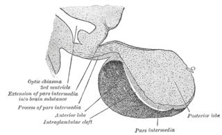
A major organ of the endocrine system, the anterior pituitary is the glandular, anterior lobe that together with the posterior lobe makes up the pituitary gland (hypophysis) which, in humans, is located at the base of the brain, protruding off the bottom of the hypothalamus.

<span class="mw-page-title-main">Parafollicular cell</span> Neuroendocrine cells in the thyroid

Parafollicular cells, also called C cells, are neuroendocrine cells in the thyroid. The primary function of these cells is to secrete calcitonin. They are located adjacent to the thyroid follicles and reside in the connective tissue. These cells are large and have a pale stain compared with the follicular cells. In teleost and avian species these cells occupy a structure outside the thyroid gland named the ultimopharyngeal body.

<span class="mw-page-title-main">Parathyroid chief cell</span>

Parathyroid chief cells are one of the two cell types of the parathyroid glands, along with oxyphil cells. The chief cells are much more prevalent in the parathyroid gland than the oxyphil cells. It is perceived that oxyphil cells may be derived from chief cells at puberty, as they are not present at birth like chief cells.

<span class="mw-page-title-main">Hypopituitarism</span> Medical condition

Hypopituitarism is the decreased (hypo) secretion of one or more of the eight hormones normally produced by the pituitary gland at the base of the brain. If there is decreased secretion of one specific pituitary hormone, the condition is known as selective hypopituitarism. If there is decreased secretion of most or all pituitary hormones, the term panhypopituitarism is used.

<span class="mw-page-title-main">Endocrine gland</span> Glands of the endocrine system that secrete hormones to blood

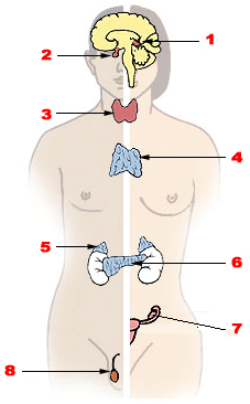
Endocrine glands are ductless glands of the endocrine system that secrete their products, hormones, directly into the blood. The major glands of the endocrine system include the pineal gland, pituitary gland, pancreas, ovaries, testicles, thyroid gland, parathyroid gland, hypothalamus and adrenal glands. The hypothalamus and pituitary glands are neuroendocrine organs.

Neuroendocrine cells are cells that receive neuronal input and, as a consequence of this input, release messenger molecules (hormones) into the blood. In this way they bring about an integration between the nervous system and the endocrine system, a process known as neuroendocrine integration. An example of a neuroendocrine cell is a cell of the adrenal medulla, which releases adrenaline to the blood. The adrenal medullary cells are controlled by the sympathetic division of the autonomic nervous system. These cells are modified postganglionic neurons. Autonomic nerve fibers lead directly to them from the central nervous system. The adrenal medullary hormones are kept in vesicles much in the same way neurotransmitters are kept in neuronal vesicles. Hormonal effects can last up to ten times longer than those of neurotransmitters. Sympathetic nerve fiber impulses stimulate the release of adrenal medullary hormones. In this way the sympathetic division of the autonomic nervous system and the medullary secretions function together.

<span class="mw-page-title-main">Hypothalamic–pituitary–gonadal axis</span> Concept of regarding the hypothalamus, pituitary gland and gonadal glands as a single entity

The hypothalamic–pituitary–gonadal axis refers to the hypothalamus, pituitary gland, and gonadal glands as if these individual endocrine glands were a single entity. Because these glands often act in concert, physiologists and endocrinologists find it convenient and descriptive to speak of them as a single system.

<span class="mw-page-title-main">Multiple endocrine neoplasia type 1</span> Medical condition

Multiple endocrine neoplasia type 1 (MEN-1) is one of a group of disorders, the multiple endocrine neoplasias, that affect the endocrine system through development of neoplastic lesions in pituitary, parathyroid gland and pancreas. Individuals suffering from this disorder are prone to developing multiple endocrine and nonendocrine tumors. It was first described by Paul Wermer in 1954.

<span class="mw-page-title-main">Endocrine disease</span> Medical condition

Endocrine diseases are disorders of the endocrine system. The branch of medicine associated with endocrine disorders is known as endocrinology.

Hypothalamic–pituitary hormones are hormones that are produced by the hypothalamus and pituitary gland. Although the organs in which they are produced are relatively small, the effects of these hormones cascade throughout the body. They can be classified as a hypothalamic–pituitary axis of which the adrenal (HPA), gonadal (HPG), thyroid (HPT), somatotropic (HPS), and prolactin (HPP) axes are branches.

Pulsatile secretion is a biochemical phenomenon observed in a wide variety of cell and tissue types, in which chemical products are secreted in a regular temporal pattern. The most common cellular products observed to be released in this manner are intercellular signaling molecules such as hormones or neurotransmitters. Examples of hormones that are secreted pulsatilely include insulin, thyrotropin, TRH, gonadotropin-releasing hormone (GnRH) and growth hormone (GH). In the nervous system, pulsatility is observed in oscillatory activity from central pattern generators. In the heart, pacemakers are able to work and secrete in a pulsatile manner. A pulsatile secretion pattern is critical to the function of many hormones in order to maintain the delicate homeostatic balance necessary for essential life processes, such as development and reproduction. Variations of the concentration in a certain frequency can be critical to hormone function, as evidenced by the case of GnRH agonists, which cause functional inhibition of the receptor for GnRH due to profound downregulation in response to constant (tonic) stimulation. Pulsatility may function to sensitize target tissues to the hormone of interest and upregulate receptors, leading to improved responses. This heightened response may have served to improve the animal's fitness in its environment and promote its evolutionary retention.

<span class="mw-page-title-main">Adrenopause</span> Decline in secretion and levels of adrenal androgens

Adrenopause is the decline in secretion and levels of adrenal androgens such as dehydroepiandrosterone (DHEA) and dehydroepiandrosterone sulfate (DHEA-S) from the zona reticularis of the adrenal glands with age. Levels of adrenal androgens start to increase around age 7 or 8 years (adrenarche), peak in early adulthood around age 20 to 25 years, and decrease at a rate of approximately 2% per year thereafter, eventually reaching levels of 10 to 20% of those of young adults by age 80 years. It is caused by the progressive apoptosis of adrenal androgen-secreting cells and hence involution of the zona reticularis. It is analogous to andropause in men and menopause in women, the abrupt or gradual decline in production of sex hormones from the gonads with age.

References

  1. Creative Commons by small.svg  This article incorporates text available under the CC BY 4.0 license.Betts, J Gordon; Desaix, Peter; Johnson, Eddie; Johnson, Jody E; Korol, Oksana; Kruse, Dean; Poe, Brandon; Wise, James; Womble, Mark D; Young, Kelly A (July 27, 2023). Anatomy & Physiology. Houston: OpenStax CNX. 17.11 Development and ageing of the endocrine system. ISBN   978-1-947172-04-3.
  2. 1 2 3 Gardner, David G.; Shoback, Dolores (2011). Greenspan's Basic & Clinical Endocrinology (9th ed.). New York: McGraw Hill. p. 562. ISBN   978-0-07-162243-1.
  3. Melmed, Shlomo; Polonsky, Kenneth S. (2011). Williams Textbook of Endocrinology (12th ed.). Saunders. p. 839. ISBN   978-1437703245.
  4. 1 2 Pescovitz, Ora H.; Eugster, Erica A. (2004). Pediatric Endocrinology: Mechanisms, Manifestations, and Management (1st ed.). Lippincott Williams & Wilkins. p. 549. ISBN   978-0781740593.
  5. Santisteban, P. (2004). Braverman LE, Utiger RD (eds.). Werner and Ingbar's The Thyroid: A Fundamental and Clinical Text (9th ed.). JB Lippincott. pp. 8–25. ISBN   978-0781750479.
  6. Melmed, Shlomo; Polonsky, Kenneth S. (2011). Williams Textbook of Endocrinology (12th ed.). Saunders. p. 844. ISBN   978-1437703245.
  7. Gardner, David G.; Shoback, Dolores (2011). Greenspan's Basic & Clinical Endocrinology (9th ed.). New York: McGraw Hill. p. 811. ISBN   978-0-07-162243-1.
  8. Fisher, Delbert A.; Brown, Rosalind S. (2012). "The maturation of thyroid function in the perinatal period and during childhood". Werner and Ingbar's The Thyroid: A Fundamental and Clinical Text (10 ed.). Philadelphia, PA: Lippincott Williams & Wilkins. pp. 775–786. ISBN   978-1451120639.
  9. 1 2 Melmed, Shlomo; Polonsky, Kenneth S. (2011). Williams Textbook of Endocrinology (12th ed.). Saunders. p. 851. ISBN   978-1437703245.
  10. Miao, D.; He B.; Karaplis C.; et al. (May 1, 2002). "Parathyroid hormone is essential for normal fetal bone formation". J Clin Invest. 109 (9): 1173–1182. doi:10.1172/JCI14817. PMC   150965 . PMID   11994406.
  11. Hochberg, Ze’ev; Tiosano, D. (2004-03-10). "Disorders of Mineral Metabolism". Pediatric Endocrinology: Mechanisms, Manifestations, and Management (1 ed.). Philadelphia, PA: Lippincott Williams & Wilkins. pp. 614–640. ISBN   978-0781740593.
  12. Bowl, M.R.; Nesbit M.A.; Harding B.; et al. (1 October 2005). "An interstitial deletion-insertion involving chromosomes 2p25.3 and Xq27.1, near SOX3, causes X-linked recessive hypoparathyroidism". J Clin Invest. 115 (10): 2822–2831. doi:10.1172/JCI24156. PMC   1201662 . PMID   16167084.
  13. Edlund, H. (July 2012). "Pancreatic organogenesis--developmental mechanisms and implications for therapy". Nat Rev Genet. 3 (7): 524–32. doi:10.1038/nrg841. PMID   12094230. S2CID   2436869.
  14. 1 2 3 4 5 Sperling, M. A.; Tulchinsky, D.; Little, A. B. (1994-05-24). "Carbohydrate metabolism: insulin and glucagons". Maternal-Fetal Endocrinology (2nd ed.). Saunders. pp. 380–400. ISBN   0721642322.
  15. 1 2 Girard, J. (November 1989). "Control of fetal and neonatal glucose metabolism by pancreatic hormones". Baillière's Clinical Endocrinology and Metabolism. 3 (3): 817–836. doi:10.1016/S0950-351X(89)80055-2. PMID   2698157.
  16. 1 2 3 Melmed, Shlomo; Polonsky, Kenneth S. (2011). Williams Textbook of Endocrinology (12th ed.). Saunders. pp. 852–4. ISBN   978-1437703245.
  17. Rahman, M. Ekhlasur; Khan, M. R. (2011-01-01). "Neonatology". Essence of Paediatrics (4th ed.). Elsevier. p. 44. ISBN   978-8131228043.
  18. Melmed, Shlomo; Polonsky, Kenneth S. (2011). Williams Textbook of Endocrinology (12th ed.). Saunders. p. 869. ISBN   978-1437703245.
  19. 1 2 Melmed, Shlomo; Polonsky, Kenneth S. (2011). Williams Textbook of Endocrinology (12th ed.). Saunders. p. 881. ISBN   978-1437703245.
  20. Nef, S; Schaad O; Stallings N; et al. (15 November 2005). "Gene expression during sex determination reveals a robust female genetic program at the onset of ovarian development". Dev. Biol. 287 (2): 361–377. doi:10.1016/j.ydbio.2005.09.008. PMID   16214126.
  21. Vainio, S; Heikkila M; Kispert A; et al. (4 February 1999). "Female development in mammals is regulated by Wnt-4 signalling". Nature. 397 (6718): 405–409. Bibcode:1999Natur.397..405V. doi:10.1038/17068. PMID   9989404. S2CID   4408910.
  22. Chassot, A; Gregoire EP; Magliano M; et al. (5 November 2008). "Genetics of ovarian differentiation: Rspo1, a major player". Nature. 2 (4–5): 219–227. doi:10.1159/000152038. PMID   18987496. S2CID   207552426.
  23. Uhlenhaut, N; Jakob S; Anlag K; et al. (11 December 2009). "Somatic sex reprogramming of adult ovaries to testes by FOXL2 ablation". Cell. 139 (6): 1130–1142. doi: 10.1016/j.cell.2009.11.021 . PMID   20005806.
  24. Couse, J; Hewitt S; Bunch D; et al. (17 December 1999). "Postnatal sex reversal of the ovaries in mice lacking estrogen receptors alpha and beta". Science. 286 (5448): 2328–2331. doi:10.1126/science.286.5448.2328. PMID   10600740.
  25. Beverdam, A; Koopman P. (1 February 2006). "Expression profiling of purified mouse gonadal somatic cells during the critical time window of sex determination reveals novel candidate genes for human sexual dysgenesis syndromes". Hum Mol Genet. 15 (3): 417–431. doi: 10.1093/hmg/ddi463 . PMID   16399799.
  26. Rathke, H (1838). "Ueber die Entsehung der glandula". Arch. Anat. Physiol. Wiss.: 482–485.
  27. 1 2 Etchevers, HC; Le Douarin NM; et al. (1 April 2001). "The cephalic neural crest provides pericytes and smooth muscle cells to all blood vessels of the face and forebrain". Development. 128 (7): 1059–1068. doi:10.1242/dev.128.7.1059. PMID   11245571.
  28. Gleiberman, AS; Fedtsova NG; Rosenfeld MG (15 September 1999). "Tissue interactions in the induction of anterior pituitary: role of the ventral diencephalon, mes- enchyme, and notochord". Dev. Biol. 213 (2): 340–353. doi: 10.1006/dbio.1999.9386 . PMID   10479452.
  29. Scully, KM; Rosenfeld MG (22 March 2002). "Pituitary development: regulatory codes in mammalian organogenesis". Science. 295 (5563): 2231–2235. Bibcode:2002Sci...295.2231S. doi:10.1126/science.1062736. PMID   11910101. S2CID   31984040.
  30. Ward, Rd; Stone BM; Raetzman LT; et al. (June 2006). "Cell proliferation and vascu larization in mouse models of pituitary hormone deficiency". Mol Endocrinol. 20 (6): 1378–1390. doi:10.1210/me.2005-0409. PMID   16556738.
  31. McCabe, MJ; Gaston-Massuet C; Tziaferi V; et al. (October 2011). "Novel FGF8 mutations associated with recessive holoprosencephaly, craniofacial defects, and hypothalamo-pituitary dysfunction". J Clin Endocrinol Metab. 96 (10): 1709–1718. doi:10.1210/jc.2011-0454. PMC   3417283 . PMID   21832120.
  32. 1 2 Zhu, X; Wang J; Ju NG; Rosenfeld MG (December 2007). "Signaling and epigenetic regulation of pituitary development". Curr Opin Cell Biol. 19 (6): 605–611. doi:10.1016/j.ceb.2007.09.011. PMC   2796608 . PMID   17988851.
  33. Treier, M; Gleiberman AS; O’Connell SM; et al. (1 June 1998). "Multistep signaling requirements for pituitary organogenesis in vivo". Genes Dev. 12 (11): 1691–1704. doi:10.1101/gad.12.11.1691. PMC   316866 . PMID   9620855.
  34. 1 2 Melmed, Shlomo; Polonsky, Kenneth S. (2011). Williams Textbook of Endocrinology (12th ed.). Saunders. p. 177. ISBN   978-1437703245.
  35. Asa, SL; Kovacs K; Laszlo FA; et al. (1986). "Human fetal adenohypophysis: histologic and immunocytochemical analysis". Neuroendocrinology. 43 (3): 308–316. doi:10.1159/000124545. PMID   3016583.