Follicle-stimulating hormone receptor

Last updated
FSHR
Structure of FSH-FSHRED.png
Available structures
PDB Ortholog search: PDBe RCSB
Identifiers
Aliases FSHR , FSHR1, FSHRO, LGR1, ODG1, follicle stimulating hormone receptor
External IDs OMIM: 136435 MGI: 95583 HomoloGene: 117 GeneCards: FSHR
Orthologs
SpeciesHumanMouse
Entrez
Ensembl
UniProt
RefSeq (mRNA)

NM_000145
NM_181446

NM_013523

RefSeq (protein)

NP_000136
NP_852111

NP_038551

Location (UCSC) Chr 2: 48.96 – 49.15 Mb Chr 17: 89.29 – 89.51 Mb
PubMed search [3] [4]
Wikidata
View/Edit Human View/Edit Mouse

The follicle-stimulating hormone receptor or FSH receptor (FSHR) is a transmembrane receptor that interacts with the follicle-stimulating hormone (FSH) and represents a G protein-coupled receptor (GPCR). Its activation is necessary for the hormonal functioning of FSH. FSHRs are found in the ovary, testis, and uterus.

Contents

FSHR gene

The gene for the FSHR is found on chromosome 2 p21 in humans. The gene sequence of the FSHR consists of about 2,080 nucleotides. [5]

Receptor structure

The seven transmembrane a-helix structure of a G protein-coupled receptor such as FSHR 7TM receptor.png
The seven transmembrane α-helix structure of a G protein-coupled receptor such as FSHR

The FSHR consists of 695 amino acids and has a molecular mass of about 76 kDa. [5] Like other GPCRs, the FSH-receptor possesses seven membrane-spanning domains or transmembrane helices.

Ligand binding and signal transduction

Upon initial binding to the LRR region of FSHR, FSH reshapes its conformation to form a new pocket. FSHR then inserts its sulfotyrosine from the hinge loop into the pockets and activates the 7-helical transmembrane domain. [6] This event leads to a transduction of the signal that activates the G protein that is bound to the receptor internally. With FSH attached, the receptor shifts conformation and, thus, mechanically activates the G protein, which detaches from the receptor and activates the cAMP system.[ citation needed ]

It is believed that a receptor molecule exists in a conformational equilibrium between active and inactive states. The binding of FSH to the receptor shifts the equilibrium between active and inactive receptors. FSH and FSH-agonists shift the equilibrium in favor of active states; FSH antagonists shift the equilibrium in favor of inactive states. For a cell to respond to FSH, only a small percentage (~1%) of receptor sites need to be activated.[ citation needed ]

Phosphorylation by cAMP-dependent protein kinases

Cyclic AMP-dependent protein kinases (protein kinase A) are activated by the signal chain coming from the G protein (that was activated by the FSH-receptor) via adenylate cyclase and cyclic AMP (cAMP).[ citation needed ]

These protein kinases are present as tetramers with two regulatory units and two catalytic units. Upon binding of cAMP to the regulatory units, the catalytic units are released and initiate the phosphorylation of proteins, leading to the physiologic action. The cyclic AMP-regulatory dimers are degraded by phosphodiesterase and release 5’AMP. DNA in the cell nucleus binds to phosphorylated proteins through the cyclic AMP response element (CRE), which results in the activation of genes. [5]

The signal is amplified by the involvement of cAMP and the resulting phosphorylation. The process is modified by prostaglandins. Other cellular regulators are participate are the intracellular calcium concentration modified by phospholipase, nitric acid, and other growth factors.

The FSH receptor can also activate the extracellular signal-regulated kinases (ERK). [9] In a feedback mechanism, these activated kinases phosphorylate the receptor. The longer the receptor remains active, the more kinases are activated, the more receptors are phosphorylated.[ citation needed ]

Action

In the ovary, the FSH receptor is necessary for follicular development and expressed on the granulosa cells. [5]

In the male, the FSH receptor has been identified on the Sertoli cells that are critical for spermatogenesis. [10]

The FSHR is expressed during the luteal phase in the secretory endometrium of the uterus. [11]

FSH receptor is selectively expressed on the surface of the blood vessels of a wide range of carcinogenic tumors. [12]

Receptor regulation

Upregulation

Upregulation refers to the increase in the number of receptor sites on the membrane. Estrogen upregulates FSH receptor sites. In turn, FSH stimulates granulosa cells to produce estrogens. This synergistic activity of estrogen and FSH allows for follicle growth and development in the ovary.[ citation needed ]

Desensitization

The FSHR become desensitized when exposed to FSH for some time. A key reaction of this downregulation is the phosphorylation of the intracellular (or cytoplasmic) receptor domain by protein kinases. This process uncouples Gs protein from the FSHR. Another way to desensitize is to uncouple the regulatory and catalytic units of the cAMP system.[ citation needed ]

Downregulation

Downregulation refers to the decrease in the number of receptor sites. This can be accomplished by metabolizing bound FSHR sites. The bound FSH-receptor complex is brought by lateral migration to a "coated pit," where such units are concentrated and then stabilized by a framework of clathrins. A pinched-off coated pit is internalized and degraded by lysosomes. Proteins may be metabolized or the receptor can be recycled. Use of long-acting agonists will downregulate the receptor population.[ citation needed ]

Modulators

Antibodies to FSHR can interfere with FSHR activity.

FSH abnormalities

Some patients with ovarian hyperstimulation syndrome may have mutations in the gene for FSHR, making them more sensitive to gonadotropin stimulation. [13]

Women with 46 XX gonadal dysgenesis experience primary amenorrhea with hypergonadotropic hypogonadism. There are forms of 46 xx gonadal dysgenesis wherein abnormalities in the FSH-receptor have been reported and are thought to be the cause of the hypogonadism. [14]

Polymorphism may affect FSH receptor populations and lead to poorer responses in infertile women receiving FSH medication for IVF. [15]

Alternative splicing of the FSHR gene may be implicated in subfertility in males [16]

Ligands

Follicle-stimulating hormone (FSH) is an agonist of the FSHR.

Small-molecule positive allosteric modulators of the FSHR have been developed. [17]

History

Alfred G. Gilman and Martin Rodbell received the 1994 Nobel Prize in Medicine and Physiology for the discovery of the G Protein System.[ citation needed ]

See also

Related Research Articles

<span class="mw-page-title-main">G protein</span> Type of proteins

G proteins, also known as guanine nucleotide-binding proteins, are a family of proteins that act as molecular switches inside cells, and are involved in transmitting signals from a variety of stimuli outside a cell to its interior. Their activity is regulated by factors that control their ability to bind to and hydrolyze guanosine triphosphate (GTP) to guanosine diphosphate (GDP). When they are bound to GTP, they are 'on', and, when they are bound to GDP, they are 'off'. G proteins belong to the larger group of enzymes called GTPases.

<span class="mw-page-title-main">Luteinizing hormone</span> Gonadotropin secreted by the adenohypophysis

Luteinizing hormone is a hormone produced by gonadotropic cells in the anterior pituitary gland. The production of LH is regulated by gonadotropin-releasing hormone (GnRH) from the hypothalamus. In females, an acute rise of LH known as an LH surge, triggers ovulation and development of the corpus luteum. In males, where LH had also been called interstitial cell–stimulating hormone (ICSH), it stimulates Leydig cell production of testosterone. It acts synergistically with follicle-stimulating hormone (FSH).

<span class="mw-page-title-main">Follicle-stimulating hormone</span> Gonadotropin that regulates the development of reproductive processes

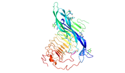
Follicle-stimulating hormone (FSH) is a gonadotropin, a glycoprotein polypeptide hormone. FSH is synthesized and secreted by the gonadotropic cells of the anterior pituitary gland and regulates the development, growth, pubertal maturation, and reproductive processes of the body. FSH and luteinizing hormone (LH) work together in the reproductive system.

A hormone receptor is a receptor molecule that binds to a specific hormone. Hormone receptors are a wide family of proteins made up of receptors for thyroid and steroid hormones, retinoids and Vitamin D, and a variety of other receptors for various ligands, such as fatty acids and prostaglandins. Hormone receptors are of mainly two classes. Receptors for peptide hormones tend to be cell surface receptors built into the plasma membrane of cells and are thus referred to as trans membrane receptors. An example of this is Actrapid. Receptors for steroid hormones are usually found within the protoplasm and are referred to as intracellular or nuclear receptors, such as testosterone. Upon hormone binding, the receptor can initiate multiple signaling pathways, which ultimately leads to changes in the behavior of the target cells.

<span class="mw-page-title-main">Gonadotropin-releasing hormone</span> Mammalian protein found in Homo sapiens

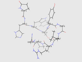
Gonadotropin-releasing hormone (GnRH) is a releasing hormone responsible for the release of follicle-stimulating hormone (FSH) and luteinizing hormone (LH) from the anterior pituitary. GnRH is a tropic peptide hormone synthesized and released from GnRH neurons within the hypothalamus. The peptide belongs to gonadotropin-releasing hormone family. It constitutes the initial step in the hypothalamic–pituitary–gonadal axis.

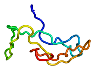
Gonadotropins are glycoprotein hormones secreted by gonadotropic cells of the anterior pituitary of vertebrates. This family includes the mammalian hormones follicle-stimulating hormone (FSH) and luteinizing hormone (LH), the placental/chorionic gonadotropins, human chorionic gonadotropin (hCG) and equine chorionic gonadotropin (eCG), as well as at least two forms of fish gonadotropins. These hormones are central to the complex endocrine system that regulates normal growth, sexual development, and reproductive function. LH and FSH are secreted by the anterior pituitary gland, while hCG and eCG are secreted by the placenta in pregnant humans and mares, respectively. The gonadotropins act on the gonads, controlling gamete and sex hormone production.

<span class="mw-page-title-main">Anti-Müllerian hormone</span> Mammalian protein found in humans

Anti-Müllerian hormone (AMH), also known as Müllerian-inhibiting hormone (MIH), is a glycoprotein hormone structurally related to inhibin and activin from the transforming growth factor beta superfamily, whose key roles are in growth differentiation and folliculogenesis. In humans, it is encoded by the AMH gene, on chromosome 19p13.3, while its receptor is encoded by the AMHR2 gene on chromosome 12.

<span class="mw-page-title-main">Estrogen receptor</span> Proteins activated by the hormone estrogen

Estrogen receptors (ERs) are a group of proteins found inside cells. They are receptors that are activated by the hormone estrogen (17β-estradiol). Two classes of ER exist: nuclear estrogen receptors, which are members of the nuclear receptor family of intracellular receptors, and membrane estrogen receptors (mERs), which are mostly G protein-coupled receptors. This article refers to the former (ER).

A biochemical cascade, also known as a signaling cascade or signaling pathway, is a series of chemical reactions that occur within a biological cell when initiated by a stimulus. This stimulus, known as a first messenger, acts on a receptor that is transduced to the cell interior through second messengers which amplify the signal and transfer it to effector molecules, causing the cell to respond to the initial stimulus. Most biochemical cascades are series of events, in which one event triggers the next, in a linear fashion. At each step of the signaling cascade, various controlling factors are involved to regulate cellular actions, in order to respond effectively to cues about their changing internal and external environments.

<span class="mw-page-title-main">Luteinizing hormone/choriogonadotropin receptor</span> Protein-coding gene in the species Homo sapiens

The luteinizing hormone/choriogonadotropin receptor (LHCGR), also lutropin/choriogonadotropin receptor (LCGR) or luteinizing hormone receptor (LHR) is a transmembrane receptor found predominantly in the ovary and testis, but also many extragonadal organs such as the uterus and breasts. The receptor interacts with both luteinizing hormone (LH) and chorionic gonadotropins and represents a G protein-coupled receptor (GPCR). Its activation is necessary for the hormonal functioning during reproduction.

<span class="mw-page-title-main">BMPR2</span> Protein-coding gene in the species Homo sapiens

Bone morphogenetic protein receptor type II or BMPR2 is a serine/threonine receptor kinase encoded by the BMPR2 gene. It binds bone morphogenetic proteins, members of the TGF beta superfamily of ligands, which are involved in paracrine signaling. BMPs are involved in a host of cellular functions including osteogenesis, cell growth and cell differentiation. Signaling in the BMP pathway begins with the binding of a BMP to the type II receptor. This causes the recruitment of a BMP type I receptor, which the type II receptor phosphorylates. The type I receptor phosphorylates an R-SMAD, a transcriptional regulator.

<span class="mw-page-title-main">Thyrotropin receptor</span> Mammalian protein found in Homo sapiens

The thyrotropin receptor is a receptor that responds to thyroid-stimulating hormone and stimulates the production of thyroxine (T4) and triiodothyronine (T3). The TSH receptor is a member of the G protein-coupled receptor superfamily of integral membrane proteins and is coupled to the Gs protein.

<span class="mw-page-title-main">ACTH receptor</span> Mammalian protein found in Homo sapiens

The adrenocorticotropic hormone receptor or ACTH receptor also known as the melanocortin receptor 2 or MC2 receptor is a type of melanocortin receptor (type 2) which is specific for ACTH. A G protein–coupled receptor located on the external cell plasma membrane, it is coupled to Gαs and upregulates levels of cAMP by activating adenylyl cyclase. The ACTH receptor plays a role in immune function and glucose metabolism.

<span class="mw-page-title-main">Glycoprotein hormones, alpha polypeptide</span> Mammalian protein found in Homo sapiens

Glycoprotein hormones, alpha polypeptide is a protein that in humans is encoded by the CGA gene.

<span class="mw-page-title-main">FSHB</span> Protein-coding gene in the species Homo sapiens

Follitropin subunit beta also known as follicle-stimulating hormone beta subunit (FSH-B) is a protein that in humans is encoded by the FSHB gene. Alternative splicing results in two transcript variants encoding the same protein.

<span class="mw-page-title-main">Activin and inhibin</span> Regulators of feedback on FSH-production

Activin and inhibin are two closely related protein complexes that have almost directly opposite biological effects. Identified in 1986, activin enhances FSH biosynthesis and secretion, and participates in the regulation of the menstrual cycle. Many other functions have been found to be exerted by activin, including roles in cell proliferation, differentiation, apoptosis, metabolism, homeostasis, immune response, wound repair, and endocrine function. Conversely, inhibin downregulates FSH synthesis and inhibits FSH secretion. The existence of inhibin was hypothesized as early as 1916; however, it was not demonstrated to exist until Neena Schwartz and Cornelia Channing's work in the mid-1970s, after which both proteins were molecularly characterized ten years later.

<span class="mw-page-title-main">Chorionic gonadotropin beta</span> Protein-coding gene in the species Homo sapiens

Choriogonadotropin subunit beta (CG-beta) also known as chorionic gonadotrophin chain beta is a protein that in humans is encoded by the CGB gene.

Follicle-stimulating hormone (FSH) insensitivity, or ovarian insensitivity to FSH in females, also referable to as ovarian follicle hypoplasia or granulosa cell hypoplasia in females, is a rare autosomal recessive genetic and endocrine syndrome affecting both females and males, with the former presenting with much greater severity of symptomatology. It is characterized by a resistance or complete insensitivity to the effects of follicle-stimulating hormone (FSH), a gonadotropin which is normally responsible for the stimulation of estrogen production by the ovaries in females and maintenance of fertility in both sexes. The condition manifests itself as hypergonadotropic hypogonadism, reduced or absent puberty, amenorrhea, and infertility in females, whereas males present merely with varying degrees of infertility and associated symptoms.

Ovarian follicle activation can be defined as primordial follicles in the ovary moving from a quiescent (inactive) to a growing phase. The primordial follicle in the ovary is what makes up the “pool” of follicles that will be induced to enter growth and developmental changes that change them into pre-ovulatory follicles, ready to be released during ovulation. The process of development from a primordial follicle to a pre-ovulatory follicle is called folliculogenesis.

Gonadotropin surge-attenuating factor (GnSAF) is a nonsteroidal ovarian hormone produced by the granulosa cells of small antral ovarian follicles in females. GnSAF is involved in regulating the secretion of luteinizing hormone (LH) from the anterior pituitary and the ovarian cycle. During the early to mid-follicular phase of the ovarian cycle, GnSAF acts on the anterior pituitary to attenuate LH release, limiting the secretion of LH to only basal levels. At the transition between follicular and luteal phase, GnSAF bioactivity declines sufficiently to permit LH secretion above basal levels, resulting in the mid-cycle LH surge that initiates ovulation. In normally ovulating women, the LH surge only occurs when the oocyte is mature and ready for extrusion. GnSAF bioactivity is responsible for the synchronised, biphasic nature of LH secretion.

References

  1. 1 2 3 GRCh38: Ensembl release 89: ENSG00000170820 - Ensembl, May 2017
  2. 1 2 3 GRCm38: Ensembl release 89: ENSMUSG00000032937 - Ensembl, May 2017
  3. "Human PubMed Reference:". National Center for Biotechnology Information, U.S. National Library of Medicine.
  4. "Mouse PubMed Reference:". National Center for Biotechnology Information, U.S. National Library of Medicine.
  5. 1 2 3 4 Simoni M, Gromoll J, Nieschlag E (Dec 1997). "The follicle-stimulating hormone receptor: biochemistry, molecular biology, physiology, and pathophysiology". Endocrine Reviews. 18 (6): 739–73. doi: 10.1210/edrv.18.6.0320 . PMID   9408742.
  6. 1 2 Jiang X, Liu H, Chen X, Chen PH, Fischer D, Sriraman V, Yu HN, Arkinstall S, He X (Jul 2012). "Structure of follicle-stimulating hormone in complex with the entire ectodomain of its receptor". Proceedings of the National Academy of Sciences of the United States of America. 109 (31): 12491–6. Bibcode:2012PNAS..10912491J. doi: 10.1073/pnas.1206643109 . PMC   3411987 . PMID   22802634.
  7. Costagliola S, Panneels V, Bonomi M, Koch J, Many MC, Smits G, Vassart G (Feb 2002). "Tyrosine sulfation is required for agonist recognition by glycoprotein hormone receptors". The EMBO Journal. 21 (4): 504–13. doi:10.1093/emboj/21.4.504. PMC   125869 . PMID   11847099.
  8. Jiang X, Dias JA, He X (Jan 2014). "Structural biology of glycoprotein hormones and their receptors: insights to signaling". Molecular and Cellular Endocrinology. 382 (1): 424–51. doi: 10.1016/j.mce.2013.08.021 . PMID   24001578.
  9. Piketty V, Kara E, Guillou F, Reiter E, Crepieux P (2006). "Follicle-stimulating hormone (FSH) activates extracellular signal-regulated kinase phosphorylation independently of beta-arrestin- and dynamin-mediated FSH receptor internalization". Reproductive Biology and Endocrinology. 4: 33. doi: 10.1186/1477-7827-4-33 . PMC   1524777 . PMID   16787538.
  10. Asatiani K, Gromoll J, Eckardstein SV, Zitzmann M, Nieschlag E, Simoni M (Jun 2002). "Distribution and function of FSH receptor genetic variants in normal men". Andrologia. 34 (3): 172–6. doi: 10.1046/j.1439-0272.2002.00493.x . PMID   12059813. S2CID   21090038.
  11. La Marca A, Carducci Artenisio A, Stabile G, Rivasi F, Volpe A (Dec 2005). "Evidence for cycle-dependent expression of follicle-stimulating hormone receptor in human endometrium". Gynecological Endocrinology. 21 (6): 303–6. doi:10.1080/09513590500402756. PMID   16390776. S2CID   24690912.
  12. Radu A, Pichon C, Camparo P, Antoine M, Allory Y, Couvelard A, Fromont G, Hai MT, Ghinea N (Oct 2010). "Expression of follicle-stimulating hormone receptor in tumor blood vessels". The New England Journal of Medicine. 363 (17): 1621–30. doi: 10.1056/NEJMoa1001283 . PMID   20961245.
  13. Delbaere A, Smits G, De Leener A, Costagliola S, Vassart G (Apr 2005). "Understanding ovarian hyperstimulation syndrome". Endocrine. 26 (3): 285–90. doi:10.1385/ENDO:26:3:285. PMID   16034183. S2CID   7607365.
  14. Aittomäki K, Lucena JL, Pakarinen P, Sistonen P, Tapanainen J, Gromoll J, Kaskikari R, Sankila EM, Lehväslaiho H, Engel AR, Nieschlag E, Huhtaniemi I, de la Chapelle A (Sep 1995). "Mutation in the follicle-stimulating hormone receptor gene causes hereditary hypergonadotropic ovarian failure". Cell. 82 (6): 959–68. doi: 10.1016/0092-8674(95)90275-9 . PMID   7553856. S2CID   14748261.
  15. Loutradis D, Patsoula E, Minas V, Koussidis GA, Antsaklis A, Michalas S, Makrigiannakis A (Apr 2006). "FSH receptor gene polymorphisms have a role for different ovarian response to stimulation in patients entering IVF/ICSI-ET programs". Journal of Assisted Reproduction and Genetics. 23 (4): 177–84. doi:10.1007/s10815-005-9015-z. PMC   3454958 . PMID   16758348.
  16. Song GJ, Park YS, Lee YS, Lee CC, Kang IS (Mar 2002). "Alternatively spliced variants of the follicle-stimulating hormone receptor gene in the testis of infertile men". Fertility and Sterility. 77 (3): 499–504. doi: 10.1016/s0015-0282(01)03221-6 . PMID   11872202.
  17. Nataraja S, Yu H, Guner J, Palmer S (2020). "Discovery and Preclinical Development of Orally Active Small Molecules that Exhibit Highly Selective Follicle Stimulating Hormone Receptor Agonism". Front Pharmacol. 11: 602593. doi: 10.3389/fphar.2020.602593 . PMC   7845544 . PMID   33519465.

Further reading