Related Research Articles

Appendicitis is inflammation of the appendix. Symptoms commonly include right lower abdominal pain, nausea, vomiting, and decreased appetite. However, approximately 40% of people do not have these typical symptoms. Severe complications of a ruptured appendix include widespread, painful inflammation of the inner lining of the abdominal wall and sepsis.
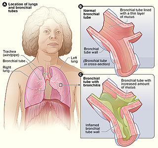
Acute bronchitis, also known as a chest cold, is short-term bronchitis – inflammation of the bronchi of the lungs. The most common symptom is a cough. Other symptoms include coughing up mucus, wheezing, shortness of breath, fever, and chest discomfort. The infection may last from a few to ten days. The cough may persist for several weeks afterward with the total duration of symptoms usually around three weeks. Some have symptoms for up to six weeks.

Clearing the cervical spine is the process by which medical professionals determine whether cervical spine injuries exist, mainly regarding cervical fracture. It is generally performed in cases of major trauma. This process can take place in the emergency department or in the field by appropriately trained EMS personnel.

A joint dislocation, also called luxation, occurs when there is an abnormal separation in the joint, where two or more bones meet. A partial dislocation is referred to as a subluxation. Dislocations are often caused by sudden trauma on the joint like an impact or fall. A joint dislocation can cause damage to the surrounding ligaments, tendons, muscles, and nerves. Dislocations can occur in any major joint or minor joint. The most common joint dislocation is a shoulder dislocation.

A cervical fracture, commonly called a broken neck, is a fracture of any of the seven cervical vertebrae in the neck. Examples of common causes in humans are traffic collisions and diving into shallow water. Abnormal movement of neck bones or pieces of bone can cause a spinal cord injury, resulting in loss of sensation, paralysis, or usually death soon thereafter, primarily via compromising neurological supply to the respiratory muscles as well as innervation to the heart.

Interstitial lung disease (ILD), or diffuse parenchymal lung disease (DPLD), is a group of respiratory diseases affecting the interstitium (the tissue and space around the alveoli of the lungs. It concerns alveolar epithelium, pulmonary capillary endothelium, basement membrane, and perivascular and perilymphatic tissues. It may occur when an injury to the lungs triggers an abnormal healing response. Ordinarily, the body generates just the right amount of tissue to repair damage, but in interstitial lung disease, the repair process is disrupted, and the tissue around the air sacs becomes scarred and thickened. This makes it more difficult for oxygen to pass into the bloodstream. The disease presents itself with the following symptoms: shortness of breath, nonproductive coughing, fatigue, and weight loss, which tend to develop slowly, over several months. The average rate of survival for someone with this disease is between three and five years. The term ILD is used to distinguish these diseases from obstructive airways diseases.

Hypophosphatasia (; also called deficiency of alkaline phosphatase, phosphoethanolaminuria, or Rathbun's syndrome; sometimes abbreviated HPP) is a rare, and sometimes fatal, inherited metabolic bone disease. Clinical symptoms are heterogeneous, ranging from the rapidly fatal, perinatal variant, with profound skeletal hypomineralization, respiratory compromise or vitamin B6 dependent seizures to a milder, progressive osteomalacia later in life. Tissue non-specific alkaline phosphatase (TNSALP) deficiency in osteoblasts and chondrocytes impairs bone mineralization, leading to rickets or osteomalacia. The pathognomonic finding is subnormal serum activity of the TNSALP enzyme, which is caused by one of 388 genetic mutations identified to date, in the gene encoding TNSALP. Genetic inheritance is autosomal recessive for the perinatal and infantile forms but either autosomal recessive or autosomal dominant in the milder forms.

A pulled elbow, also known as nursemaid's elbow or a radial head subluxation, is when the ligament that wraps around the radial head slips off. Often a child will hold their arm against their body with the elbow slightly bent. They will not move the arm as this results in pain. Touching the arm, without moving the elbow, is usually not painful.

In medicine, the Ottawa ankle rules are a set of guidelines for clinicians to help decide if a patient with foot or ankle pain should be offered X-rays to diagnose a possible bone fracture. Before the introduction of the rules most patients with ankle injuries would have been imaged. However the vast majority of patients with unclear ankle injuries do not have bone fractures. As a result, many unnecessary X-rays were taken, which was costly, time-consuming and a slight health risk due to radiation exposure.
The knee examination, in medicine and physiotherapy, is performed as part of a physical examination, or when a patient presents with knee pain or a history that suggests a pathology of the knee joint.

The fat pad sign, also known as the sail sign, is a potential finding on elbow radiography which suggests a fracture of one or more bones at the elbow. It is may indicate an occult fracture that is not directly visible. Its name derives from the fact that it has the shape of a spinnaker (sail). It is caused by displacement of the fat pad around the elbow joint. Both anterior and posterior fat pad signs exist, and both can be found on the same X-ray.

Projectional radiography, also known as conventional radiography, is a form of radiography and medical imaging that produces two-dimensional images by x-ray radiation. The image acquisition is generally performed by radiographers, and the images are often examined by radiologists. Both the procedure and any resultant images are often simply called "X-ray". Plain radiography or roentgenography generally refers to projectional radiography. Plain radiography can also refer to radiography without a radiocontrast agent or radiography that generates single static images, as contrasted to fluoroscopy, which are technically also projectional.

An abdominal x-ray is an x-ray of the abdomen. It is sometimes abbreviated to AXR, or KUB.

Radial tunnel syndrome (RTS) is caused by increased pressure on the radial nerve as it travels from the upper arm to the hand and wrist.

The Taylor Spatial Frame (TSF) is an external fixator used by paediatric and orthopaedic surgeons to treat complex fractures and bone deformities. The medical device shares a number of components and features of the Ilizarov apparatus. The Taylor Spatial Frame is a hexapod device based on a Stewart platform, and was invented by orthopaedic surgeon Charles Taylor. The device consists of two or more aluminum or carbon fibre rings connected by six struts. Each strut can be independently lengthened or shortened to achieve the desired result, e.g. compression at the fracture site, lengthening, etc. Connected to a bone by tensioned wires or half pins, the attached bone can be manipulated in three dimensions and 9 degrees of freedom. Angular, translational, rotational, and length deformities can all be corrected simultaneously with the TSF.

The elbow is the region between the upper arm and the forearm that surrounds the elbow joint. The elbow includes prominent landmarks such as the olecranon, the cubital fossa, and the lateral and the medial epicondyles of the humerus. The elbow joint is a hinge joint between the arm and the forearm; more specifically between the humerus in the upper arm and the radius and ulna in the forearm which allows the forearm and hand to be moved towards and away from the body. The term elbow is specifically used for humans and other primates, and in other vertebrates forelimb plus joint is used.

A supracondylar humerus fracture is a fracture of the distal humerus just above the elbow joint. The fracture is usually transverse or oblique and above the medial and lateral condyles and epicondyles. This fracture pattern is relatively rare in adults, but is the most common type of elbow fracture in children. In children, many of these fractures are non-displaced and can be treated with casting. Some are angulated or displaced and are best treated with surgery. In children, most of these fractures can be treated effectively with expectation for full recovery. Some of these injuries can be complicated by poor healing or by associated blood vessel or nerve injuries with serious complications.
Posterolateral corner injuries of the knee are injuries to a complex area formed by the interaction of multiple structures. Injuries to the posterolateral corner can be debilitating to the person and require recognition and treatment to avoid long term consequences. Injuries to the PLC often occur in combination with other ligamentous injuries to the knee; most commonly the anterior cruciate ligament (ACL) and posterior cruciate ligament (PCL). As with any injury, an understanding of the anatomy and functional interactions of the posterolateral corner is important to diagnosing and treating the injury.

Pain in the hip is the experience of pain in the muscles or joints in the hip/ pelvic region, a condition commonly arising from any of a number of factors. Sometimes it is closely associated with lower back pain.

A medial epicondyle fracture is an avulsion injury to the medial epicondyle of the humerus; the prominence of bone on the inside of the elbow. Medial epicondyle fractures account for 10% elbow fractures in children. 25% of injuries are associated with a dislocation of the elbow.
References
- ↑ Elbow-Extension Test Is Useful for Ruling Out Elbow Fracture, Journal Watch General Medicine January 13, 2009
- ↑ Appelboam A et al. Elbow extension test to rule out elbow fracture: Multicentre, prospective validation and observational study of diagnostic accuracy in adults and children. BMJ 2008 Dec 9; 337:a2428.