Unhappy triad

Last updated
Unhappy triad
Other namesTerrible triad, horrible triangle, O'Donoghue's triad, the three musketeers [1] blown knee
Knee diagram.svg
Knee
Specialty Emergency medicine   OOjs UI icon edit-ltr-progressive.svg

The unhappy triad, also known as a blown knee among other names, is an injury to the anterior cruciate ligament, medial collateral ligament, and meniscus. Analysis during the 1990s indicated that this 'classic' O'Donoghue triad is actually an unusual clinical entity among athletes with knee injuries. Some authors mistakenly believe that in this type of injury, "combined anterior cruciate and medial collateral ligament (ACL- MCL) disruptions that were incurred during athletic endeavors" always present with concomitant medial meniscus injury. However, the 1990 analysis showed that lateral meniscus tears are more common than medial meniscus tears in conjunction with sprains of the ACL. [1]

Contents

Symptoms

Cause

The unhappy triad occurs due to a lateral blow to the knee causing a rupture in the anterior cruciate ligament, medial collateral ligament, and meniscus. Injury is most often sustained when a lateral (from the outside) force impacts the knee while the foot is fixed on the ground. The strong valgus or rotary force to the knee tears the ACL, MCL, and medial meniscus all together. This type of injury occurs often in contact sports such as football, rugby, or motocross. During the injury, the leg is laterally rotated and over-abducted. In about 10% of cases, the force is applied to the opposite side of the knee, and the lateral and posterolateral ligaments are torn.

Anatomy

Capsule of right knee-joint (distended). Posterior aspect. Gray352.png
Capsule of right knee-joint (distended). Posterior aspect.

Skeletal components involved in the unhappy triad include: patella, femur, tibia. No muscles are directly involved in this injury, only ligaments; However, strengthening the hip flexor and hip extensor muscles may help alleviate the injury.

The medial collateral ligament, posterior cruciate ligament, anterior cruciate ligament, and lateral collateral ligament are the four primary ligaments of the knee. The medial and lateral collateral ligaments primarily provide support to varus and valgus forces whereas the anterior and posterior cruciate ligaments prevent anterior and posterior translation of the tibia on the femur. [2]

Structures in triad

Superior view of the structures in triad. Gray349.png
Superior view of the structures in triad.

The classic O'Donoghue triad is characterized by an injury to three knee structures (in order):

Component injuries

The anterior cruciate ligament

The anterior cruciate ligament is one of the four crucial ligaments in the knee. It originates from the lateral condyle of the femur and goes to the intercondyloid eminence of the tibia. Its function is to provide stability in the knee and minimize stress across the knee joint. It also restrains excessive forward movement in the leg and limits rotational movements in the knee.

Injury

Anterior cruciate ligament tear seen on MRI. VKB-Riss MRT T1 PDW sag.jpg
Anterior cruciate ligament tear seen on MRI.

An anterior cruciate ligament injury results from excess tension on the ligament. This can come from a sudden stop or twisting motion of the knee. A few initial symptoms include swelling, knee instability, and pain. A popping sound or sensation may or may not be heard when the ACL first tears. A following symptom usually includes the feeling of the knee "giving out". Tearing of the ACL is the most significant injury because it leaves the knee unstable, which also causes the knee to lose its normal function.

Epidemiology

It is estimated that 100,000 new anterior cruciate ligament injuries occur in the U.S. each year. [6] Approximately half of the new ACL injuries involve injuries to the ligament, tendon, and or muscle of that affected knee. [7] Women are at greater risk for ACL injuries than men due to their greater Q angle. The Q angle is the angle formed by a line drawn from the anterior superior iliac spine to central patella and a second line drawn from central patella to tibial tubercle.

Meniscus

Tear of medial meniscus Tear of medial meniscus.jpg
Tear of medial meniscus

The medial meniscus is a C-wedge shaped piece of cartilage that acts as a"shock absorber" between the femur and the tibia. Each knee has two menisci, one at the outer edge and one at the inner edge of the knee. The menisci are tough and rubber-like to help cushion the joint and help keep it stable.

The lateral meniscus is also known as the external semilunar fibrocartilage. It is a fibrocartilage band on the lateral side of the knee joint and can easily be injured with torsional stress or direct force.

Each knee has a medial and lateral meniscus, consisting of connective tissue and collagen fibers. Menisci are needed to distribute the body weight across the knee. Without them, the body weight is distributed unevenly on the femur and tibia, possibly leading to early arthritis in the knee joint. The menisci are nourished by small blood vessels, but each has a large central section which is avascular and does not get a direct blood supply. This poses a problem in a meniscus injury, as blood flow is diminished and the avascular areas tend to not heal.

Injury

The tear of meniscus is among the most common knee injuries. It is usually caused by torsional stress; twisting or turning of the knee too quickly with the foot planted on the ground while the knee is flexed. The feeling of a "pop" in the knee is usually felt when the meniscus is torn. Athletes, particularly those who participate in contact sports, are at a greater risk for meniscal tears. Sports-related meniscal tears often occur with other knee injuries, such as an anterior cruciate ligament tear.

A torn meniscus is commonly referred to as torn cartilage in the knee. Menisci tear in different ways and are noted by how they look, as well as where the tear occurs in the meniscus. Two types of tears include minor, which includes stiffness and swelling within two to three days but usually goes away in two to three weeks. Then there is severe, which without treatment, a piece of meniscus may come loose and drift into the joint space. Tears include longitudinal, parrot-beak, flap, bucket handle, and mixed/complex.

Epidemiology

Injury to the medial meniscus is about five times greater than injury to the lateral meniscus due to its anatomical attachment to the MCL. [8] Lateral meniscal tears are more common in acute ACL injuries, whereas medial meniscal injuries are more common in chronic ACL-deficient knees and more amenable to repair. [9]

Meta-analysis shows that in acute injuries of the ACL associated with a meniscus tear, 44% were of the medial meniscus, whereas 56% were of the lateral meniscus; in chronic ACL insufficiency, 70% were medial whereas 30% were lateral. [10]

Medial collateral ligament

Left knee-joint from behind, showing interior ligaments Gray348.png
Left knee-joint from behind, showing interior ligaments

Injury

Because the medial collateral ligament resists widening of the inside of the knee joint, the ligament is usually injured when the outside of the knee joint is struck. This force causes the outside of the knee to buckle, and the inside to widen. When the MCL is stretched too far, it is susceptible to tearing and injury. This is the injury seen by the action of "clipping" in a football game.

An injury to the MCL may occur as an isolated injury, or it may be part of a complex injury to the knee. Other ligaments ACL, or meniscus, may be torn along with a MCL injury.

Symptoms

The most common symptom following an MCL injury is pain directly over the ligament. Swelling over the torn ligament may appear, and bruising and generalized joint swelling are common 1 to 2 days after the injury. In more severe injuries, patients may complain that the knee feels unstable.

Treatment

Treatment of an MCL tear depends on the severity of the injury. Treatment always begins with allowing the pain to subside, beginning work on mobility, followed by strengthening the knee to return to sports and activities. Bracing can often be useful for treatment of MCL injuries. Fortunately, most often surgery is not necessary for the treatment of an MCL tear.

Treatments

A patellar autograft is performed to the replace damaged ligament.The grafts are inserted through a tunnel that is drilled through the tibia and femur LCA-RX2.jpg
A patellar autograft is performed to the replace damaged ligament.The grafts are inserted through a tunnel that is drilled through the tibia and femur

Treatment of the unhappy triad usually requires surgery. An ACL surgery is common and the meniscus can be treated during the surgery as well. The MCL is rehabilitated through time and immobilization. Physical therapy after the surgery and the use of a knee brace help speed up the healing process. A typical surgery for a blown knee includes:

The goal of reconstruction surgery is to prevent instability and restore the function of the torn ligament, creating a stable knee. There are certain factors that the patient must consider when deciding for or against surgery.

An important post-surgical treatment of unhappy triad is Physical Therapy (PT). PT includes exercise ambulatory programs, mobilizations, and modalities to help ease symptoms and speed up the recovery process. The purpose of physical therapy is strengthening muscle and increasing the knee's range of motion without damaging the new grafts. [11] Exercise ambulatory programs are prescribed by a Physical Therapist and should be used during recovery. Physical Therapist will provide immediate knee mobilization manually or with continuous passive motion (CPM) within the first week. Neuromuscular electrical stimulation (NMES) should be used for a span of 6-8 weeks postoperative. Immediate cryotherapy could be used for treatment as well. A combination of functional tests and validated patient-reports are used to determine a patient’s readiness to return to activities. [12]

Epidemiology

A study containing 100 consecutive patients with a recent anterior cruciate ligament injury were examined with respect to type of sports activity that caused the injury. Of the 100 consecutive ACL injuries, there were also 53 medial collateral ligament injuries, 12 medial, 35 lateral and 11 bicompartmental meniscal lesions. 59/100 patients were injured during contact sports, 30/100 in downhill skiing and 11/100 in other recreational activities, traffic accidents or at work. [13]

An associated medial collateral ligament tear was more common in skiing (22/30) than during contact sports (23/59), whereas a bicompartmental meniscal lesion was found more frequently in contact sports (9/59) than in skiing (0/30). Weightbearing was reported by 56/59 of the patients with contact sports injuries whereas 8/30 of those with skiing injuries. Non-weightbearing in the injury situation led to the same rate of MCL tears (18/28) as weightbearing (35/72) but significantly more intact menisci (19/28 vs 23/72). Thus, contact sports injuries were more often sustained during weightbearing, with a resultant joint compression of both femuro-tibial compartments as shown by the higher incidence of bicompartmental meniscal lesions. The classic "unhappy triad" was a rare finding (8/100) and Fridén T, Erlandsson T, Zätterström R, Lindstrand A, and Moritz U. suggest that this entity should be replaced by the "unhappy compression injury". [13]

History

MRI images of Unhappy triad from left to right: ACL, MCL, medial meniscus. Unhappy triad - MRT PDW SPIR sag cor sag 001.jpg
MRI images of Unhappy triad from left to right: ACL, MCL, medial meniscus.

In 1936, Cambell stated that an "impairment of the anterior crucial and medial ligaments is associated with injuries of the internal cartilage". In 1950, O'Donoghue described the unhappy triad as: (1) rupture of the medial collateral ligament, (2) damage to the medial meniscus and (3) rupture of the anterior cruciate ligament. O'Donoghue estimated the incidence rate in the traumatic sports knee to be 25%. [3]

In 1991, Shelbourne and Nitz questioned the validity of O'Donoghue's terrible triad study. A review of all arthroscopically confirmed acute injuries of second degree or worse to the ACL and MCL was performed. Of the 52 knees reviewed, 80% of group 1 had lateral meniscus tears and 29% had associated medial meniscus tears. None of the medial meniscus tears were isolated; medial meniscus tears were not present in the absence of a lateral meniscus tear. [3] From this study, it was concluded that the structures more typically involved in a triad were the anterior cruciate ligament, medial collateral ligament, and the lateral (not medial) meniscus.

Terminology

The term "unhappy triad" was coined by O'Donoghue in 1950. [3] [14] [15] However, since then, this term and the term "terrible triad" have also been used to describe several other combinations of joint injuries, including the terrible triad of the elbow [16] and shoulder. [17]

The term "terrible triad" is also sometimes used in the popular press to describe conditions relating to pain, or even to refer to the MacDonald triad of sociopathic behavior.

See also

Related Research Articles

<span class="mw-page-title-main">Knee</span> Region around the kneecap

In humans and other primates, the knee joins the thigh with the leg and consists of two joints: one between the femur and tibia, and one between the femur and patella. It is the largest joint in the human body. The knee is a modified hinge joint, which permits flexion and extension as well as slight internal and external rotation. The knee is vulnerable to injury and to the development of osteoarthritis.

<span class="mw-page-title-main">Posterior cruciate ligament</span> One of four major ligaments of the knee

The posterior cruciate ligament (PCL) is a ligament in each knee of humans and various other animals. It works as a counterpart to the anterior cruciate ligament (ACL). It connects the posterior intercondylar area of the tibia to the medial condyle of the femur. This configuration allows the PCL to resist forces pushing the tibia posteriorly relative to the femur.

<span class="mw-page-title-main">Anterior cruciate ligament</span> Type of cruciate ligament in the human knee

The anterior cruciate ligament (ACL) is one of a pair of cruciate ligaments in the human knee. The two ligaments are also called "cruciform" ligaments, as they are arranged in a crossed formation. In the quadruped stifle joint, based on its anatomical position, it is also referred to as the cranial cruciate ligament. The term cruciate translates to cross. This name is fitting because the ACL crosses the posterior cruciate ligament to form an “X”. It is composed of strong, fibrous material and assists in controlling excessive motion. This is done by limiting mobility of the joint. The anterior cruciate ligament is one of the four main ligaments of the knee, providing 85% of the restraining force to anterior tibial displacement at 30 and 90° of knee flexion. The ACL is the most injured ligament of the four located in the knee.

<span class="mw-page-title-main">Segond fracture</span> Avulsion fracture of the lateral tibial condyle of the knee

The Segond fracture is a type of avulsion fracture from the lateral tibial plateau of the knee, immediately below the articular surface of the tibia.

<span class="mw-page-title-main">Anterior cruciate ligament reconstruction</span> Surgical process

Anterior cruciate ligament reconstruction is a surgical tissue graft replacement of the anterior cruciate ligament, located in the knee, to restore its function after an injury. The torn ligament can either be removed from the knee, or preserved before reconstruction through an arthroscopic procedure. ACL repair is also a surgical option. This involves repairing the ACL by re-attaching it, instead of performing a reconstruction. Theoretical advantages of repair include faster recovery and a lack of donor site morbidity, but randomised controlled trials and long-term data regarding re-rupture rates using contemporary surgical techniques are lacking.

<span class="mw-page-title-main">Medial collateral ligament</span> Ligament on the inner side of the knee joint

The medial collateral ligament (MCL), also called the superficial medial collateral ligament (sMCL) or tibial collateral ligament (TCL), is one of the major ligaments of the knee. It is on the medial (inner) side of the knee joint and occurs in humans and other primates. Its primary function is to resist valgus forces on the knee.

<span class="mw-page-title-main">Meniscus (anatomy)</span> Fibrocartilaginous part of a bone joint

A meniscus is a crescent-shaped fibrocartilaginous anatomical structure that, in contrast to an articular disc, only partly divides a joint cavity. In humans they are present in the knee, wrist, acromioclavicular, sternoclavicular, and temporomandibular joints; in other animals they may be present in other joints.

A meniscus transplant or meniscal transplant is a transplant of the meniscus of the knee, which separates the thigh bone (femur) from the lower leg bone (tibia). The worn or damaged meniscus is removed and is replaced with a new one from a donor. The meniscus to be transplanted is taken from a cadaver, and, as such, is known as an allograft. Meniscal transplantation is technically difficult, as it must be sized accurately for each person, positioned properly and secured to the tibial plateau. As of 2012, only a few surgeons have significant volume of experience in meniscus transplantation worldwide.

<span class="mw-page-title-main">Medial meniscus</span>

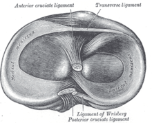
The medial meniscus is a fibrocartilage semicircular band that spans the knee joint medially, located between the medial condyle of the femur and the medial condyle of the tibia. It is also referred to as the internal semilunar fibrocartilage. The medial meniscus has more of a crescent shape while the lateral meniscus is more circular. The anterior aspects of both menisci are connected by the transverse ligament. It is a common site of injury, especially if the knee is twisted.

<span class="mw-page-title-main">Lateral meniscus</span>

The lateral meniscus is a fibrocartilaginous band that spans the lateral side of the interior of the knee joint. It is one of two menisci of the knee, the other being the medial meniscus. It is nearly circular and covers a larger portion of the articular surface than the medial. It can occasionally be injured or torn by twisting the knee or applying direct force, as seen in contact sports.

The knee examination, in medicine and physiotherapy, is performed as part of a physical examination, or when a patient presents with knee pain or a history that suggests a pathology of the knee joint.

<span class="mw-page-title-main">Anterior cruciate ligament injury</span> Ligament injury near the knee

An anterior cruciate ligament injury occurs when the anterior cruciate ligament (ACL) is either stretched, partially torn, or completely torn. The most common injury is a complete tear. Symptoms include pain, an audible cracking sound during injury, instability of the knee, and joint swelling. Swelling generally appears within a couple of hours. In approximately 50% of cases, other structures of the knee such as surrounding ligaments, cartilage, or meniscus are damaged.

<span class="mw-page-title-main">Patellar tendon</span> Tendon in the human knee

The patellar tendon is the distal portion of the common tendon of the quadriceps femoris, which is continued from the patella to the tibial tuberosity. It is also sometimes called the patellar ligament as it forms a bone to bone connection when the patella is fully ossified.

<span class="mw-page-title-main">Meniscus tear</span> Rupturing of the fibrocartilage strips in the knee called menisci

A tear of a meniscus is a rupturing of one or more of the fibrocartilage strips in the knee called menisci. When doctors and patients refer to "torn cartilage" in the knee, they actually may be referring to an injury to a meniscus at the top of one of the tibiae. Menisci can be torn during innocuous activities such as walking or squatting. They can also be torn by traumatic force encountered in sports or other forms of physical exertion. The traumatic action is most often a twisting movement at the knee while the leg is bent. In older adults, the meniscus can be damaged following prolonged 'wear and tear'. Especially acute injuries can lead to displaced tears which can cause mechanical symptoms such as clicking, catching, or locking during motion of the joint. The joint will be in pain when in use, but when there is no load, the pain goes away.

<span class="mw-page-title-main">Transverse ligament of knee</span>

The transverse or [anterior] meniscomeniscal ligament is a ligament in the knee joint that connects the anterior convex margin of the lateral meniscus to the anterior end of the medial meniscus.

The pivot-shift test is one of the three major tests for assessing anterior cruciate injury or laxity, the other two being the anterior drawer and Lachman test. However, unlike the other two, it tests for instability, an important determinant as to how the knee will function. In fact, it is instability, not simply the injury to the anterior cruciate ligament itself, that places the menisci at future risk, and gives rise to the feeling that the "knee is not secure" or "may give out".

<span class="mw-page-title-main">Discoid meniscus</span>

Discoid meniscus is a rare human anatomic variant that usually affects the lateral meniscus of the knee. Usually a person with this anomaly has no complaints; however, it may present as pain, swelling, or a snapping sound heard from the affected knee. Strong suggestive findings on magnetic resonance imaging includes a thickened meniscal body seen on more than two contiguous sagittal slices.

<span class="mw-page-title-main">Knee pain</span> Medical condition

Knee pain is pain in or around the knee.

Posterolateral corner injuries of the knee are injuries to a complex area formed by the interaction of multiple structures. Injuries to the posterolateral corner can be debilitating to the person and require recognition and treatment to avoid long term consequences. Injuries to the PLC often occur in combination with other ligamentous injuries to the knee; most commonly the anterior cruciate ligament (ACL) and posterior cruciate ligament (PCL). As with any injury, an understanding of the anatomy and functional interactions of the posterolateral corner is important to diagnosing and treating the injury.

<span class="mw-page-title-main">Medial knee injuries</span> Medical condition

Medial knee injuries are the most common type of knee injury. The medial ligament complex of the knee consists of:

References

  1. 1 2 3 Shelbourne K, Nitz P (1991). "The O'Donoghue triad revisited: Combined knee injuries involving anterior cruciate and medial collateral ligament tears". Am J Sports Med. 19 (5): 474–7. doi:10.1177/036354659101900509. PMID   1962712. S2CID   45964892.
  2. Winterstein, Andrew P. (2009). Athletic training student primer : a foundation for success (2nd ed.). Thorofare, NJ: SLACK. pp. 109–110. ISBN   978-1-55642-804-3.
  3. 1 2 3 4 Barber, FA (1992). "What is the terrible triad?". Arthroscopy. 8 (1): 19–22. doi:10.1016/0749-8063(92)90130-4. PMID   1550645.
  4. Balmer K, Gunst M, Rüedi T (January 1983). "[The "unhappy triad" (UT): slogan or reality in fresh knee injuries?]". Helv Chim Acta (in German). 49 (5): 675–7. PMID   6687587.
  5. Staron RB, Haramati N, Feldman F, et al. (November 1994). "O'Donoghue's triad: magnetic resonance imaging evidence". Skeletal Radiol. 23 (8): 633–6. doi:10.1007/BF02580384. PMID   7886473. S2CID   19514336.
  6. Nedeff DD, Bach BR (2001). "Arthroscopic anterior cruciate ligament reconstruction using patellar tendon autografts: a comprehensive review of contemporary literature". Am J Knee Surg. 14 (4): 243–58. PMID   11703038.
  7. Alford, J. Winslow; Bernard R. Bach Jr (July 2001). "Managing ACL tears: evaluation and diagnosis". The Journal of Musculoskeletal Medicine: 381–390.
  8. Imaging of Orthopedic Sports Injuries (Medical Radiology / Diagnostic Imaging). Berlin: Springer. 2006. ISBN   3-540-26014-5.
  9. Johnson, Glen O.; Starkey, Chad (2005). Athletic training and sports medicine. Boston: Jones and Bartlett Publishers. ISBN   0-7637-0536-5.
  10. Bellabarba C, Bush-Joseph CA, Bach BR (January 1997). "Patterns of meniscal injury in the anterior cruciate-deficient knee: a review of the literature". Am. J. Orthop. 26 (1): 18–23. PMID   9021030.
  11. "A Complex Injury of the Knee - The Unhappy Triad". Orthopaedic and Spine Center of Newport News. 2020-09-01. Retrieved 2021-07-20.
  12. Andrade, Renato; Pereira, Rogério; Van Cingel, Robert; Staal, J Bart; Espregueira-Mendes, João (May 2020). "How should clinicians rehabilitate patients after ACL reconstruction? A systematic review of clinical practice guidelines (CPGs) with a focus on quality appraisal (AGREE II)". British Journal of Sports Medicine. 54 (9): 512–519. doi: 10.1136/bjsports-2018-100310 . PMID   31175108.
  13. 1 2 Fridén T, Erlandsson T, Zätterström R, Lindstrand A, Moritz U (1995). "Compression or distraction of the anterior cruciate injured knee. Variations in injury pattern in contact sports and downhill skiing". Knee Surg Sports Traumatol Arthrosc. 3 (3): 144–7. doi:10.1007/bf01565473. PMID   8821269. S2CID   25692188.
  14. O'Donoghue D (1950). "Surgical treatment of fresh injuries to the major ligaments of the knee". J Bone Joint Surg Am. 32 (A:4): 721–38. doi:10.2106/00004623-195032040-00001. PMID   14784482.
  15. O'Donoghue D (1964). "The unhappy triad: Etiology, diagnosis and treatment". Am J Orthop. 6: 242–7 PASSIM. PMID   14237439.
  16. Pugh D, McKee M (2002). "The "terrible triad" of the elbow". Tech Hand up Extrem Surg. 6 (1): 21–9. doi: 10.1097/00130911-200203000-00005 . PMID   16520629.
  17. Simonich S, Wright T (2003). "Terrible triad of the shoulder". J Shoulder Elbow Surg. 12 (6): 566–8. doi:10.1016/S1058-2746(03)00216-7. PMID   14671520.