Knee examination

Last updated

The knee examination, in medicine and physiotherapy, is performed as part of a physical examination, or when a patient presents with knee pain or a history that suggests a pathology of the knee joint.

Contents

The exam includes several parts:

The latter three steps are often remembered with the saying look, feel, move.

History taking

Before performing a physical examination of the knee joint, a comprehensive history should be asked. A thorough history can be helpful in locating the possible pathological site during the physical examination. The mechanism of injury, location, character of the knee pain, the presence of a "pop" sound at the time of the injury (indicates ligamentous tear or fracture), swelling, infections, ability to stand or walk, sensation of instability (suggestive of subluxation), or any previous traumatic injuries to the joint are all important historical features. The most common knee problems are: soft tissue inflammation, injury, or osteoarthritis. The mechanism of the knee injury can give a clue of the possible structures that can be injured. For example, applying valgus stress on the knee can cause medial collateral ligament rupture, meanwhile a varus force can cause lateral collateral ligament rupture. When a person suddenly slows down during running, twisting, or pivoting with valgus force applying on the knee, the anterior cruciate ligament can rupture. Posterior dislocation of the tibia can cause posterior cruciate ligament injury. Twisting and pivoting while bearing weight can cause tearing of the meniscus. Fractures of the knee are less common but should be considered if direct trauma to the knee has occurred such as during a fall. Examples of fractures involving knee joints are: tibial plateau fractures, fractures of the lateral condyle of femur, medial condyle of femur, and patellar fractures. [1]

For non-traumatic causes of knee pain, history such as fever, morning stiffness, pain after exercise, infections, history of gout or psoriasis, and previous activities that contributes to long-term overuse of the knee joint should be asked. Knee pain due to long-term overuse are reproducible. For example, repetitive jumping can cause inflammation of patellar tendon. Repetitive kneeling can cause prepatellar inflammation of synovial bursa. [1]

General examination

Physical examination of the knee begins by observing the person's gait to assess for any abnormalities seen while walking. Gait assessment can be used to differentiate genuine knee pain or pain which referred from hip, lower back or the foot. A person can be asked to perform a duckwalk. This requires the person to squat and walk in that position. In order to perform a duckwalk, the person has to be free of ligamentous tear, knee effusions, and meniscal tears. The person can also be asked to stand with both feet stuck together. This position is useful to observe for valgus or varus deformity of the knees which is suggestive of osteoarthritis. The circumference of each thigh can be measured to look for wasting of quadriceps muscles. Skin around the knee can also be observed for psoriasis, hematoma, rash, abrasions, lacerations, or cellulitis which could be important causes of the knee pathology. [1]

Palpation

Palpation of the knee should begin from the unaffected side first. This will reassure the patient and is useful for comparison with the affected knee. The back of the hand can be used to assess the warmth of the knee. The knee is then flexed 90 degrees and the anterior structures are assessed. Inflammation of the patellar tendon is present if the patellar tendon is painful upon palpation. Radiographic imaging should be done if the examination findings fulfills the Ottawa rules: age 55 years and older, pain at the head of fibula, patellar pain, unable to flex the knee to 90 degrees, and inability to stand and walk at least four steps. If anterior cruciate ligament injury is suspected, radiographic imaging should also be ordered because it is frequently associated with lateral tibial plateau fracture. If there is a painful, reddish, and warm swelling in front of the patella, acute prepatellar bursitis should be considered which may require aspiration or drainage. Those presented with these features usually had history of frequent kneeling and direct trauma over the knee. [1]

Pain, swelling, and a defect of the insertion of the quadriceps tendon into the superior part of the patella is suggestive of quadriceps tendon rupture. A "pop" sound may be associated with this injury, followed by the loss of the ability to straighten the knee (knee extension). Pain at the medial joint line (medial to the inferior border of the patella) indicates medial compartment osteoarthritis, injury to the medial collateral ligament, or a medial meniscal tear. Pain at the midpoint between the anterior part of the medial joint line and tibial tuberosity is suggestive of Pes anserine bursitis (inflammation of anserine bursa. Lateral joint line tenderness is associated with lateral compartment osteoarthritis, lateral collateral ligament injury, and lateral meniscal tear. Pain at the lateral femoral condyle is suggestive of iliotibial band syndrome. Swelling at the popliteal fossa may reveal a Baker's cyst. [1]

Motion

Assessment of effusion

The absence of normal grooves around patella may indicates a patellar intra-articular effusion. There are two ways to confirm the effusion. The knee is extended fully before the examination begins. This first way is the patellar tap. It is to squeeze the fluid between the patella and the femur by pressing at the medial patella using a non-dominant hand. Then, using the dominant hand to press on the patella vertically. If the patella is ballotable, then patellar intra-articular effusion is present. Another way is the milking of the patella. First, the effusion is milked at the medial border of the patella from inferior to superior aspect. Then, using another hand, the effusion is milked at the lateral border of the patella from superior to inferior aspect. If effusion is present, a bulge will be appear at the medial border of the patella because the effusion is milked back to the medial patella. [1]

Assessment of range of motion

Both the active and passive range of motion should be assessed. The normal knee extension is between 0 and 10 degrees. The normal knee flexion is between 130 and 150 degrees. Any pain, abnormal movement, or crepitus of the patella should be noted. If there is pain or crepitus during active extension of the knee, while the patella is being compressed against the patellofemoral groove, patellofemoral pain syndrome or chondromalacia patellae should be suspected. Pain with active range of motion but no pain during passive range of motion is suggestive of inflammation of the tendon. Pain during active and passive range of motion is suggestive of pathology in the knee joint. [1]

Assessment of collateral ligaments

Valgus stress test can be performed with the examined knee in 25 degrees flexion to determine the integrity of the medial collateral ligament. Similarly, varus stress test can be performed to access the integrity of the lateral collateral ligament. The degree of collateral ligament sprain can also be assessed during the valgus and varus tests. In a first degree tear, the ligament has less than 5 mm laxity with a definite resistance when the knee is pulled. In a second degree sprain, there is laxity when the knee is tested at 25 degrees of flexion, but no laxity at extension with a definite resistance when the knee is pulled. In a third degree tear, there will be 10 mm laxity with no definite resistance either with knee with full extension or flexion. [1]

Assessment of anterior cruciate ligament

The anterior drawer and Lachman tests can be used to access the integrity of the anterior cruciate ligament. In the anterior drawer test, the person being examined should lie down on their back (supine position) with the knee in 90 degrees flexion. The foot is secured on the bed with the examiner sitting on the foot. The tibia is then pulled forward by using both hands. If the anterior movement of the affected knee is greater than the unaffected knee, then the anterior drawer test is positive. The Lachman test is more sensitive than the anterior drawer test. For the Lachman test, the person lies down in supine position with the knee flexed at 20 degrees and the heel touching the bed. The tibia is then pulled forward. If there is 6 to 8 millimeters of laxity, with no definitive resistance when the knee is pulled, then the test is positive thus raising concern for a torn anterior cruciate ligament. A large collection of blood in the knee can be associated with bony fractures and cruciate ligament tear. [1]

Assessment of posterior cruciate ligament

Posterior drawer test and tibial sag tests can determine the integrity of the posterior cruciate ligament. Similar to anterior drawer test, the knee should be flexed 90 degrees and the tibia is pushed backwards. If the tibia can be pushed posteriorly, then the posterior drawer test is positive. In tibial sag test, both knees are flexed at 90 degrees with the person in supine position and bilateral feet touching the bed. Bilateral knees are then watch for posterior displacement of tibia. If the affected tibia slowly displaced posteriorly, the posterior cruciate ligament is affected. [1]

Assessment of meniscus

Those with meniscal injuries may report symptoms such as clicking, catching, or locking of knees. Apart from joint line tenderness, there are three other methods of accessing meniscus tear: the McMurray test, the Thessaly test, and the Apley grind test. In McMurray test, the person should lie down in supine position with the knee should in 90 degrees flexion. the examiner put one hand with the thumb and the index finger on the medial and lateral joint lines respectively. Another hand is used to control the heel. To test the medial meniscus, the hand at the heel applies a valgus force and external rotates the leg while extending the knee. To test for the lateral meniscus, the varus force, internal rotation are applied to the leg while extending the knee. Any clicking, popping, or catching at the respective joint line indicates the corresponding meniscal tear. [1]

In Apley compression test, the person lie down in prone position with the knee flexed at 90 degrees. One hand is used to stabilise the hip and another hand grasp the foot and apply a downward compression force while external and internal rotates the leg. Pain during compression indicates meniscal tear. Examination for anterior cruciate ligament tear should be done for those with meniscal tear because these two conditions often occurs together. [1]

Additional Tests

See also

Related Research Articles
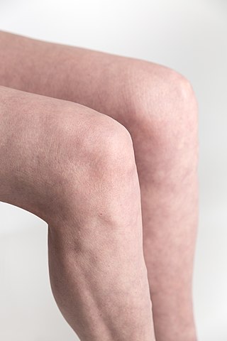
Knee Region around the kneecap

In humans and other primates, the knee joins the thigh with the leg and consists of two joints: one between the femur and tibia, and one between the femur and patella. It is the largest joint in the human body. The knee is a modified hinge joint, which permits flexion and extension as well as slight internal and external rotation. The knee is vulnerable to injury and to the development of osteoarthritis.

Tibia Long bone of the lower leg

The tibia, also known as the shinbone or shankbone, is the larger, stronger, and anterior (frontal) of the two bones in the leg below the knee in vertebrates, and it connects the knee with the ankle bones. The tibia is found on the medial side of the leg next to the fibula and closer to the median plane or centre-line. The tibia is connected to the fibula by the interosseous membrane of leg, forming a type of fibrous joint called a syndesmosis with very little movement. The tibia is named for the flute tibia. It is the second largest bone in the human body next to the femur. The leg bones are the strongest long bones as they support the rest of the body.

Posterior cruciate ligament One of four major ligaments of the knee

The posterior cruciate ligament (PCL) is a ligament in each knee of humans and various other animals. It works as a counterpart to the anterior cruciate ligament (ACL). It connects the posterior intercondylar area of the tibia to the medial condyle of the femur. This configuration allows the PCL to resist forces pushing the tibia posteriorly relative to the femur.

Anterior cruciate ligament Type of cruciate ligament in the human knee

The anterior cruciate ligament (ACL) is one of a pair of cruciate ligaments in the human knee. The two ligaments are also called "cruciform" ligaments, as they are arranged in a crossed formation. In the quadruped stifle joint, based on its anatomical position, it is also referred to as the cranial cruciate ligament. The term cruciate translates to cross. This name is fitting because the ACL crosses the posterior cruciate ligament to form an “X”. It is composed of strong, fibrous material and assists in controlling excessive motion. This is done by limiting mobility of the joint. The anterior cruciate ligament is one of the four main ligaments of the knee, providing 85% of the restraining force to anterior tibial displacement at 30 and 90° of knee flexion. The ACL is the most injured ligament of the four located in the knee.

Segond fracture Avulsion fracture of the lateral tibial condyle of the knee

The Segond fracture is a type of avulsion fracture from the lateral tibial plateau of the knee, immediately below the articular surface of the tibia.

The drawer test is used in the initial clinical assessment of suspected rupture of the cruciate ligaments in the knee. The patient should be supine with the hips flexed to 45 degrees, the knees flexed to 90 degrees and the feet flat on table. The examiner positions himself by sitting on the examination table in front of the involved knee and grasping the tibia just below the joint line of the knee. The thumbs are placed along the joint line on either side of the patellar tendon. The tibia is then drawn forward anteriorly. An increased amount of anterior tibial translation compared with the opposite limb or lack of a firm end-point may indicate either a sprain of the anteromedial bundle or complete tear of the ACL. If the tibia pulls forward or backward more than normal, the test is considered positive. Excessive displacement of the tibia anteriorly suggests that the anterior cruciate ligament is injured, whereas excessive posterior displacement of the tibia may indicate injury of the posterior cruciate ligament.

Medial collateral ligament Ligament on the inner side of the knee joint

The medial collateral ligament (MCL), or tibial collateral ligament (TCL), is one of the four major ligaments of the knee. It is on the medial (inner) side of the knee joint in humans and other primates. Its primary function is to resist outward turning forces on the knee.

Medial meniscus

The medial meniscus is a fibrocartilage semicircular band that spans the knee joint medially, located between the medial condyle of the femur and the medial condyle of the tibia. It is also referred to as the internal semilunar fibrocartilage. The medial meniscus has more of a crescent shape while the lateral meniscus is more circular. The anterior aspects of both menisci are connected by the transverse ligament. It is a common site of injury, especially if the knee is twisted.

Lateral meniscus

The lateral meniscus is a fibrocartilaginous band that spans the lateral side of the interior of the knee joint. It is one of two menisci of the knee, the other being the medial meniscus. It is nearly circular and covers a larger portion of the articular surface than the medial. It can occasionally be injured or torn by twisting the knee or applying direct force, as seen in contact sports.
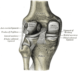
Lower extremity of femur

The lower extremity of femur is the lower end of the femur in human and other animals, closer to the knee. It is larger than the upper extremity of femur, is somewhat cuboid in form, but its transverse diameter is greater than its antero-posterior; it consists of two oblong eminences known as the lateral condyle and medial condyle.

Unhappy triad Medical condition of the knee

The unhappy triad, also known as a blown knee among other names, is an injury to the anterior cruciate ligament, medial collateral ligament, and meniscus. Analysis during the 1990s indicated that this 'classic' O'Donoghue triad is actually an unusual clinical entity among athletes with knee injuries. Some authors mistakenly believe that in this type of injury, "combined anterior cruciate and medial collateral ligament disruptions that were incurred during athletic endeavors" always present with concomitant medial meniscus injury. However, the 1990 analysis showed that lateral meniscus tears are more common than medial meniscus tears in conjunction with sprains of the ACL.

Articular capsule of the knee joint

The articular capsule of the knee joint is the wide and lax joint capsule of the knee. It is thin in front and at the side, and contains the patella, ligaments, menisci, and bursae of the knee. The capsule consists of an inner synovial membrane, and an outer fibrous membrane separated by fatty deposits anteriorly and posteriorly.

Meniscus tear Rupturing of the fibrocartilage strips in the knee called menisci

A tear of a meniscus is a rupturing of one or more of the fibrocartilage strips in the knee called menisci. When doctors and patients refer to "torn cartilage" in the knee, they actually may be referring to an injury to a meniscus at the top of one of the tibiae. Menisci can be torn during innocuous activities such as walking or squatting. They can also be torn by traumatic force encountered in sports or other forms of physical exertion. The traumatic action is most often a twisting movement at the knee while the leg is bent. In older adults, the meniscus can be damaged following prolonged 'wear and tear'. Especially acute injuries can lead to displaced tears which can cause mechanical symptoms such as clicking, catching, or locking during motion of the joint. The joint will be in pain when in use, but when there is no load, the pain goes away.

The pivot-shift test is one of the three major tests for assessing anterior cruciate injury or laxity, the other two being the anterior drawer and Lachman test. However, unlike the other two, it tests for instability, an important determinant as to how the knee will function. In fact, it is instability, not simply the injury to the anterior cruciate ligament itself, that places the menisci at future risk, and gives rise to the feeling that the "knee is not secure" or "may give out".

Patellar dislocation Medical condition

A patellar dislocation is a knee injury in which the patella (kneecap) slips out of its normal position. Often the knee is partly bent, painful and swollen. The patella is also often felt and seen out of place. Complications may include a patella fracture or arthritis.

Knee pain Medical condition

Knee pain is pain in or around the knee.

Posterolateral corner injuries of the knee are injuries to a complex area formed by the interaction of multiple structures. Injuries to the posterolateral corner can be debilitating to the person and require recognition and treatment to avoid long term consequences. Injuries to the PLC often occur in combination with other ligamentous injuries to the knee; most commonly the anterior cruciate ligament (ACL) and posterior cruciate ligament (PCL). As with any injury, an understanding of the anatomy and functional interactions of the posterolateral corner is important to diagnosing and treating the injury.

Tibial plateau fracture Medical condition

A tibial plateau fracture is a break of the upper part of the tibia (shinbone) that involves the knee joint. Symptoms include pain, swelling, and a decreased ability to move the knee. People are generally unable to walk. Complication may include injury to the artery or nerve, arthritis, and compartment syndrome.

Medial knee injuries Medical condition

Medial knee injuries are the most common type of knee injury. The medial ligament complex of the knee is composed of the superficial medial collateral ligament (sMCL), deep medial collateral ligament (dMCL), and the posterior oblique ligament (POL). These ligaments have also been called the medial collateral ligament (MCL), tibial collateral ligament, mid-third capsular ligament, and oblique fibers of the sMCL, respectively. This complex is the major stabilizer of the medial knee. Injuries to the medial side of the knee are most commonly isolated to these ligaments. A thorough understanding of the anatomy and function of the medial knee structures, along with a detailed history and physical exam, are imperative to diagnosing and treating these injuries.

Posterior cruciate ligament injury Medical condition

The function of the posterior cruciate ligament (PCL) is to prevent the femur from sliding off the anterior edge of the tibia and to prevent the tibia from displacing posterior to the femur. Common causes of PCL injuries are direct blows to the flexed knee, such as the knee hitting the dashboard in a car accident or falling hard on the knee, both instances displacing the tibia posterior to the femur.

References

  1. 1 2 3 4 5 6 7 8 9 10 11 12 Teresa L., Schraeder; Richard M., Terek; C. Christopher, Smith (22 July 2010). "Clinical Evaluation of the Knee". The New England Journal of Medicine. 363 (4): 5. doi:10.1056/NEJMvcm0803821.
  2. Jacobs, Brian (21 March 2011). "eMedicine – Knee Osteochondritis Dissecans". Mediscape. Retrieved 2 April 2011.
  3. Wittke R (November 2004). "Acute and chronic injuries to the knee in the doctor's office". MMW Fortschr Med (in German). 146 (45): 46–49. PMID   15581106.