Iliotibial band syndrome

Last updated
Iliotibial band syndrome
Other namesIliotibial band friction syndrome (ITBFS) [1]
Iliotibial band syndrome-en.svg
Specialty Sports medicine, orthopedics

Iliotibial band syndrome (ITBS) is the second most common knee injury, and is caused by inflammation located on the lateral aspect of the knee due to friction between the iliotibial band and the lateral epicondyle of the femur. [2] Pain is felt most commonly on the lateral aspect of the knee and is most intensive at 30 degrees of knee flexion. [2] Risk factors in women include increased hip adduction and knee internal rotation. [2] [3] Risk factors seen in men are increased hip internal rotation and knee adduction. [2] ITB syndrome is most associated with long-distance running, cycling, weight-lifting, and with military training. [4] [5]

Contents

Signs and symptoms

ITBS symptoms range from a stinging sensation just above the knee and outside of the knee (lateral side of the knee) joint, to swelling or thickening of the tissue in the area where the band moves over the femur. The stinging sensation just above the knee joint is felt on the outside of the knee or along the entire length of the iliotibial band. At initial symptom onset pain typically occurs following activity, but as the condition progresses pain is frequently felt during activities and may be present at rest. [6] Pain may also be present above and below the knee, where the ITB attaches to the tibia. [7] Pain is frequently worsened by running up or downhill or by stride lengthening. [8]

Risk Factors

ITBS is associated with various risk factors including training habits, anatomical abnormalities, or muscular imbalances: [9]

Anatomical mechanism

Iliotibial band syndrome is one of the leading causes of lateral knee pain in runners. The iliotibial band is a thick band of fascia composing the tendon of the tensor fascia latae muscle. It is located on the lateral aspect of the knee, extending from the outside of the pelvis, over the hip and knee, and inserting just below the knee. The band serves to stabilizing the knee. It has been proposed that during activity such as running and cycling the iliotibial band slides back and forth over the lateral femoral epicondyle, which causes friction and inflammation of the band. [6] It has also been suggested that symptoms are caused by impingement of the iliotibial band in the knee during 30 degree flexion, which is a position common in running and cycling. [11] Additional proposed mechanisms causing the symptoms of ITBS include compression of the fat and soft tissues beneath the iliotibial band, and chronic iliotibial band bursitis. [6]

Diagnosis

Diagnosis of iliotibial band syndrome is primarily based on history and physical exam findings, including tenderness at the lateral femoral epicondyle, where the iliotibial band passes over the bone. [12]

Differential Diagnosis

Other conditions that may present with knee pain similar to ITBS that must be differentiated include a lateral meniscus tear, degerative joint disease, tendinopathy of the biceps femoris, stress fracture, patellofemoral pain syndrome, and injury to the lateral collateral ligament. [6]

Exam Maneuvers

There are several physical exam maneuvers used to test iliotibial band function and provoke symptoms diagnostic of ITBS. The Noble test is used to assess for iliotibial band disfuction, in which the examiner extends the patient's knee from the 90 degree position with pain over the lateral femoral epicondyle occurring at 30 degrees of flexion. Additional tests include the Ober test to detect iliotibial band contracture, where the patient lies on their side and the examiner attempts to abduct, extend, and then adduct the leg. A positive test occurs with inability to adduct the leg due to iliotibial band shortening. The Thomas test is used to detect excessive tightness of the iliotibial band. In this test the patient holds the unaffected leg to their chest while the examiner straightens and lowers the other leg to a horizonal position, inability to fully straighten and lower the leg indicates excessive band tightness. [13] [14] [15]

The iliotibial band may be assessed by MRI in severe cases. Wrap around artifact MRI right knee on proton density fat saturation sequence.jpg
The iliotibial band may be assessed by MRI in severe cases.

Imaging

Imaging studies are generally not needed for diagnosis of ITBS, as characteristic symptoms and physical exam findings are sufficient for diagnosis. However, in severe or persistent cases MRI may be used to confirm the diagnosis as well as rule out other causes of lateral knee pain. Ultrasonography may also be used to evaluate disease progression by measuring iliotibial band thickness. [6]

Treatment

Conservative treatments

While ITBS pain can be acute, the iliotibial band can be treated conservatively with rest, ice, compression and elevation (RICE) to reduce pain and inflammation, followed by stretching. [16] Utilization of corticosteroid injections and the use of oral nonsteroidal anti-inflammatory drugs (NSAIDs) or topical NSAIDs on the painful area are possible treatments for ITB syndrome. Corticosteroid injections have been shown to decrease running pains significantly 7 days after the initial treatment. [17] Similar results can be found with the use of anti-inflammatory medication, analgesic/anti-inflammatory medication, specifically. [17] Physical therapy is an effective treatment modality, with the goal of stretching the iliotibial band, tensor fascia latae, and gluteus medius. [6] Other non-invasive treatments include modalities such as flexibility and strength training, neuromuscular/gait training, manual therapy, training volume reduction, myofascial release, or changes in running shoe. [2] [17] [3] [18]   Muscular training of the gluteus maximus and hip external rotators is stressed highly as those muscles are associated with many of the risk factors of ITBS. [2] For runners specifically, neuromuscular/gait training may be needed for success in muscular training interventions to ensure that those trained muscles are used properly in the mechanics of running. [2] Strength training alone will not result in decrease in pain due to ITBS, however, gait training, on its own can result in running form modification that reduces the prevalence of risk factors. [3]

Surgical treatments

Surgery treatments are utilized if several conservative approaches fail to produce results. [17] 6 months of conservative treatments are generally used before surgical intervention. [3] Surgery typically involves removal of a small piece of the iliotibial band to release excessive tension. Other procedures that have been utilized include resection of the iliotibial band bursa and z-lengthening. In the z-lengthening procedure, two horizontal incisions are made in the band and connected by a vertical incision, forming a z. The resulting sections are reattached together in a lengthened position, increasing the length of the band. [6]

Prognosis

ITBS symptoms typically improve with treatment. An estimated 50-90% of patients have symptom resolution with 4-8 weeks of conservative treatment, while surgical patients also generally have good outcomes. [19] Complications of ITBS include recurrence and exacerbation by return to activity following treatment, as well as possible progression to patellofemoral pain syndrome. [19]

Epidemiology

Occupation

ITBS commonly affects athletes and has been reported in runners, cyclists, rowers, skiers, and triathletes, as well as basketball, soccer, and field hockey players. [6]

Significant association between the diagnosis of ITBS and occupational background of the patients has been thoroughly determined. Occupations that require extensive use of iliotibial band are more susceptible to develop ITBS due to continuum of their iliotibial band repeatedly abrading against lateral epicondyle prominence, thereby inducing inflammatory response. Professional or amateur runners are at high clinical risk of ITBS in which shows particularly greater risk in long-distance. Study suggests ITBS alone makes up 12% of all running-related injuries and 1.6% to 12% of runners are affected by ITBS. [20]

The relationship between ITBS and mortality/morbidity is claimed to be absent. A study showed that coordination variability did not vary significantly between runners with no injury and runners with ITBS. [21] This result elucidates that the runner's ability to coordinate themselves toward direction of their intention (motor coordination) is not, or very minorly affected by the pain of ITBS. [22]

Additionally, military trainee in marine boot camps displayed high incidence rate of ITBS. Varying incidence rate of 5.3–22% in basic training was reported in a case study. A report from the U.S. Marine Corps announces that running/overuse-related injuries accounted for >12% of all injuries. [23]

Studies suggest that there is not a difference in ITBS incidence rate between patients of different race, gender, or age. However, there has been a claim that females are more prone to ITBS due to their anatomical differences in the pelvis and lower extremities. Males with a larger lateral epicondyle prominence may also be more susceptible to ITBS. [24]   Higher incidence rate of ITBS has been reported between the ages of 15 and 50, which generally includes most active athletes. [25]

Other professions that had noticeable association with ITBS include cyclists, heavy weightlifters, et cetera. One observational study discovered 24% of 254 cyclists were diagnosed with ITBS within 6 years. [26] A study provided data that shows more than half (50%) of professional cyclists complain of knee pain. [27] Additional studies have shown that ITBS makes up 15-24% of all overuse injuries in cyclists. [6]

History

ITBS was originally described by Lieutenant Commander James W. Renne in 1975 after observing frequent lateral knee pain in recruits participating in intensive military training. He initially named the syndrome iliotibial band friction syndrome. [28]

See also

Related Research Articles

<span class="mw-page-title-main">Knee</span> Leg joint in primates

In humans and other primates, the knee joins the thigh with the leg and consists of two joints: one between the femur and tibia, and one between the femur and patella. It is the largest joint in the human body. The knee is a modified hinge joint, which permits flexion and extension as well as slight internal and external rotation. The knee is vulnerable to injury and to the development of osteoarthritis.

<span class="mw-page-title-main">Tendinopathy</span> Inflammation of the tendon

Tendinopathy is a type of tendon disorder that results in pain, swelling, and impaired function. The pain is typically worse with movement. It most commonly occurs around the shoulder, elbow, wrist, hip, knee, or ankle.

<span class="mw-page-title-main">Sprain</span> Damage to one or more ligaments in a joint

A sprain is a soft tissue injury of the ligaments within a joint, often caused by a sudden movement abruptly forcing the joint to exceed its functional range of motion. Ligaments are tough, inelastic fibers made of collagen that connect two or more bones to form a joint and are important for joint stability and proprioception, which is the body's sense of limb position and movement. Sprains may be mild, moderate, or severe, with the latter two classes involving some degree of tearing of the ligament. Sprains can occur at any joint but most commonly occur in the ankle, knee, or wrist. An equivalent injury to a muscle or tendon is known as a strain.

<span class="mw-page-title-main">Tennis elbow</span> Condition in which the outer part of the elbow becomes sore and tender

Tennis elbow, also known as lateral epicondylitis or enthesopathy of the extensor carpi radialis origin, is an enthesopathy of the origin of the extensor carpi radialis brevis on the lateral epicondyle. The outer part of the elbow becomes painful and tender. The pain may also extend into the back of the forearm. Onset of symptoms is generally gradual, although they can seem sudden and be misinterpreted as an injury. Golfer's elbow is a similar condition that affects the inside of the elbow.

<span class="mw-page-title-main">Osgood–Schlatter disease</span> Inflammation of the patellar ligament

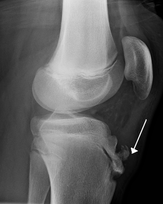
Osgood–Schlatter disease (OSD) is inflammation of the patellar ligament at the tibial tuberosity (apophysitis) usually affecting adolescents during growth spurts. It is characterized by a painful bump just below the knee that is worse with activity and better with rest. Episodes of pain typically last a few weeks to months. One or both knees may be affected and flares may recur.

<span class="mw-page-title-main">Chondromalacia patellae</span> Medical condition

Chondromalacia patellae is an inflammation of the underside of the patella and softening of the cartilage.

<span class="mw-page-title-main">Plantaris muscle</span> One of the superficial muscles of the superficial posterior compartment of the leg

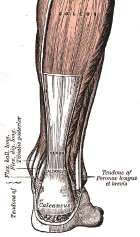
The plantaris is one of the superficial muscles of the superficial posterior compartment of the leg, one of the fascial compartments of the leg.

<span class="mw-page-title-main">Snapping hip syndrome</span> Medical condition

Snapping hip syndrome, also referred to as dancer's hip, is a medical condition characterized by a snapping sensation felt when the hip is flexed and extended. This may be accompanied by a snapping or popping noise and pain or discomfort. Pain often decreases with rest and diminished activity. Snapping hip syndrome is commonly classified by the location of the snapping as either extra-articular or intra-articular.

<span class="mw-page-title-main">Iliotibial tract</span> Longitudinal fibrous reinforcement of the fascia lata

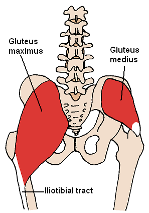
The iliotibial tract or iliotibial band is a longitudinal fibrous reinforcement of the fascia lata. The action of the muscles associated with the ITB flex, extend, abduct, and laterally and medially rotate the hip. The ITB contributes to lateral knee stabilization. During knee extension the ITB moves anterior to the lateral condyle of the femur, while ~30 degrees knee flexion, the ITB moves posterior to the lateral condyle. However, it has been suggested that this is only an illusion due to the changing tension in the anterior and posterior fibers during movement. It originates at the anterolateral iliac tubercle portion of the external lip of the iliac crest and inserts at the lateral condyle of the tibia at Gerdy's tubercle. The figure shows only the proximal part of the iliotibial tract.

The knee examination, in medicine and physiotherapy, is performed as part of a physical examination, or when a patient presents with knee pain or a history that suggests a pathology of the knee joint.

<span class="mw-page-title-main">Knee taping</span>

Knee taping is a procedure performed by physiotherapists or physicians to alleviate the symptoms of patellofemoral pain. Though knee taping has been shown to offer short-term pain relief, its long-term efficacy is confounded by several studies. The mechanism of action by which it alleviates pain is unknown, though it has been suggested by physicians that it could correct patella position, facilitate/inhibit quadriceps components or bear stress associated with peripatellar tissues or patellar compression. Evidence for these suggestions, however, has been contradictory or absent.

Greater trochanteric pain syndrome (GTPS), a form of bursitis, is inflammation of the trochanteric bursa, a part of the hip.

<span class="mw-page-title-main">Patellofemoral pain syndrome</span> Medical condition

Patellofemoral pain syndrome is knee pain as a result of problems between the kneecap and the femur. The pain is generally in the front of the knee and comes on gradually. Pain may worsen with sitting, excessive use, or climbing and descending stairs.

Patellar subluxation syndrome, is an injury that is concerned with the kneecap. Patellar subluxation is more common than patellar dislocation and is just as disabling.

<span class="mw-page-title-main">Patellar dislocation</span> Medical condition

A patellar dislocation is a knee injury in which the patella (kneecap) slips out of its normal position. Often the knee is partly bent, painful and swollen. The patella is also often felt and seen out of place. Complications may include a patella fracture or arthritis.

The medial patellofemoral ligament (MPFL) is one of several ligaments on the medial aspect of the knee. It originates in the superomedial aspect of the patella and inserts in the space between the adductor tubercle and the medial femoral epicondyle. The ligament itself extends from the femur to the superomedial patella, and its shape is similar to a trapezoid. It keeps the patella in place, but its main function is to prevent lateral displacement of the patella.

<span class="mw-page-title-main">Knee pain</span> Medical condition

Knee pain is pain in or around the knee.

<span class="mw-page-title-main">Medial knee injuries</span> Medical condition

Medial knee injuries are the most common type of knee injury. The medial ligament complex of the knee consists of:

Muscle strain is one of the most common injuries in tennis. When an isolated large-energy appears during the muscle contraction and at the same time, bodyweight applies huge amounts of pressure to the lengthened muscle, which can result in the occurrence of muscle strain. Inflammation and bleeding are triggered when muscle strain occur which resulted in redness, pain and swelling. Overuse is also common in tennis players from all levels. Muscle, cartilage, nerves, bursae, ligaments and tendons may be damaged from overuse. The repetitive use of a particular muscle without time for repair and recover in the most common case among the injury.

Running injuries affect about half of runners annually. The frequencies of various RRI depend on the type of running, such as speed and mileage. Some injuries are acute, caused by sudden overstress, such as side stitch, strains, and sprains. Many of the common injuries that affect runners are chronic, developing over longer periods as the result of overuse. Common overuse injuries include shin splints, stress fractures, Achilles tendinitis, Iliotibial band syndrome, Patellofemoral pain, and plantar fasciitis.

References

  1. Ellis, R; Hing, W; Reid, D (August 2007). "Iliotibial band friction syndrome—A systematic review". Manual Therapy. 12 (3): 200–8. doi:10.1016/j.math.2006.08.004. PMID   17208506.
  2. 1 2 3 4 5 6 7 Baker, Rober L.; Fredericson, Michael (2016). "ClinicalKey". www.clinicalkey.com. Retrieved 2019-11-17.
  3. 1 2 3 4 Neal, Bradley (2016). "Iliotibial Band Syndrome: A Narrative Review". Co-Kinetic Journal. 67: 16–20 via EBSCO host.
  4. "Iliotibial Band Syndrome: Background, Epidemiology, Functional Anatomy". 2019-11-10.{{cite journal}}: Cite journal requires |journal= (help)
  5. Hadeed, Andrew; Tapscott, David C. (2019), "Iliotibial Band Friction Syndrome", StatPearls, StatPearls Publishing, PMID   31194342 , retrieved 2019-11-17
  6. 1 2 3 4 5 6 7 8 9 Strauss, Eric J.; Kim, Suezie; Calcei, Jacob G.; Park, Daniel (December 2011). "Iliotibial Band Syndrome: Evaluation and Management:". American Academy of Orthopaedic Surgeon. 19 (12): 728–736. doi:10.5435/00124635-201112000-00003. ISSN   1067-151X.
  7. "Iliotibial band syndrome - aftercare: MedlinePlus Medical Encyclopedia". medlineplus.gov. Retrieved 2022-02-22.
  8. Khaund, Razib; Flynn, Sharon H. (2005-04-15). "Iliotibial band syndrome: a common source of knee pain". American Family Physician. 71 (8): 1545–1550. ISSN   0002-838X. PMID   15864895.
  9. "Iliotibial Band Syndrome Specialist Clinic | Singapore Sports and Orthopaedic Clinic - Neurosurgeon" . Retrieved 2022-06-24.
  10. Farrell, Kevin C.; Reisinger, Kim D.; Tillman, Mark D. (March 2003). "Force and repetition in cycling: possible implications for iliotibial band friction syndrome". The Knee. 10 (1): 103–109. doi:10.1016/S0968-0160(02)00090-X. PMID   12649036.
  11. Fairclough, John; Hayashi, Koji; Toumi, Hechmi; Lyons, Kathleen; Bydder, Graeme; Phillips, Nicola; Best, Thomas M; Benjamin, Mike (March 2006). "The functional anatomy of the iliotibial band during flexion and extension of the knee: implications for understanding iliotibial band syndrome". Journal of Anatomy. 208 (3): 309–316. doi:10.1111/j.1469-7580.2006.00531.x. ISSN   0021-8782. PMC   2100245 . PMID   16533314.
  12. Flynn, Sharon H.; Khaund, Razib (2005-04-15). "Iliotibial Band Syndrome: A Common Source of Knee Pain". American Family Physician. 71 (8): 1545–1550. PMID   15864895.
  13. Hariri, Sanaz; Savidge, Edgar T.; Reinold, Michael M.; Zachazewski, James; Gill, Thomas J. (July 2009). "Treatment of Recalcitrant Iliotibial Band Friction Syndrome with Open Iliotibial Band Bursectomy: Indications, Technique, and Clinical Outcomes". The American Journal of Sports Medicine. 37 (7): 1417–1424. doi:10.1177/0363546509332039. ISSN   0363-5465.
  14. Fredericson, Michael; Weir, Adam (May 2006). "Practical Management of Iliotibial Band Friction Syndrome in Runners". Clinical Journal of Sport Medicine. 16 (3): 261–268. doi:10.1097/00042752-200605000-00013. ISSN   1050-642X.
  15. West, R; Irrgang, J (2009). "Overuse injuries of the lower extremity". Orthopaedic Knowledge Update: Sports Medicine 4.
  16. Barber, F. Alan; Sutker, Allan N. (August 1992). "Iliotibial Band Syndrome". Sports Medicine. 14 (2): 144–148. doi:10.2165/00007256-199214020-00005. PMID   1509227.
  17. 1 2 3 4 Beals, Corey; Flanigan, David (2013). "A Review of Treatments for Iliotibial Band Syndrome in the Athletic Population". Journal of Sports Medicine. 2013: 367169. doi: 10.1155/2013/367169 . ISSN   2356-7651. PMC   4590904 . PMID   26464876.
  18. Weckström, Kristoffer; Söderström, Johan (2016). "Radial extracorporeal shockwave therapy compared with manual therapy in runners with iliotibial band syndrome". Journal of Back and Musculoskeletal Rehabilitation. 29 (1): 161–170. doi:10.3233/BMR-150612. PMID   26406193.
  19. 1 2 Hadeed, Andrew; Tapscott, David C. (2023), "Iliotibial Band Friction Syndrome", StatPearls, Treasure Island (FL): StatPearls Publishing, PMID   31194342 , retrieved 2023-11-06
  20. Richards, David P.; Alan Barber, F.; Troop, Randal L. (March 2003). "Iliotibial band Z-lengthening". Arthroscopy: The Journal of Arthroscopic & Related Surgery. 19 (3): 326–329. doi:10.1053/jars.2003.50081. ISSN   0749-8063. PMID   12627161.
  21. Hafer, Jocelyn F.; Brown, Allison M.; Boyer, Katherine A. (August 2017). "Exertion and pain do not alter coordination variability in runners with iliotibial band syndrome". Clinical Biomechanics. 47: 73–78. doi:10.1016/j.clinbiomech.2017.06.006. ISSN   0268-0033. PMID   28618309. S2CID   4007166.
  22. "Physical Medicine and Rehabilitation for Iliotibial Band Syndrome: Practice Essentials, Pathophysiology, Epidemiology". 2022-04-04.{{cite journal}}: Cite journal requires |journal= (help)
  23. Jensen, Andrew E; Laird, Melissa; Jameson, Jason T; Kelly, Karen R (2019-03-01). "Prevalence of Musculoskeletal Injuries Sustained During Marine Corps Recruit Training". Military Medicine. 184 (Supplement_1): 511–520. doi: 10.1093/milmed/usy387 . ISSN   0026-4075. PMID   30901397.
  24. Everhart, Joshua S.; Kirven, James C.; Higgins, John; Hair, Andrew; Chaudhari, Ajit A.M.W.; Flanigan, David C. (August 2019). "The relationship between lateral epicondyle morphology and iliotibial band friction syndrome: A matched case–control study". The Knee. 26 (6): 1198–1203. doi:10.1016/j.knee.2019.07.015. PMID   31439366. S2CID   201616794.
  25. "Physical Medicine and Rehabilitation for Iliotibial Band Syndrome: Practice Essentials, Pathophysiology, Epidemiology". 2022-04-04.{{cite journal}}: Cite journal requires |journal= (help)
  26. Farrell, Kevin C.; Reisinger, Kim D.; Tillman, Mark D. (March 2003). "Force and repetition in cycling: possible implications for iliotibial band friction syndrome". The Knee. 10 (1): 103–109. doi:10.1016/s0968-0160(02)00090-x. ISSN   0968-0160. PMID   12649036.
  27. Holmes, James C.; Pruitt, Andrew L.; Whalen, Nina J. (May 1993). "Iliotibial band syndrome in cyclists". The American Journal of Sports Medicine. 21 (3): 419–424. doi:10.1177/036354659302100316. ISSN   0363-5465. PMID   8166785. S2CID   21010647.
  28. Renne, James (December 1975). "The Iliotibial Band Friction Syndrome". The Journal of Bone & Joint Surgery.

Further reading

van der Worp, Maarten P.; van der Horst, Nick; de Wijer, Anton; Backx, Frank J. G.; Nijhuis-van der Sanden, Maria W. G. (23 December 2012). "Iliotibial Band Syndrome in Runners". Sports Medicine. 42 (11): 969–992. doi:10.1007/BF03262306. S2CID   73959693.