Greater trochanteric pain syndrome

Last updated
Greater trochanteric pain syndrome
Other namesTrochanteric bursitis
Treatment Amplified musculoskeletal pain syndrome

Greater trochanteric pain syndrome (GTPS), a form of bursitis, is inflammation of the trochanteric bursa, a part of the hip.

Contents

This bursa is at the top, outer side of the femur, between the insertion of the gluteus medius and gluteus minimus muscles into the greater trochanter of the femur and the femoral shaft. It has the function, in common with other bursae, of working as a shock absorber and as a lubricant for the movement of the muscles adjacent to it.[ citation needed ]

Occasionally, this bursa can become inflamed and clinically painful and tender. This condition can be a manifestation of an injury (often resulting from a twisting motion or from overuse), but sometimes arises for no obviously definable cause. The symptoms are pain in the hip region on walking, and tenderness over the upper part of the femur, which may result in the inability to lie in comfort on the affected side.[ citation needed ]

More often the lateral hip pain is caused by disease of the gluteal tendons that secondarily inflames the bursa. This is most common in middle-aged women and is associated with a chronic and debilitating pain which does not respond to conservative treatment. Other causes of trochanteric bursitis include uneven leg length, iliotibial band syndrome, and weakness of the hip abductor muscles. [1]

Greater trochanteric pain syndrome can remain incorrectly diagnosed for years, because it shares the same pattern of pain with many other musculoskeletal conditions. Thus people with this condition may be labeled malingerers, or may undergo many ineffective treatments due to misdiagnosis. [2] It may also coexist with low back pain, arthritis, and obesity. [3]

Signs and symptoms

The primary symptom is hip pain, especially hip pain on the outer (lateral) side of the joint. This pain may appear when the affected person is walking or lying down on that side.[ citation needed ]

Diagnosis

A doctor may begin the diagnosis by asking the patient to stand on one leg and then the other, while observing the effect on the position of the hips. Palpating the hip and leg may reveal the location of the pain, and range-of-motion tests can help to identify its source.[ citation needed ]

X-rays, ultrasound and magnetic resonance imaging may reveal tears or swelling. But often these imaging tests do not reveal any obvious abnormality in patients with documented[ clarification needed ] GTPS. [2] [ better source needed ]

Prevention

Because wear on the hip joint traces to the structures that support it (the posture of the legs, and ultimately, the feet), proper fitting shoes with adequate support are important to preventing GTPS. For someone who has flat feet, wearing proper orthotic inserts and replacing them as often as recommended are also important preventive measures.[ citation needed ]

Strength in the core and legs is also important to posture, so physical training also helps to prevent GTPS. But it is equally important to avoid exercises that damage the hip. [4]

Treatment

Conservative treatments have a 90% success rate and can include any or a combination of the following: pain relief medication, NSAIDs, physiotherapy, shockwave therapy (SWT) and corticosteroid injection. Surgery is usually for cases that are non-respondent to conservative treatments and is often a combination of bursectomy, iliotibial band (ITB) release, trochanteric reduction osteotomy or gluteal tendon repair. [5] A 2011 review found that traditional nonoperative treatment helped most patients, low-energy SWT was a good alternative, and surgery was effective in refractory cases and superior to corticosteroid therapy and physical therapy. [6] There are numerous case reports in which surgery has relieved GTPS, but its effectiveness is not documented in clinical trials as of 2009. [3]

The primary treatment is rest. This does not mean bed rest or immobilizing the area but avoiding actions which result in aggravation of the pain. Icing the joint may help. A non-steroidal anti-inflammatory drug may relieve pain and reduce the inflammation. If these are ineffective, the definitive treatment is steroid injection into the inflamed area.

Physical therapy to strengthen the hip muscles and stretch the iliotibial band can relieve tension in the hip and reduce friction. The use of point ultrasound may be helpful, and is undergoing clinical trials. [7]

In extreme cases, where the pain does not improve after physical therapy, cortisone shots, and anti-inflammatory medication, the inflamed bursa can be removed surgically. The procedure is known as a bursectomy. Tears in the muscles may also be repaired, and loose material from arthritic degeneration of the hip removed. [4] At the time of bursal surgery, a very close examination of the gluteal tendons will reveal sometimes subtle and sometimes very obvious degeneration and detachment of the gluteal tendons. If this detachment is not repaired, removal of the bursa alone will make little or no difference to the symptoms.[ citation needed ]

The bursa is not required, so the main potential complication is potential reaction to anaesthetic. The surgery can be performed arthroscopically and, consequently, on an outpatient basis. Patients often have to use crutches for a few days following surgery up to a few weeks for more involved procedures.[ citation needed ]

See also

Related Research Articles

Iliotibial band syndrome (ITBS) is the second most common knee injury, and is caused by inflammation located on the lateral aspect of the knee due to friction between the iliotibial band and the lateral epicondyle of the femur. Pain is felt most commonly on the lateral aspect of the knee and is most intensive at 30 degrees of knee flexion. Risk factors in women include increased hip adduction and knee internal rotation. Risk factors seen in men are increased hip internal rotation and knee adduction. ITB syndrome is most associated with long-distance running, cycling, weight-lifting, and with military training.

<span class="mw-page-title-main">Knee</span> Leg joint in primates

In humans and other primates, the knee joins the thigh with the leg and consists of two joints: one between the femur and tibia, and one between the femur and patella. It is the largest joint in the human body. The knee is a modified hinge joint, which permits flexion and extension as well as slight internal and external rotation. The knee is vulnerable to injury and to the development of osteoarthritis.

<span class="mw-page-title-main">Shoulder problem</span> Medical condition

Shoulder problems including pain, are one of the more common reasons for physician visits for musculoskeletal symptoms. The shoulder is the most movable joint in the body. However, it is an unstable joint because of the range of motion allowed. This instability increases the likelihood of joint injury, often leading to a degenerative process in which tissues break down and no longer function well.

<span class="mw-page-title-main">Gluteus maximus</span> Largest and most superficial of the three gluteal muscles

The gluteus maximus is the main extensor muscle of the hip in humans. It is the largest and outermost of the three gluteal muscles and makes up a large part of the shape and appearance of each side of the hips. It is the single largest muscle in the human body. Its thick fleshy mass, in a quadrilateral shape, forms the prominence of the buttocks. The other gluteal muscles are the medius and minimus, and sometimes informally these are collectively referred to as the glutes.

<span class="mw-page-title-main">Gluteus medius</span> One of the three gluteal muscles

The gluteus medius, one of the three gluteal muscles, is a broad, thick, radiating muscle. It is situated on the outer surface of the pelvis.

<span class="mw-page-title-main">Bursitis</span> Human disease

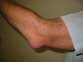
Bursitis is the inflammation of one or more bursae of synovial fluid in the body. They are lined with a synovial membrane that secretes a lubricating synovial fluid. There are more than 150 bursae in the human body. The bursae rest at the points where internal functionaries, such as muscles and tendons, slide across bone. Healthy bursae create a smooth, almost frictionless functional gliding surface making normal movement painless. When bursitis occurs, however, movement relying on the inflamed bursa becomes difficult and painful. Moreover, movement of tendons and muscles over the inflamed bursa aggravates its inflammation, perpetuating the problem. Muscle can also be stiffened.

<span class="mw-page-title-main">Greater trochanter</span> Quadrilateral part of the thigh bone

The greater trochanter of the femur is a large, irregular, quadrilateral eminence and a part of the skeletal system.

<span class="mw-page-title-main">Piriformis muscle</span> One of six small hip muscles in the lateral rotator group

The piriformis muscle is a flat, pyramidally-shaped muscle in the gluteal region of the lower limbs. It is one of the six muscles in the lateral rotator group.

<span class="mw-page-title-main">Piriformis syndrome</span> Medical condition

Piriformis syndrome is a condition which is believed to result from compression of the sciatic nerve by the piriformis muscle. The largest and most bulky nerve in the human body is the sciatic nerve. Starting at its origin it is 2 cm wide and 0.5 cm thick. The sciatic nerve forms the roots of L4-S3 segments of the lumbosacral plexus. The nerve will pass inferiorly to the piriformis muscle, in the direction of the lower limb where it divides into common tibial and fibular nerves. Symptoms may include pain and numbness in the buttocks and down the leg. Often symptoms are worsened with sitting or running.

<span class="mw-page-title-main">Synovial bursa</span> Fluid-filled sacs which cushion the bones and muscles around joints

A synovial bursa, usually simply bursa, is a small fluid-filled sac lined by synovial membrane with an inner capillary layer of viscous synovial fluid. It provides a cushion between bones and tendons and/or muscles around a joint. This helps to reduce friction between the bones and allows free movement. Bursae are found around most major joints of the body.

<span class="mw-page-title-main">Obturator internus muscle</span> One of six small hip muscles in the lateral rotator group

The internal obturator muscle or obturator internus muscle originates on the medial surface of the obturator membrane, the ischium near the membrane, and the rim of the pubis.

<span class="mw-page-title-main">Tensor fasciae latae muscle</span> Muscle of the thigh

The tensor fasciae latae is a muscle of the thigh. Together with the gluteus maximus, it acts on the iliotibial band and is continuous with the iliotibial tract, which attaches to the tibia. The muscle assists in keeping the balance of the pelvis while standing, walking, or running.

<span class="mw-page-title-main">Gluteal muscles</span> Group of three muscles which make up the buttocks

The gluteal muscles, often called glutes, are a group of three muscles which make up the gluteal region commonly known as the buttocks: the gluteus maximus, gluteus medius and gluteus minimus. The three muscles originate from the ilium and sacrum and insert on the femur. The functions of the muscles include extension, abduction, external rotation, and internal rotation of the hip joint.

Trendelenburg gait, named after Friedrich Trendelenburg, is an abnormal gait. It is caused by weakness or ineffective action of the gluteus medius muscle and the gluteus minimus muscle.

<span class="mw-page-title-main">Snapping hip syndrome</span> Medical condition

Snapping hip syndrome, also referred to as dancer's hip, is a medical condition characterized by a snapping sensation felt when the hip is flexed and extended. This may be accompanied by a snapping or popping noise and pain or discomfort. Pain often decreases with rest and diminished activity. Snapping hip syndrome is commonly classified by the location of the snapping as either extra-articular or intra-articular.

A bursectomy is the removal of a bursa, which is a small sac filled with synovial fluid that cushions adjacent bone structures and reduces friction in joint movement. This procedure is usually carried out to relieve chronic inflammation (bursitis) or infection, when conservative management has failed to improve patient outcomes.

<span class="mw-page-title-main">Subacromial bursitis</span> Medical condition

Subacromial bursitis is a condition caused by inflammation of the bursa that separates the superior surface of the supraspinatus tendon from the overlying coraco-acromial ligament, acromion, and coracoid and from the deep surface of the deltoid muscle. The subacromial bursa helps the motion of the supraspinatus tendon of the rotator cuff in activities such as overhead work.

<span class="mw-page-title-main">Extracorporeal shockwave therapy</span> Ultrasonic, non-invasive, outpatient treatment

Extracorporeal shockwave therapy (ESWT) is a non-invasive, out-patient alternative to surgery for those with many joint and tendon disorders. ESWT sends acoustic shock waves into bone or soft tissue, in effect reinjuring the area on a cellular level and breaking up the scarring that has penetrated tendons and ligaments. The controlled reinjuring of tissue allows the body to regenerate blood vessels and bone cells. The resulting revascularization leads to faster healing and often a return to pre-injury activity levels. ESWT is mostly used for kidney stones removal, in physical therapy and orthopedics.

<span class="mw-page-title-main">Pes anserine bursitis</span> Medical condition

Pes anserine bursitis is an inflammatory condition of the medial (inner) knee at the anserine bursa, a sub muscular bursa, just below the pes anserinus.

<span class="mw-page-title-main">Knee pain</span> Medical condition

Knee pain is pain in or around the knee.

References

  1. Trochanteric Bursitis at eMedicine
  2. 1 2 Dougherty C, Dougherty JJ (August 27, 2008). "Evaluating hip pathology in trochanteric pain syndrome". The Journal of Musculoskeletal Medicine.
  3. 1 2 Williams BS, Cohen SP (2009). "Greater Trochanteric Pain Syndrome: A Review of Anatomy, Diagnosis and Treatment". Anesthesia & Analgesia. 108 (5): 1662–1670. doi: 10.1213/ane.0b013e31819d6562 . PMID   19372352. S2CID   5521326.
  4. 1 2 Dougherty C, Dougherty JJ (November 1, 2008). "Managing and preventing hip pathology in trochanteric pain syndrome".{{cite journal}}: Cite journal requires |journal= (help)
  5. Reid, Diane (March 2016). "The management of greater trochanteric pain syndrome: A systematic literature review". Journal of Orthopaedics. 13 (1): 15–28. doi:10.1016/j.jor.2015.12.006. PMC   4761624 . PMID   26955229.
  6. Lustenberger, David P; Ng, Vincent Y; Best, Thomas M; Ellis, Thomas J (September 2011). "Efficacy of Treatment of Trochanteric Bursitis: A Systematic Review". Clinical Journal of Sport Medicine. 21 (5): 447–453. doi:10.1097/JSM.0b013e318221299c. PMC   3689218 . PMID   21814140.
  7. Clinical trial number NCT01642043 for "Point-of-Care Ultrasound in Greater Trochanteric Pain Syndrome" at ClinicalTrials.gov