Euchromatin

Last updated
Distinction between Euchromatin and Heterochromatin Sha-Boyer-Fig1-CCBy3.0.jpg
Distinction between Euchromatin and Heterochromatin

Euchromatin (also called "open chromatin") is a lightly packed form of chromatin (DNA, RNA, and protein) that is enriched in genes, and is often (but not always) under active transcription. Euchromatin stands in contrast to heterochromatin, which is tightly packed and less accessible for transcription. 92% of the human genome is euchromatic. [1]

Contents

In eukaryotes, euchromatin comprises the most active portion of the genome within the cell nucleus. In prokaryotes, euchromatin is the only form of chromatin present; this indicates that the heterochromatin structure evolved later along with the nucleus, possibly as a mechanism to handle increasing genome size.

Structure

Euchromatin is composed of repeating subunits known as nucleosomes, reminiscent of an unfolded set of beads on a string, that are approximately 11 nm in diameter. [2] At the core of these nucleosomes are a set of four histone protein pairs: H3, H4, H2A, and H2B. [2] Each core histone protein possesses a 'tail' structure, which can vary in several ways; it is thought that these variations act as "master control switches" through different methylation and acetylation states, which determine the overall arrangement of the chromatin. [2] Approximately 147 base pairs of DNA are wound around the histone octamers, or a little less than 2 turns of the helix. [3] Nucleosomes along the strand are linked together via the histone, H1, [4] and a short space of open linker DNA, ranging from around 0-80 base pairs. The key distinction between the structure of euchromatin and heterochromatin is that the nucleosomes in euchromatin are much more widely spaced, which allows for easier access of different protein complexes to the DNA strand and thus increased gene transcription. [2]

Appearance

Microscopy of heterochromatic versus euchromatic nuclei (H&E stain). Heterochromatic versus euchromatic nuclei.jpg
Microscopy of heterochromatic versus euchromatic nuclei (H&E stain).

Euchromatin resembles a set of beads on a string at large magnifications. [2] From farther away, it can resemble a ball of tangled thread, such as in some electron microscope visualizations. [5] In both optical and electron microscopic visualizations, euchromatin appears lighter in color than heterochromatin - which is also present in the nucleus and appears darkly [6] - due to its less compact structure. [5] When visualizing chromosomes, such as in a karyogram, cytogenetic banding is used to stain the chromosomes. Cytogenetic banding allows us to see which parts of the chromosome are made up of euchromatin or heterochromatin in order to differentiate chromosomal subsections, irregularities or rearrangements. [7] One such example is G banding, otherwise known as Giemsa staining where euchromatin appears lighter than heterochromatin. [8]

Appearance of Heterochromatin and Euchromatin Under Various Visualization Techniques [8] [9] [10] [11] [12] [2]
Giemsa (G-) BandingReverse (R-) BandingConstitutive Heterochromatin (C-) bandingQuinacrine (Q-) bandingTelomeric R (T-) banding
EuchromatinLighterDarkerLighterDullLight
HeterochromatinDarkerLighterDarkerBright (Fluorescent)Darker (Faint)

Function

Schematic karyogram of a human, showing an overview of the human genome using G banding, which is a method that includes Giemsa staining, wherein the lighter staining regions are generally more euchromatic, whereas darker regions generally are more heterochromatic.
.mw-parser-output .hatnote{font-style:italic}.mw-parser-output div.hatnote{padding-left:1.6em;margin-bottom:0.5em}.mw-parser-output .hatnote i{font-style:normal}.mw-parser-output .hatnote+link+.hatnote{margin-top:-0.5em}
Further information: Karyotype Human karyotype with bands and sub-bands.png
Schematic karyogram of a human, showing an overview of the human genome using G banding, which is a method that includes Giemsa staining, wherein the lighter staining regions are generally more euchromatic, whereas darker regions generally are more heterochromatic.

Transcription

Euchromatin participates in the active transcription of DNA to mRNA products. The unfolded structure allows gene regulatory proteins and RNA polymerase complexes to bind to the DNA sequence, which can then initiate the transcription process. [2] While not all euchromatin is necessarily transcribed, as the euchromatin is divided into transcriptionally active and inactive domains, [13] euchromatin is still generally associated with active gene transcription. There is therefore a direct link to how actively productive a cell is and the amount of euchromatin that can be found in its nucleus.

It is thought that the cell uses transformation from euchromatin into heterochromatin as a method of controlling gene expression and replication, since such processes behave differently on densely compacted chromatin. This is known as the 'accessibility hypothesis'. [14] One example of constitutive euchromatin that is 'always turned on' is housekeeping genes, which code for the proteins needed for basic functions of cell survival. [15]

Epigenetics

Epigenetics involves changes in the phenotype that can be inherited without changing the DNA sequence. This can occur through many types of environmental interactions. [16] Regarding euchromatin, post-translational modifications of the histones can alter the structure of chromatin, resulting in altered gene expression without changing the DNA. [17] Additionally, a loss of heterochromatin and increase in euchromatin has been shown to correlate with an accelerated aging process, especially in diseases known to resemble premature aging. [18] Research has shown epigenetic markers on histones for a number of additional diseases. [19] [20]

Regulation

Euchromatin is primarily regulated by post-translational modifications to its nucleosomes' histones, conducted by many histone-modifying enzymes. These modifications occur on the histones' N-terminal tails that protrude from the nucleosome structure, and are thought of to recruit enzymes to either keep the chromatin in its open form, as euchromatin, or in its closed form, as heterochromatin. [21] Histone acetylation, for instance, is typically associated with euchromatin structure, whereas histone methylation promotes heterochromatin remodeling. [22] Acetylation makes the histone group more negatively charged, which in turn disrupts its interactions with the DNA strand, essentially "opening" the strand for easier access. [21] Acetylation can occur on multiple lysine residues of a histone's N-terminal tail and in different histones of the same nucleosome, which is thought to further increase DNA accessibility for transcription factors. [21]

Phosphorylation of histones is another method by which euchromatin is regulated. [21] This tends to occur on the N-terminal tails of the histones, however some sites are present in the core. [21] Phosphorylation is controlled by kinases and phosphatases, which add and remove the phosphate groups respectively. This can occur at serine, threonine, or tyrosine residues present in euchromatin. [21] [22] Since the phosphate groups added to the structure will incorporate a negative charge, it will promote the more relaxed "open" form, similar to acetylation. [22] In regards to functionality, histone phosphorylation is involved with gene expression, DNA damage repair, and chromatin remodeling. [22]

Another method of regulation that incorporates a negative charge, thereby favoring the "open" form, is ADP ribosylation. [22] This process adds one or more ADP-ribose units to the histone, and is involved in the DNA damage response pathway. [22]

See also

Related Research Articles

Chromatin is a complex of DNA and protein found in eukaryotic cells. The primary function is to package long DNA molecules into more compact, denser structures. This prevents the strands from becoming tangled and also plays important roles in reinforcing the DNA during cell division, preventing DNA damage, and regulating gene expression and DNA replication. During mitosis and meiosis, chromatin facilitates proper segregation of the chromosomes in anaphase; the characteristic shapes of chromosomes visible during this stage are the result of DNA being coiled into highly condensed chromatin.

<span class="mw-page-title-main">Histone</span> Family proteins package and order the DNA into structural units called nucleosomes.

In biology, histones are highly basic proteins abundant in lysine and arginine residues that are found in eukaryotic cell nuclei. They act as spools around which DNA winds to create structural units called nucleosomes. Nucleosomes in turn are wrapped into 30-nanometer fibers that form tightly packed chromatin. Histones prevent DNA from becoming tangled and protect it from DNA damage. In addition, histones play important roles in gene regulation and DNA replication. Without histones, unwound DNA in chromosomes would be very long. For example, each human cell has about 1.8 meters of DNA if completely stretched out; however, when wound about histones, this length is reduced to about 90 micrometers (0.09 mm) of 30 nm diameter chromatin fibers.

<span class="mw-page-title-main">Nucleosome</span> Basic structural unit of DNA packaging in eukaryotes

A nucleosome is the basic structural unit of DNA packaging in eukaryotes. The structure of a nucleosome consists of a segment of DNA wound around eight histone proteins and resembles thread wrapped around a spool. The nucleosome is the fundamental subunit of chromatin. Each nucleosome is composed of a little less than two turns of DNA wrapped around a set of eight proteins called histones, which are known as a histone octamer. Each histone octamer is composed of two copies each of the histone proteins H2A, H2B, H3, and H4.

<span class="mw-page-title-main">Constitutive heterochromatin</span>

Constitutive heterochromatin domains are regions of DNA found throughout the chromosomes of eukaryotes. The majority of constitutive heterochromatin is found at the pericentromeric regions of chromosomes, but is also found at the telomeres and throughout the chromosomes. In humans there is significantly more constitutive heterochromatin found on chromosomes 1, 9, 16, 19 and Y. Constitutive heterochromatin is composed mainly of high copy number tandem repeats known as satellite repeats, minisatellite and microsatellite repeats, and transposon repeats. In humans these regions account for about 200Mb or 6.5% of the total human genome, but their repeat composition makes them difficult to sequence, so only small regions have been sequenced.

<span class="mw-page-title-main">Histone H4</span> One of the five main histone proteins involved in the structure of chromatin

Histone H4 is one of the five main histone proteins involved in the structure of chromatin in eukaryotic cells. Featuring a main globular domain and a long N-terminal tail, H4 is involved with the structure of the nucleosome of the 'beads on a string' organization. Histone proteins are highly post-translationally modified. Covalently bonded modifications include acetylation and methylation of the N-terminal tails. These modifications may alter expression of genes located on DNA associated with its parent histone octamer. Histone H4 is an important protein in the structure and function of chromatin, where its sequence variants and variable modification states are thought to play a role in the dynamic and long term regulation of genes.

Histone H2B is one of the 5 main histone proteins involved in the structure of chromatin in eukaryotic cells. Featuring a main globular domain and long N-terminal and C-terminal tails, H2B is involved with the structure of the nucleosomes.

Histone methylation is a process by which methyl groups are transferred to amino acids of histone proteins that make up nucleosomes, which the DNA double helix wraps around to form chromosomes. Methylation of histones can either increase or decrease transcription of genes, depending on which amino acids in the histones are methylated, and how many methyl groups are attached. Methylation events that weaken chemical attractions between histone tails and DNA increase transcription because they enable the DNA to uncoil from nucleosomes so that transcription factor proteins and RNA polymerase can access the DNA. This process is critical for the regulation of gene expression that allows different cells to express different genes.

The histone code is a hypothesis that the transcription of genetic information encoded in DNA is in part regulated by chemical modifications to histone proteins, primarily on their unstructured ends. Together with similar modifications such as DNA methylation it is part of the epigenetic code. Histones associate with DNA to form nucleosomes, which themselves bundle to form chromatin fibers, which in turn make up the more familiar chromosome. Histones are globular proteins with a flexible N-terminus that protrudes from the nucleosome. Many of the histone tail modifications correlate very well to chromatin structure and both histone modification state and chromatin structure correlate well to gene expression levels. The critical concept of the histone code hypothesis is that the histone modifications serve to recruit other proteins by specific recognition of the modified histone via protein domains specialized for such purposes, rather than through simply stabilizing or destabilizing the interaction between histone and the underlying DNA. These recruited proteins then act to alter chromatin structure actively or to promote transcription. For details of gene expression regulation by histone modifications see table below.

<span class="mw-page-title-main">Histone acetylation and deacetylation</span>

Histone acetylation and deacetylation are the processes by which the lysine residues within the N-terminal tail protruding from the histone core of the nucleosome are acetylated and deacetylated as part of gene regulation.

<span class="mw-page-title-main">Histone-modifying enzymes</span> Type of enzymes

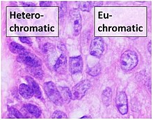
Histone-modifying enzymes are enzymes involved in the modification of histone substrates after protein translation and affect cellular processes including gene expression. To safely store the eukaryotic genome, DNA is wrapped around four core histone proteins, which then join to form nucleosomes. These nucleosomes further fold together into highly condensed chromatin, which renders the organism's genetic material far less accessible to the factors required for gene transcription, DNA replication, recombination and repair. Subsequently, eukaryotic organisms have developed intricate mechanisms to overcome this repressive barrier imposed by the chromatin through histone modification, a type of post-translational modification which typically involves covalently attaching certain groups to histone residues. Once added to the histone, these groups elicit either a loose and open histone conformation, euchromatin, or a tight and closed histone conformation, heterochromatin. Euchromatin marks active transcription and gene expression, as the light packing of histones in this way allows entry for proteins involved in the transcription process. As such, the tightly packed heterochromatin marks the absence of current gene expression.

Chromatin remodeling is the dynamic modification of chromatin architecture to allow access of condensed genomic DNA to the regulatory transcription machinery proteins, and thereby control gene expression. Such remodeling is principally carried out by 1) covalent histone modifications by specific enzymes, e.g., histone acetyltransferases (HATs), deacetylases, methyltransferases, and kinases, and 2) ATP-dependent chromatin remodeling complexes which either move, eject or restructure nucleosomes. Besides actively regulating gene expression, dynamic remodeling of chromatin imparts an epigenetic regulatory role in several key biological processes, egg cells DNA replication and repair; apoptosis; chromosome segregation as well as development and pluripotency. Aberrations in chromatin remodeling proteins are found to be associated with human diseases, including cancer. Targeting chromatin remodeling pathways is currently evolving as a major therapeutic strategy in the treatment of several cancers.

H3K27ac is an epigenetic modification to the DNA packaging protein histone H3. It is a mark that indicates acetylation of the lysine residue at N-terminal position 27 of the histone H3 protein.

H3K36me3 is an epigenetic modification to the DNA packaging protein Histone H3. It is a mark that indicates the tri-methylation at the 36th lysine residue of the histone H3 protein and often associated with gene bodies.

H4K16ac is an epigenetic modification to the DNA packaging protein Histone H4. It is a mark that indicates the acetylation at the 16th lysine residue of the histone H4 protein.

H4K5ac is an epigenetic modification to the DNA packaging protein histone H4. It is a mark that indicates the acetylation at the 5th lysine residue of the histone H4 protein. H4K5 is the closest lysine residue to the N-terminal tail of histone H4. It is enriched at the transcription start site (TSS) and along gene bodies. Acetylation of histone H4K5 and H4K12ac is enriched at centromeres.

H4K12ac is an epigenetic modification to the DNA packaging protein histone H4. It is a mark that indicates the acetylation at the 12th lysine residue of the histone H4 protein. H4K12ac is involved in learning and memory. It is possible that restoring this modification could reduce age-related decline in memory.

H3K56ac is an epigenetic modification to the DNA packaging protein Histone H3. It is a mark that indicates the acetylation at the 56th lysine residue of the histone H3 protein.

H3K36me is an epigenetic modification to the DNA packaging protein Histone H3, specifically, the mono-methylation at the 36th lysine residue of the histone H3 protein.

H3S28P is an epigenetic modification to the DNA packaging protein histone H3. It is a mark that indicates the phosphorylation the 28th serine residue of the histone H3 protein.

H3Y41P is an epigenetic modification to the DNA packaging protein histone H3. It is a mark that indicates the phosphorylation the 41st tyrosine residue of the histone H3 protein.

References

  1. International Human Genome Sequencing Consortium (October 2004). "Finishing the euchromatic sequence of the human genome". Nature. 431 (7011): 931–945. Bibcode:2004Natur.431..931H. doi: 10.1038/nature03001 . PMID   15496913. S2CID   186242248.
  2. 1 2 3 4 5 6 7 Babu A, Verma RS (January 1987). Bourne GH, Jeon KW, Friedlander M (eds.). "Chromosome structure: euchromatin and heterochromatin". International Review of Cytology. Academic Press. 108: 1–60. doi:10.1016/s0074-7696(08)61435-7. ISBN   978-0-12-364508-1. PMID   2822591.
  3. "Definition: nucleosome/nucleosomes". Scitable Nature Education. Retrieved 2021-10-06.
  4. Mobley AS (January 2019). "Chapter 4 - Induced Pluripotent Stem Cells". In Mobley AS (ed.). Neural Stem Cells and Adult Neurogenesis. Academic Press. pp. 67–94. ISBN   978-0-12-811014-0.
  5. 1 2 "The cell. 4. Nucleus. Chromatin. Atlas of plant and animal histology". mmegias.webs.uvigo.es. Retrieved 2021-12-02.
  6. Enukashvily NI (January 2013). "Chapter Two - Mammalian Satellite DNA: A Speaking Dumb". In Donev R, Ponomartsev NV (eds.). Advances in Protein Chemistry and Structural Biology. Organisation of Chromosomes. Vol. 90. Academic Press. pp. 31–65. doi:10.1016/B978-0-12-410523-2.00002-X. ISBN   978-0-12-410523-2. PMID   23582201.
  7. Shen CH (January 2019). "Chapter 13 - Molecular Diagnosis of Chromosomal Disorders". In Shen CH (ed.). Diagnostic Molecular Biology. Academic Press. pp. 331–358. doi:10.1016/B978-0-12-802823-0.00013-4. ISBN   978-0-12-802823-0. S2CID   131915096.
  8. 1 2 "Giemsa banding". Biology Articles, Tutorials & Dictionary Online. 2019-10-07. Retrieved 2021-12-02.
  9. "Reverse banding - Definition and Examples - Biology Online Dictionary". Biology Articles, Tutorials & Dictionary Online. 2020-09-18. Retrieved 2021-12-02.
  10. "Constitutive heterochromatin banding". Biology Articles, Tutorials & Dictionary Online. 2019-10-07. Retrieved 2021-12-02.
  11. "Quinacrine banding". Biology Articles, Tutorials & Dictionary Online. 2019-10-07. Retrieved 2021-12-02.
  12. "T-banding". Biology Articles, Tutorials & Dictionary Online. 2019-10-07. Retrieved 2021-12-02.
  13. Verschure PJ, van Der Kraan I, Manders EM, van Driel R (October 1999). "Spatial relationship between transcription sites and chromosome territories". The Journal of Cell Biology. 147 (1): 13–24. doi:10.1083/jcb.147.1.13. PMC   2164981 . PMID   10508851.
  14. Muegge K (2003-04-01). "Modifications of histone cores and tails in V(D)J recombination". Genome Biology. 4 (4): 211. doi:10.1186/gb-2003-4-4-211. PMC   154571 . PMID   12702201.
  15. Eisenberg E, Levanon EY (October 2013). "Human housekeeping genes, revisited". Trends in Genetics. 29 (10): 569–574. doi:10.1016/j.tig.2013.05.010. PMID   23810203.
  16. Arney KL, Fisher AG (September 2004). "Epigenetic aspects of differentiation". Journal of Cell Science. 117 (Pt 19): 4355–4363. doi:10.1242/jcs.01390. PMID   15331660. S2CID   24376600.
  17. Singh NP, Madabhushi SR, Srivastava S, Senthilkumar R, Neeraja C, Khosla S, Mishra RK (May 2011). "Epigenetic profile of the euchromatic region of human Y chromosome". Nucleic Acids Research. 39 (9): 3594–3606. doi:10.1093/nar/gkq1342. PMC   3089472 . PMID   21252296.
  18. Wang J, Jia ST, Jia S (May 2016). "New Insights into the Regulation of Heterochromatin". Trends in Genetics. 32 (5): 284–294. doi:10.1016/j.tig.2016.02.005. PMC   4842111 . PMID   27005444.
  19. Simmons D (2008). "Epigenetic Influences and Disease". Nature Education. 1 (1): 6. Retrieved 2021-12-02.
  20. Alaskhar Alhamwe B, Khalaila R, Wolf J, von Bülow V, Harb H, Alhamdan F, et al. (2018-05-23). "Histone modifications and their role in epigenetics of atopy and allergic diseases". Allergy, Asthma, and Clinical Immunology. 14 (1): 39. doi:10.1186/s13223-018-0259-4. PMC   5966915 . PMID   29796022.
  21. 1 2 3 4 5 6 Bannister AJ, Kouzarides T (March 2011). "Regulation of chromatin by histone modifications". Cell Research. 21 (3): 381–395. doi:10.1038/cr.2011.22. PMC   3193420 . PMID   21321607.
  22. 1 2 3 4 5 6 Singh D, Nishi K, Khambata K, Balasinor NH (January 2020). "Introduction to epigenetics: basic concepts and advancements in the field". In Tollefsbol T (ed.). Epigenetics and Reproductive Health. Translational Epigenetics. Vol. 21. Academic Press. pp. xxv–xliv. doi:10.1016/B978-0-12-819753-0.02001-8. ISBN   978-0-12-819753-0. S2CID   235031860.

Further reading