Centromere

Last updated
In this diagram of a duplicated chromosome, (2) identifies the centromere--the region that joins the two sister chromatids, or each half of the chromosome. In prophase of mitosis, specialized regions on centromeres called kinetochores attach chromosomes to spindle fibers. Chromosome.svg
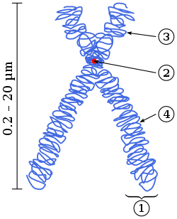
In this diagram of a duplicated chromosome, (2) identifies the centromere—the region that joins the two sister chromatids, or each half of the chromosome. In prophase of mitosis, specialized regions on centromeres called kinetochores attach chromosomes to spindle fibers.

The centromere links a pair of sister chromatids together during cell division. This constricted region of chromosome connects the sister chromatids, creating a short arm (p) and a long arm (q) on the chromatids. During mitosis, spindle fibers attach to the centromere via the kinetochore.

Contents

The physical role of the centromere is to act as the site of assembly of the kinetochores – a highly complex multiprotein structure that is responsible for the actual events of chromosome segregation – i.e. binding microtubules and signaling to the cell cycle machinery when all chromosomes have adopted correct attachments to the spindle, so that it is safe for cell division to proceed to completion and for cells to enter anaphase.

There are, broadly speaking, two types of centromeres. "Point centromeres" bind to specific proteins that recognize particular DNA sequences with high efficiency. Any piece of DNA with the point centromere DNA sequence on it will typically form a centromere if present in the appropriate species. The best characterized point centromeres are those of the budding yeast, Saccharomyces cerevisiae . "Regional centromeres" is the term coined to describe most centromeres, which typically form on regions of preferred DNA sequence, but which can form on other DNA sequences as well. The signal for formation of a regional centromere appears to be epigenetic. Most organisms, ranging from the fission yeast Schizosaccharomyces pombe to humans, have regional centromeres.

Regarding mitotic chromosome structure, centromeres represent a constricted region of the chromosome (often referred to as the primary constriction) where two identical sister chromatids are most closely in contact. When cells enter mitosis, the sister chromatids (the two copies of each chromosomal DNA molecule resulting from DNA replication in chromatin form) are linked along their length by the action of the cohesin complex. It is now believed that this complex is mostly released from chromosome arms during prophase, so that by the time the chromosomes line up at the mid-plane of the mitotic spindle (also known as the metaphase plate), the last place where they are linked with one another is in the chromatin in and around the centromere.

Position

Classifications of Chromosomes

I
Telocentric
Centromere placement very close to the top, p arms barely visible if visible at all.
II
Acrocentric
q arms are still much longer than the p arms, but the p arms are longer than those in telocentric.
III
Submetacentric
p and q arms are very close in length but not equal.
IV
Metacentric
p and q arms are equal in length.
A: Short arm (p arm)
B: Centromere
C: Long arm (q arm)
D: Sister Chromatids Centromere Placement.svg
Classifications of Chromosomes
ITelocentricCentromere placement very close to the top, p arms barely visible if visible at all.
IIAcrocentricq arms are still much longer than the p arms, but the p arms are longer than those in telocentric.
IIISubmetacentricp and q arms are very close in length but not equal.
IVMetacentricp and q arms are equal in length.
A: Short arm (p arm)
B: Centromere
C: Long arm (q arm)
D: Sister Chromatids

In humans, centromere positions define the chromosomal karyotype, in which each chromosome has two arms, p (the shorter of the two) and q (the longer). The short arm 'p' is reportedly named for the French word "petit" meaning 'small'. [1] The position of the centromere relative to any particular linear chromosome is used to classify chromosomes as metacentric, submetacentric, acrocentric, telocentric, or holocentric. [2] [3]

Categorization of chromosomes according to the relative arms length [3]
Centromere positionArms length ratioSignDescription
Medial sensu stricto1.0 – 1.6M Metacentric
Medial region1.7m Metacentric
Submedial3.0sm Submetacentric
Subterminal3.1 – 6.9st Subtelocentric
Terminal region7.0t Acrocentric
Terminal sensu strictoT Telocentric
Notes Metacentric : M+m Atelocentric : M+m+sm+st+t

Metacentric

Metacentric means that the centromere is positioned midway between the chromosome ends, resulting in the arms being approximately equal in length. When the centromeres are metacentric, the chromosomes appear to be "x-shaped."

Submetacentric

Submetacentric means that the centromere is positioned below the middle, with one chromosome arm shorter than the other, often resulting in an L shape.

Acrocentric

An acrocentric chromosome's centromere is situated so that one of the chromosome arms is much shorter than the other. The "acro-" in acrocentric refers to the Greek word for "peak." The human genome has six acrocentric chromosomes, including five autosomal chromosomes (13, 14, 15, 21, 22) and the Y chromosome.

Short acrocentric p-arms contain little genetic material and can be translocated without significant harm, as in a balanced Robertsonian translocation. In addition to some protein coding genes, human acrocentric p-arms also contain Nucleolus organizer regions (NORs), from which ribosomal RNA is transcribed. However, a proportion of acrocentric p-arms in cell lines and tissues from normal human donors do not contain detectable NORs. [4] The domestic horse genome includes one metacentric chromosome that is homologous to two acrocentric chromosomes in the conspecific but undomesticated Przewalski's horse. This may reflect either fixation of a balanced Robertsonian translocation in domestic horses or, conversely, fixation of the fission of one metacentric chromosome into two acrocentric chromosomes in Przewalski's horses. A similar situation exists between the human and great ape genomes, with a reduction of two acrocentric chromosomes in the great apes to one metacentric chromosome in humans (see aneuploidy and the human chromosome 2).

Many diseases from the result of unbalanced translocations more frequently involve acrocentric chromosomes than other non-acrocentric chromosomes. Acrocentric chromosomes are usually located in and around the nucleolus. As a result these chromosomes tend to be less densely packed than chromosomes in the nuclear periphery. Consistently, chromosomal regions that are less densely packed are also more prone to chromosomal translocations in cancers.

Telocentric

Telocentric chromosomes have a centromere at one end of the chromosome and therefore exhibit only one arm at the cytological (microscopic) level. They are not present in humans but can form through cellular chromosomal errors. Telocentric chromosomes occur naturally in many species, such as the house mouse, in which all chromosomes except the Y are telocentric.

Subtelocentric

Subtelocentric chromosomes' centromeres are located between the middle and the end of the chromosomes, but reside closer to the end of the chromosomes.

Centromere types

Acentric

An acentric chromosome is fragment of a chromosome that lacks a centromere. Since centromeres are the attachment point for spindle fibers in cell division, acentric fragments are not evenly distributed to daughter cells during cell division. As a result, a daughter cell will lack the acentric fragment and deleterious consequences could occur.

Chromosome-breaking events can also generate acentric chromosomes or acentric fragments.   

Dicentric

A dicentric chromosome is an abnormal chromosome with two centromeres, which can be unstable through cell divisions. It can form through translocation between or fusion of two chromosome segments, each with a centromere. Some rearrangements produce both dicentric chromosomes and acentric fragments which can not attach to spindles at mitosis. [5] The formation of dicentric chromosomes has been attributed to genetic processes, such as Robertsonian translocation [6] and paracentric inversion. [7] Dicentric chromosomes can have a variety of fates, including mitotic stability. [8] In some cases, their stability comes from inactivation of one of the two centromeres to make a functionally monocentric chromosome capable of normal transmission to daughter cells during cell division. [9]

For example, human chromosome 2, which is believed to be the result of a Robertsonian translocation at some point in the evolution between the great apes and Homo, has a second, vestigial, centromere near the middle of its long arm. [10]

Monocentric

The monocentric chromosome is a chromosome that has only one centromere in a chromosome and forms a narrow constriction.

Monocentric centromeres are the most common structure on highly repetitive DNA in plants and animals. [11]

Holocentric

Unlike monocentric chromosomes, holocentric chromosomes have no distinct primary constriction when viewed at mitosis. Instead, spindle fibers attach along almost the entire (Greek: holo-) length of the chromosome. In holocentric chromosomes centromeric proteins, such as CENPA (CenH3) are spread over the whole chromosome. [12] The nematode, Caenorhabditis elegans, is a well-known example of an organism with holocentric chromosomes, [13] but this type of centromere can be found in various species, plants, and animals, across eukaryotes. Holocentromeres are actually composed of multiple distributed centromere units that form a line-like structure along the chromosomes during mitosis. [14] Alternative or nonconventional strategies are deployed at meiosis to achieve the homologous chromosome pairing and segregation needed to produce viable gametes or gametophytes for sexual reproduction.

Different types of holocentromeres exist in different species, namely with or without centromeric repetitive DNA sequences and with or without CenH3. Holocentricity has evolved at least 13 times independently in various green algae, protozoans, invertebrates, and different plant families. [15] Contrary to monocentric species where acentric fragments usually become lost during cell division, the breakage of holocentric chromosomes creates fragments with normal spindle fiber attachment sites. [16] Because of this, organisms with holocentric chromosomes can more rapidly evolve karyotype variation, able to heal fragmented chromosomes through subsequent addition of telomere caps at the sites of breakage. [17]

Polycentric

Human chromosomes

Human karyogram, with each row vertically aligned at centromere level, and with annotated bands and sub-bands. It is a graphical representation of the idealized human diploid karyotype. It shows dark and white regions on G banding. It shows both the female (XX) and male (XY) versions of the sex chromosome.
Further information: Karyotype Human karyotype with bands and sub-bands.png
Human karyogram, with each row vertically aligned at centromere level, and with annotated bands and sub-bands. It is a graphical representation of the idealized human diploid karyotype. It shows dark and white regions on G banding. It shows both the female (XX) and male (XY) versions of the sex chromosome.
Table of human chromosomes with data on centromeres and sizes.
ChromosomeCentromere
position (Mbp)
CategoryChromosome
Size (Mbp)
Centromere
size (Mbp)
1 125.0metacentric247.27.4
2 93.3submetacentric242.86.3
3 91.0metacentric199.46.0
4 50.4submetacentric191.3
5 48.4submetacentric180.8
6 61.0submetacentric170.9
7 59.9submetacentric158.8
8 45.6submetacentric146.3
9 49.0submetacentric140.4
10 40.2submetacentric135.4
11 53.7submetacentric134.5
12 35.8submetacentric132.3
13 17.9acrocentric114.1
14 17.6acrocentric106.3
15 19.0acrocentric100.3
16 36.6metacentric88.8
17 24.0submetacentric78.7
18 17.2submetacentric76.1
19 26.5metacentric63.8
20 27.5metacentric62.4
21 13.2acrocentric46.9
22 14.7acrocentric49.5
X 60.6submetacentric154.9
Y 12.5acrocentric57.7

Based on the micrographic characteristics of size, position of the centromere and sometimes the presence of a chromosomal satellite, the human chromosomes are classified into the following groups: [18]

GroupChromosomesFeatures
Group AChromosome 1-3Large, metacentric and submetacentric
Group BChromosome 4-5Large, submetacentric
Group CChromosome 6-12, XMedium-sized, submetacentric
Group DChromosome 13-15Medium-sized, acrocentric, with satellite
Group EChromosome 16-18Small, metacentric and submetacentric
Group FChromosome 19-20Very small, metacentric
Group GChromosome 21-22, YVery small, acrocentric, with satellite

Sequence

There are two types of centromeres. [19] In regional centromeres, DNA sequences contribute to but do not define function. Regional centromeres contain large amounts of DNA and are often packaged into heterochromatin. In most eukaryotes, the centromere's DNA sequence consists of large arrays of repetitive DNA (e.g. satellite DNA) where the sequence within individual repeat elements is similar but not identical. In humans, the primary centromeric repeat unit is called α-satellite (or alphoid), although a number of other sequence types are found in this region. [20] Centromere satellites are hypothesized to evolve by a process called layered expansion. They evolve rapidly between species, and analyses in wild mice show that satellite copy number and heterogeneity relates to population origins and subspecies. [21] Additionally, satellite sequences may be affected by inbreeding. [21]

Point centromeres are smaller and more compact. DNA sequences are both necessary and sufficient to specify centromere identity and function in organisms with point centromeres. In budding yeasts, the centromere region is relatively small (about 125 bp DNA) and contains two highly conserved DNA sequences that serve as binding sites for essential kinetochore proteins. [20]

Inheritance

Since centromeric DNA sequence is not the key determinant of centromeric identity in metazoans, it is thought that epigenetic inheritance plays a major role in specifying the centromere. [22] The daughter chromosomes will assemble centromeres in the same place as the parent chromosome, independent of sequence. It has been proposed that histone H3 variant CENP-A (Centromere Protein A) is the epigenetic mark of the centromere. [23] The question arises whether there must be still some original way in which the centromere is specified, even if it is subsequently propagated epigenetically. If the centromere is inherited epigenetically from one generation to the next, the problem is pushed back to the origin of the first metazoans.

On the other hand, thanks to comparisons of the centromeres in the X chromosomes, epigenetic and structural variations have been seen in these regions. In addition, a recent assembly of the human genome has detected a possible mechanism of how pericentromeric and centromeric structures evolve, through a layered expansion model for αSat sequences. This model proposes that different αSat sequence repeats emerge periodically and expand within an active vector, displacing old sequences, and becoming the site of kinetochore assembly. The αSat can originate from the same, or from different vectors. As this process is repeated over time, the layers that flank the active centromere shrink and deteriorate. This process raises questions about the relationship between this dynamic evolutionary process and the position of the centromere. [24]

Structure

The centromeric DNA is normally in a heterochromatin state, which is essential for the recruitment of the cohesin complex that mediates sister chromatid cohesion after DNA replication as well as coordinating sister chromatid separation during anaphase. In this chromatin, the normal histone H3 is replaced with a centromere-specific variant, CENP-A in humans. [25] The presence of CENP-A is believed to be important for the assembly of the kinetochore on the centromere. CENP-C has been shown to localise almost exclusively to these regions of CENP-A associated chromatin. In human cells, the histones are found to be most enriched for H4K20me3 and H3K9me3 [26] which are known heterochromatic modifications. In Drosophila, Islands of retroelements are major components of the centromeres. [27]

In the yeast Schizosaccharomyces pombe (and probably in other eukaryotes), the formation of centromeric heterochromatin is connected to RNAi. [28] In nematodes such as Caenorhabditis elegans , some plants, and the insect orders Lepidoptera and Hemiptera, chromosomes are "holocentric", indicating that there is not a primary site of microtubule attachments or a primary constriction, and a "diffuse" kinetochore assembles along the entire length of the chromosome.

Centromeric aberrations

In rare cases, neocentromeres can form at new sites on a chromosome as a result of a repositioning of the centromere. This phenomenon is most well known from human clinical studies and there are currently over 90 known human neocentromeres identified on 20 different chromosomes. [29] [30] The formation of a neocentromere must be coupled with the inactivation of the previous centromere, since chromosomes with two functional centromeres (Dicentric chromosome) will result in chromosome breakage during mitosis. In some unusual cases human neocentromeres have been observed to form spontaneously on fragmented chromosomes. Some of these new positions were originally euchromatic and lack alpha satellite DNA altogether. Neocentromeres lack the repetitive structure seen in normal centromeres which suggest that centromere formation is mainly controlled epigenetically. [31] [32] Over time a neocentromere can accumulate repetitive elements and mature into what is known as an evolutionary new centromere. There are several well known examples in primate chromosomes where the centromere position is different from the human centromere of the same chromosome and is thought to be evolutionary new centromeres. [31] Centromere repositioning and the formation of evolutionary new centromeres has been suggested to be a mechanism of speciation. [33]

Centromere proteins are also the autoantigenic target for some anti-nuclear antibodies, such as anti-centromere antibodies.

Dysfunction and disease

It has been known that centromere misregulation contributes to mis-segregation of chromosomes, which is strongly related to cancer and miscarriage. Notably, overexpression of many centromere genes have been linked to cancer malignant phenotypes. Overexpression of these centromere genes can increase genomic instability in cancers. Elevated genomic instability on one hand relates to malignant phenotypes; on the other hand, it makes the tumor cells more vulnerable to specific adjuvant therapies such as certain chemotherapies and radiotherapy. [34] Instability of centromere repetitive DNA was recently shown in cancer and aging. [35]

Repair of centromeric DNA

When DNA breaks occur at centromeres in the G1 phase of the cell cycle, the cells are able to recruit the homologous recombinational repair machinery to the damaged site, even in the absence of a sister chromatid. [36] It appears that homologous recombinational repair can occur at centromeric breaks throughout the cell cycle in order to prevent the activation of inaccurate mutagenic DNA repair pathways and to preserve centromeric integrity. [36]

Etymology and pronunciation

The word centromere ( /ˈsɛntrəˌmɪər/ [37] [38] ) uses combining forms of centro- and -mere , yielding "central part", describing the centromere's location at the center of the chromosome.

See also

Related Research Articles

<span class="mw-page-title-main">Spindle checkpoint</span> Cell cycle checkpoint

The spindle checkpoint, also known as the metaphase-to-anaphase transition, the spindle assembly checkpoint (SAC), the metaphase checkpoint, or the mitotic checkpoint, is a cell cycle checkpoint during metaphase of mitosis or meiosis that prevents the separation of the duplicated chromosomes (anaphase) until each chromosome is properly attached to the spindle. To achieve proper segregation, the two kinetochores on the sister chromatids must be attached to opposite spindle poles. Only this pattern of attachment will ensure that each daughter cell receives one copy of the chromosome. The defining biochemical feature of this checkpoint is the stimulation of the anaphase-promoting complex by M-phase cyclin-CDK complexes, which in turn causes the proteolytic destruction of cyclins and proteins that hold the sister chromatids together.

<span class="mw-page-title-main">Kinetochore</span> Protein complex that allows microtubules to attach to chromosomes during cell division

A kinetochore is a disc-shaped protein structure associated with duplicated chromatids in eukaryotic cells where the spindle fibers attach during cell division to pull sister chromatids apart. The kinetochore assembles on the centromere and links the chromosome to microtubule polymers from the mitotic spindle during mitosis and meiosis. The term kinetochore was first used in a footnote in a 1934 Cytology book by Lester W. Sharp and commonly accepted in 1936. Sharp's footnote reads: "The convenient term kinetochore has been suggested to the author by J. A. Moore", likely referring to John Alexander Moore who had joined Columbia University as a freshman in 1932.

<span class="mw-page-title-main">Robertsonian translocation</span> Human chromosomal abnormality

Robertsonian translocation (ROB) is a chromosomal abnormality where the entire long arms of two different chromosomes become fused to each other. It is the most common form of chromosomal translocation in humans, affecting 1 out of every 1,000 babies born. It does not usually cause medical problems, though some people may produce gametes with an incorrect number of chromosomes, resulting in a risk of miscarriage. In rare cases this translocation results in Down syndrome and Patau syndrome. Robertsonian translocations result in a reduction in the number of chromosomes. A Robertsonian evolutionary fusion, which may have occurred in the common ancestor of humans and other great apes, is the reason humans have 46 chromosomes while all other primates have 48. Detailed DNA studies of chimpanzee, orangutan, gorilla and bonobo apes has determined that where human chromosome 2 is present in our DNA in all four great apes this is split into two separate chromosomes typically numbered 2a and 2b. Similarly, the fact that horses have 64 chromosomes and donkeys 62, and that they can still have common, albeit usually infertile, offspring, may be due to a Robertsonian evolutionary fusion at some point in the descent of today's donkeys from their common ancestor.

<span class="mw-page-title-main">Isochromosome</span>

An isochromosome is an unbalanced structural abnormality in which the arms of the chromosome are mirror images of each other. The chromosome consists of two copies of either the long (q) arm or the short (p) arm because isochromosome formation is equivalent to a simultaneous duplication and deletion of genetic material. Consequently, there is partial trisomy of the genes present in the isochromosome and partial monosomy of the genes in the lost arm.

<span class="mw-page-title-main">Micronucleus</span> Small nucleus in the cells of some organisms

A micronucleus is a small nucleus that forms whenever a chromosome or a fragment of a chromosome is not incorporated into one of the daughter nuclei during cell division. It usually is a sign of genotoxic events and chromosomal instability. Micronuclei are commonly seen in cancerous cells and may indicate genomic damage events that can increase the risk of developmental or degenerative diseases.

An acentric fragment is a segment of a chromosome that lacks a centromere.

A dicentric chromosome is an abnormal chromosome with two centromeres. It is formed through the fusion of two chromosome segments, each with a centromere, resulting in the loss of acentric fragments and the formation of dicentric fragments. The formation of dicentric chromosomes has been attributed to genetic processes, such as Robertsonian translocation and paracentric inversion. Dicentric chromosomes have important roles in the mitotic stability of chromosomes and the formation of pseudodicentric chromosomes. Their existence has been linked to certain natural phenomena such as irradiation and have been documented to underlie certain clinical syndromes, notably Kabuki syndrome. The formation of dicentric chromosomes and their implications on centromere function are studied in certain clinical cytogenetics laboratories.

<span class="mw-page-title-main">Aurora kinase B</span> Protein

Aurora kinase B is a protein that functions in the attachment of the mitotic spindle to the centromere.

<span class="mw-page-title-main">Centromere protein B</span> Protein-coding gene in the species Homo sapiens

Centromere protein B also known as major centromere autoantigen B is an autoantigen protein of the cell nucleus. In humans, centromere protein B is encoded by the CENPB gene.

<span class="mw-page-title-main">CENPA</span> Protein-coding gene in the species Homo sapiens

Centromere protein A, also known as CENPA, is a protein which in humans is encoded by the CENPA gene. CENPA is a histone H3 variant which is the critical factor determining the kinetochore position(s) on each chromosome in most eukaryotes including humans.

<span class="mw-page-title-main">CENPC1</span> Protein-coding gene in the species Homo sapiens

Centromere protein C 1 is a protein that in humans is encoded by the CENPC1 gene.

<span class="mw-page-title-main">CENPH</span> Protein-coding gene in the species Homo sapiens

Centromere protein H is a protein that in humans is encoded by the CENPH gene. It is involved in the assembly of kinetochore proteins, mitotic progression and chromosome segregation.

<span class="mw-page-title-main">Shugoshin 1</span> Protein-coding gene in the species Homo sapiens

Shugoshin 1 or Shugoshin-like 1, is a protein that in humans is encoded by the SGO1 gene.

<span class="mw-page-title-main">CENPI</span> Protein-coding gene in humans

Centromere protein I is a protein that in humans is encoded by the CENPI gene.

In molecular biology, the protein domain named the Shugoshin N-terminal coiled-coil region is a domain found on the N-terminal region of the Shugoshin protein in eukaryotes. It has a role in attaching to the kinetochores, structures on the chromatids where microtubules attach. Shugoshin has a conserved coiled-coil N-terminal domain and a highly conserved C-terminal region. Shugoshin is a crucial target of Bub1 kinase that plays a central role in the cohesion of chromosomes during cell division.

Chromosomal instability (CIN) is a type of genomic instability in which chromosomes are unstable, such that either whole chromosomes or parts of chromosomes are duplicated or deleted. More specifically, CIN refers to the increase in rate of addition or loss of entire chromosomes or sections of them. The unequal distribution of DNA to daughter cells upon mitosis results in a failure to maintain euploidy leading to aneuploidy. In other words, the daughter cells do not have the same number of chromosomes as the cell they originated from. Chromosomal instability is the most common form of genetic instability and cause of aneuploidy.

<span class="mw-page-title-main">Neocentromere</span>

Neocentromeres are new centromeres that form at a place on the chromosome that is usually not centromeric. They typically arise due to disruption of the normal centromere. These neocentromeres should not be confused with “knobs”, which were also described as “neocentromeres” in maize in the 1950s. Unlike most normal centromeres, neocentromeres do not contain satellite sequences that are highly repetitive but instead consist of unique sequences. Despite this, most neocentromeres are still able to carry out the functions of normal centromeres in regulating chromosome segregation and inheritance. This raises many questions on what is necessary versus what is sufficient for constituting a centromere.

<span class="mw-page-title-main">Kaustuv Sanyal</span>

Kaustuv Sanyal is an Indian molecular biologist, mycologist and a professor at the Molecular Biology and Genetics Unit of the Jawaharlal Nehru Centre for Advanced Scientific Research (JNCASR). He is known for his molecular and genetic studies of pathogenic yeasts such as Candida and Cryptococcus). An alumnus of Bidhan Chandra Krishi Viswavidyalaya and Madurai Kamaraj University from where he earned a BSc in agriculture and MSc in biotechnology respectively, Sanyal did his doctoral studies at Bose Institute to secure a PhD in Yeast genetics. He moved to the University of California, Santa Barbara, USA to work in the laboratory of John Carbon on the discovery of centromeres in Candida albicans. He joined JNCASR in 2005. He is a member of the Faculty of 1000 in the disciplines of Microbial Evolution and Genomics and has delivered invited speeches which include the Gordon Research Conference, EMBO conferences on comparative genomics and kinetochores. The Department of Biotechnology of the Government of India awarded him the National Bioscience Award for Career Development, one of the highest Indian science awards, for his contributions to biosciences, in 2012. He has also been awarded with the prestigious Tata Innovation Fellowship in 2017. The National Academy of Sciences, India elected him as a fellow in 2014. He is also an elected fellow of Indian Academy of Sciences (2017), and the Indian National Science Academy (2018). In 2019, he has been elected to Fellowship in the American Academy of Microbiology (AAM), the honorific leadership group within the American Society for Microbiology.

Holocentric chromosomes are chromosomes that possess multiple kinetochores along their length rather than the single centromere typical of other chromosomes. They were first described in cytogenetic experiments in 1935. Since this first observation, the term holocentric chromosome has referred to chromosomes that: i) lack the primary constriction corresponding to the centromere observed in monocentric chromosomes; and ii) possess multiple kinetochores dispersed along the entire chromosomal axis, such that microtubules bind to the chromosome along its entire length and move broadside to the pole from the metaphase plate. Holocentric chromosomes are also termed holokinetic, because, during cell division, the sister chromatids move apart in parallel and do not form the classical V-shaped figures typical of monocentric chromosomes.

<span class="mw-page-title-main">Monocentric chromosome</span> Chromosome that has only one centromere in a chromosome and forms a narrow constriction.

The monocentric chromosome is a chromosome that has only one centromere in a chromosome and forms a narrow constriction.

References

  1. "p + q = Solved, Being the True Story of How the Chromosome Got Its Name". 2011-05-03.
  2. "What different types of chromosomes exist?", Nikolay's Genetics Lessons, YouTube, 2013-10-12, archived from the original on 2021-12-11, retrieved 2017-05-28
  3. 1 2 Levan A, Fredga K, Sandberg AA (December 1964). "Nomenclature for centromeric position on chromosomes". Hereditas. 52 (2): 201–220. doi:10.1111/j.1601-5223.1964.tb01953.x.
  4. van Sluis M, van Vuuren C, Mangan H, McStay B (May 2020). "NORs on human acrocentric chromosome p-arms are active by default and can associate with nucleoli independently of rDNA". Proceedings of the National Academy of Sciences of the United States of America. 117 (19): 10368–10377. Bibcode:2020PNAS..11710368V. doi: 10.1073/pnas.2001812117 . PMC   7229746 . PMID   32332163.
  5. Nussbaum R, McInnes R, Willard H, Hamosh A (2007). Thompson & Thompson Genetics in Medicine. Philadelphia(PA): Saunders. p. 72. ISBN   978-1-4160-3080-5.
  6. Thompson & Thompson Genetics in Medicine (7th ed.). p. 62.
  7. Hartwell L, Hood L, Goldberg M, Reynolds A, Lee S (2011). Genetics From Genes to Genomes (4th ed.). New York: McGraw-Hill. ISBN   9780073525266.
  8. Lynch SA, Ashcroft KA, Zwolinski S, Clarke C, Burn J (March 1995). "Kabuki syndrome-like features in monozygotic twin boys with a pseudodicentric chromosome 13". Journal of Medical Genetics. 32 (3): 227–230. doi:10.1136/jmg.32.3.227. PMC   1050324 . PMID   7783176.
  9. Dicentric chromosomes: unique models to study centromere function and inactivation at springer.com
  10. Avarello; et al. (1992). "Evidence for an ancestral alphoid domain on the long arm of human chromosome 2". Human Genetics. 89 (2): 247–9. doi:10.1007/BF00217134. PMID   1587535. S2CID   1441285.
  11. Barra V, Fachinetti D (October 2018). "The dark side of centromeres: types, causes and consequences of structural abnormalities implicating centromeric DNA". Nature Communications. 9 (1): 4340. Bibcode:2018NatCo...9.4340B. doi:10.1038/s41467-018-06545-y. PMC   6194107 . PMID   30337534.
  12. Neumann P, Navrátilová A, Schroeder-Reiter E, Koblížková A, Steinbauerová V, Chocholová E, et al. (2012). "Stretching the rules: monocentric chromosomes with multiple centromere domains". PLOS Genetics. 8 (6): e1002777. doi: 10.1371/journal.pgen.1002777 . PMC   3380829 . PMID   22737088.
  13. Dernburg AF (June 2001). "Here, there, and everywhere: kinetochore function on holocentric chromosomes". The Journal of Cell Biology. 153 (6): F33–F38. doi:10.1083/jcb.153.6.F33. PMC   2192025 . PMID   11402076.
  14. Marques A, Ribeiro T, Neumann P, Macas J, Novák P, Schubert V, et al. (November 2015). "Holocentromeres in Rhynchospora are associated with genome-wide centromere-specific repeat arrays interspersed among euchromatin". Proceedings of the National Academy of Sciences of the United States of America. 112 (44): 13633–13638. Bibcode:2015PNAS..11213633M. doi: 10.1073/pnas.1512255112 . PMC   4640781 . PMID   26489653.
  15. Melters DP, Paliulis LV, Korf IF, Chan SW (July 2012). "Holocentric chromosomes: convergent evolution, meiotic adaptations, and genomic analysis". Chromosome Research. 20 (5): 579–593. doi: 10.1007/s10577-012-9292-1 . PMID   22766638. S2CID   3351527.
  16. Hughes-Schrader S; Ris H (August 1941). "The diffuse spindle attachment of coccids, verified by the mitotic behavior of induced chromosome fragments". Journal of Experimental Zoology. 87 (3): 429–456. doi:10.1002/jez.1400870306. ISSN   0022-104X.
  17. Jankowska M, Fuchs J, Klocke E, Fojtová M, Polanská P, Fajkus J, et al. (December 2015). "Holokinetic centromeres and efficient telomere healing enable rapid karyotype evolution". Chromosoma. 124 (4): 519–528. doi:10.1007/s00412-015-0524-y. PMID   26062516. S2CID   2530401.
  18. Erwinsyah, R., Riandi, & Nurjhani, M. (2017). "Relevance of human chromosome analysis activities against mutation concept in genetics course. IOP Conference Series". Materials Science and Engineering. doi: 10.1088/1757-899x/180/1/012285 . S2CID   90739754.{{cite journal}}: CS1 maint: multiple names: authors list (link)
  19. Pluta AF, Mackay AM, Ainsztein AM, Goldberg IG, Earnshaw WC (December 1995). "The centromere: hub of chromosomal activities". Science. 270 (5242): 1591–1594. Bibcode:1995Sci...270.1591P. doi:10.1126/science.270.5242.1591. PMID   7502067. S2CID   44632550.
  20. 1 2 Mehta GD, Agarwal MP, Ghosh SK (August 2010). "Centromere identity: a challenge to be faced". Molecular Genetics and Genomics. 284 (2): 75–94. doi:10.1007/s00438-010-0553-4. PMID   20585957. S2CID   24881938.
  21. 1 2 Arora UP, Charlebois C, Lawal RA, Dumont BL (April 2021). "Population and subspecies diversity at mouse centromere satellites". BMC Genomics. 22 (1): 279. doi: 10.1186/s12864-021-07591-5 . PMC   8052823 . PMID   33865332.
  22. Dalal Y (February 2009). "Epigenetic specification of centromeres". Biochemistry and Cell Biology. 87 (1): 273–282. doi:10.1139/O08-135. PMID   19234541.
  23. Bernad R, Sánchez P, Losada A (November 2009). "Epigenetic specification of centromeres by CENP-A". Experimental Cell Research. 315 (19): 3233–3241. doi:10.1016/j.yexcr.2009.07.023. PMID   19660450.
  24. Altemose, Nicolas; Logsdon, Glennis A.; Bzikadze, Andrey V.; Sidhwani, Pragya; Langley, Sasha A.; Caldas, Gina V.; Hoyt, Savannah J.; Uralsky, Lev; Ryabov, Fedor D.; Shew, Colin J.; Sauria, Michael E. G.; Borchers, Matthew; Gershman, Ariel; Mikheenko, Alla; Shepelev, Valery A. (April 2022). "Complete genomic and epigenetic maps of human centromeres". Science. 376 (6588): eabl4178. doi:10.1126/science.abl4178. ISSN   0036-8075. PMC   9233505 . PMID   35357911.
  25. Chueh AC, Wong LH, Wong N, Choo KH (January 2005). "Variable and hierarchical size distribution of L1-retroelement-enriched CENP-A clusters within a functional human neocentromere". Human Molecular Genetics. 14 (1): 85–93. doi: 10.1093/hmg/ddi008 . PMID   15537667.
  26. Rosenfeld JA, Wang Z, Schones DE, Zhao K, DeSalle R, Zhang MQ (March 2009). "Determination of enriched histone modifications in non-genic portions of the human genome". BMC Genomics. 10: 143. doi: 10.1186/1471-2164-10-143 . PMC   2667539 . PMID   19335899.
  27. Chang CH, Chavan A, Palladino J, Wei X, Martins NM, Santinello B, et al. (May 2019). "Islands of retroelements are major components of Drosophila centromeres". PLOS Biology. 17 (5): e3000241. doi: 10.1371/journal.pbio.3000241 . PMC   6516634 . PMID   31086362.
  28. Volpe TA, Kidner C, Hall IM, Teng G, Grewal SI, Martienssen RA (September 2002). "Regulation of heterochromatic silencing and histone H3 lysine-9 methylation by RNAi". Science. 297 (5588): 1833–1837. Bibcode:2002Sci...297.1833V. doi: 10.1126/science.1074973 . PMID   12193640. S2CID   2613813.
  29. Marshall OJ, Chueh AC, Wong LH, Choo KH (February 2008). "Neocentromeres: new insights into centromere structure, disease development, and karyotype evolution". American Journal of Human Genetics. 82 (2): 261–282. doi:10.1016/j.ajhg.2007.11.009. PMC   2427194 . PMID   18252209.
  30. Warburton PE (2004). "Chromosomal dynamics of human neocentromere formation". Chromosome Research. 12 (6): 617–626. doi:10.1023/B:CHRO.0000036585.44138.4b. PMID   15289667. S2CID   29472338.
  31. 1 2 Rocchi M, Archidiacono N, Schempp W, Capozzi O, Stanyon R (January 2012). "Centromere repositioning in mammals". Heredity. 108 (1): 59–67. doi:10.1038/hdy.2011.101. PMC   3238114 . PMID   22045381.
  32. Tolomeo D, Capozzi O, Stanyon RR, Archidiacono N, D'Addabbo P, Catacchio CR, et al. (February 2017). "Epigenetic origin of evolutionary novel centromeres". Scientific Reports. 7 (1): 41980. Bibcode:2017NatSR...741980T. doi:10.1038/srep41980. PMC   5290474 . PMID   28155877.
  33. Brown JD, O'Neill RJ (September 2010). "Chromosomes, conflict, and epigenetics: chromosomal speciation revisited". Annual Review of Genomics and Human Genetics. 11 (1): 291–316. doi:10.1146/annurev-genom-082509-141554. PMID   20438362.
  34. Zhang W, Mao JH, Zhu W, Jain AK, Liu K, Brown JB, Karpen GH (August 2016). "Centromere and kinetochore gene misexpression predicts cancer patient survival and response to radiotherapy and chemotherapy". Nature Communications. 7: 12619. Bibcode:2016NatCo...712619Z. doi:10.1038/ncomms12619. PMC   5013662 . PMID   27577169.
  35. Giunta S, Funabiki H (February 2017). "Integrity of the human centromere DNA repeats is protected by CENP-A, CENP-C, and CENP-T". Proceedings of the National Academy of Sciences of the United States of America. 114 (8): 1928–1933. Bibcode:2017PNAS..114.1928G. doi: 10.1073/pnas.1615133114 . PMC   5338446 . PMID   28167779.
  36. 1 2 Yilmaz D, Furst A, Meaburn K, Lezaja A, Wen Y, Altmeyer M, Reina-San-Martin B, Soutoglou E (December 2021). "Activation of homologous recombination in G1 preserves centromeric integrity". Nature. 600 (7890): 748–753. Bibcode:2021Natur.600..748Y. doi:10.1038/s41586-021-04200-z. PMID   34853474. S2CID   244800481.
  37. "Centromere". Merriam-Webster.com Dictionary .
  38. "Centromere". Dictionary.com Unabridged (Online). n.d.

Further reading