Ferrochelatase

Last updated

Protoporphyrin ferrochelatase
1hrk.jpg
Ferrochelatase homodimer, Human
Identifiers
EC no. 4.98.1.1
CAS no. 9012-93-5
Databases
IntEnz IntEnz view
BRENDA BRENDA entry
ExPASy NiceZyme view
KEGG KEGG entry
MetaCyc metabolic pathway
PRIAM profile
PDB structures RCSB PDB PDBe PDBsum
Gene Ontology AmiGO / QuickGO
Search
PMC articles
PubMed articles
NCBI proteins
Ferrochelatase
Human Ferrochelatase 2 angstrom crystal structure.png
Human ferrochelatase
Identifiers
SymbolFerrochelatase
Pfam PF00762
InterPro IPR001015
PROSITE PDOC00462
SCOP2 1ak1 / SCOPe / SUPFAM
OPM superfamily 129
OPM protein 1hrk
Available protein structures:
Pfam   structures / ECOD  
PDB RCSB PDB; PDBe; PDBj
PDBsum structure summary

Protoporphyrin ferrochelatase (EC 4.98.1.1, formerly EC 4.99.1.1, or ferrochelatase; systematic name protoheme ferro-lyase (protoporphyrin-forming)) is an enzyme encoded by the FECH gene in humans. [1] Ferrochelatase catalyses the eighth and terminal step in the biosynthesis of heme, converting protoporphyrin IX into heme B. It catalyses the reaction:

Contents

protoheme + 2 H+ = protoporphyrin + Fe2+
Hemeb-formation.svg

Function

Summary of heme B biosynthesis--note that some reactions occur in the cytoplasm and some in the mitochondrion (yellow) Heme synthesis.png
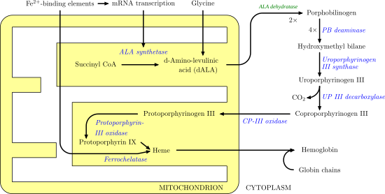
Summary of heme B biosynthesis—note that some reactions occur in the cytoplasm and some in the mitochondrion (yellow)

Ferrochelatase catalyzes the insertion of ferrous iron into protoporphyrin IX in the heme biosynthesis pathway to form heme B. The enzyme is localized to the matrix-facing side of the inner mitochondrial membrane. Ferrochelatase is the best known member of a family of enzymes that add divalent metal cations to tetrapyrrole structures. [2] For example, magnesium chelatase adds magnesium to protoporphyrin IX in the first step of bacteriochlorophyll biosynthesis. [3]

Heme B is an essential cofactor in many proteins and enzymes. In particular, heme b plays a key role as the oxygen carrier in hemoglobin in red blood cells and myoglobin in muscle cells. Furthermore, heme B is found in cytochrome b, a key component in Q-cytochrome c oxidoreductase (complex III) in oxidative phosphorylation. [4]

Structure

Human ferrochelatase is a homodimer composed of two 359 amino acid polypeptide chains. It has a total molecular weight of 85.07 kDa. [5] Each subunit is composed of five regions: a mitochondrial localization sequence, the N terminal domain, two folded domains, and a C terminal extension. Residues 1–62 form a mitochondrial localization domain that is cleaved in post-translational modification. The folded domains contain a total of 17 α-helices and 8 β-sheets. The C terminal extension contains three of the four cysteine residues (Cys403, Cys406, Cys411) that coordinate the catalytic iron–sulfur cluster (2Fe-2S). The fourth coordinating cysteine resides in the N-terminal domain (Cys196). [6]

The active pocket of ferrocheltase consists of two hydrophobic "lips" and a hydrophilic interior. The hydrophobic lips, consisting of the highly conserved residues 300–311, face the inner mitochondrial membrane and facilitate the passage of the poorly soluble protoporphyrin IX substrate and the heme product via the membrane. The interior of the active site pocket contains a highly conserved acidic surface that facilitates proton extraction from protoporphyrin. Histidine and aspartate residues roughly 20 angstroms from the center of the active site on the mitochondrial matrix side of the enzyme coordinate metal binding. [6]

Mechanism

Protoporphyrin IX with pyrrole rings lettered. Protoporphyrin IX with letters.svg
Protoporphyrin IX with pyrrole rings lettered.

The mechanism of human protoporphyrin metalation remains under investigation. Many researchers have hypothesized distortion of the porphyrin macrocycle as key to catalysis. Researchers studying Bacillus subtilis ferrochelatase propose a mechanism for iron insertion into protoporphyrin in which the enzyme tightly grips rings B, C, and D while bending ring A 36o. Normally planar, this distortion exposes the lone pair of electrons on the nitrogen in ring A to the Fe+2 ion. [2] Subsequent investigation revealed a 100o distortion in protoporphyrin bound to human ferrochelatase. A highly conserved histidine residue (His183 in B. subtilis, His263 in humans) is essential for determining the type of distortion, as well as acting as the initial proton acceptor from protoporphyrin. [6] [7] Anionic residues form a pathway facilitating proton movement away from the catalytic histidine. [6] Frataxin chaperones iron to the matrix side of ferrochelatase, where aspartate and histidine residues on both proteins coordinate iron transfer into ferrochelatase. [8] Two arginine and tyrosine residues in the active site (Arg164, Tyr165) may perform the final metalation. [6]

Ferrochelatase active site with protoporphyrin IX substrate in green. Residues shown are: hydrophobic groups holding protoporphyrin IX (yellow), anionic proton transfer path (dark blue), metalation residues (cyan), catalytic histidine (red). Fech-2qd1-activesite.png
Ferrochelatase active site with protoporphyrin IX substrate in green. Residues shown are: hydrophobic groups holding protoporphyrin IX (yellow), anionic proton transfer path (dark blue), metalation residues (cyan), catalytic histidine (red).

Clinical significance

Defects in ferrochelatase create a buildup of protoporphyrin IX, causing erythropoietic protoporphyria (EPP). [9] The disease can result from a variety of mutations in FECH, most of which behave in an autosomal dominant manner with low clinical penetrance. Clinically, patients with EPP present with a range of symptoms, from asymptomatic to suffering from an extremely painful photosensitivity. In less than five percent of cases, accumulation of protoporphyrin in the liver results in cholestasis (blockage of bile flow from the liver to the small intestine) and terminal liver failure. [10]

In cases of lead poisoning, lead inhibits ferrochelatase activity, in part resulting in porphyria. [11]

Interactions

Ferrochelatase interacts with numerous other enzymes involved in heme biosynthesis, catabolism, and transport, including protoporphyrinogen oxidase, 5-aminolevulinate synthase, ABCB10, ABCB7, succinyl-CoA synthetase, [12] and mitoferrin-1. [13] Multiple studies have suggested the existence of an oligomeric complex that enables substrate channeling and coordination of overall iron and porphyrin metabolism throughout the cell. [12] [13] N-methylmesoporphyrin (N-MeMP) is a competitive inhibitor with protoporphyrin IX and is thought to be a transition state analog. As such, N-MeMP has been used extensively as a stabilizing ligand for x-ray crystallography structure determination. [14] Frataxin acts as the Fe+2 chaperone and complexes with ferrochelatase on its mitochondrial matrix side. [8] Ferrochelatase can also insert other divalent metal ions into protoporphyrin. Some ions, such as Zn +2, Ni, and Co form other metalloporphyrins while heavier metal ions such as Mn, Pb, Hg, and Cd inhibit product release after metallation. [15]

See also

Related Research Articles

<span class="mw-page-title-main">Porphyria</span> Group of inherited metabolic disorders

Porphyria is a group of liver disorders in which substances called porphyrins build up in the body, negatively affecting the skin or nervous system. The types that affect the nervous system are also known as acute porphyria, as symptoms are rapid in onset and short in duration. Symptoms of an attack include abdominal pain, chest pain, vomiting, confusion, constipation, fever, high blood pressure, and high heart rate. The attacks usually last for days to weeks. Complications may include paralysis, low blood sodium levels, and seizures. Attacks may be triggered by alcohol, smoking, hormonal changes, fasting, stress, or certain medications. If the skin is affected, blisters or itching may occur with sunlight exposure.

<span class="mw-page-title-main">Heme</span> Chemical coordination complex of an iron ion chelated to a porphyrin

Heme, or haem, is a precursor to hemoglobin, which is necessary to bind oxygen in the bloodstream. Heme is biosynthesized in both the bone marrow and the liver.

<span class="mw-page-title-main">Hereditary coproporphyria</span> Medical condition

Hereditary coproporphyria (HCP) is a disorder of heme biosynthesis, classified as an acute hepatic porphyria. HCP is caused by a deficiency of the enzyme coproporphyrinogen oxidase, coded for by the CPOX gene, and is inherited in an autosomal dominant fashion, although homozygous individuals have been identified. Unlike acute intermittent porphyria, individuals with HCP can present with cutaneous findings similar to those found in porphyria cutanea tarda in addition to the acute attacks of abdominal pain, vomiting and neurological dysfunction characteristic of acute porphyrias. Like other porphyrias, attacks of HCP can be induced by certain drugs, environmental stressors or diet changes. Biochemical and molecular testing can be used to narrow down the diagnosis of a porphyria and identify the specific genetic defect. Overall, porphyrias are rare diseases. The combined incidence for all forms of the disease has been estimated at 1:20,000. The exact incidence of HCP is difficult to determine, due to its reduced penetrance.

<span class="mw-page-title-main">Porphyria cutanea tarda</span> Medical condition

Porphyria cutanea tarda is the most common subtype of porphyria. The disease is named because it is a porphyria that often presents with skin manifestations later in life. The disorder results from low levels of the enzyme responsible for the fifth step in heme production. Heme is a vital molecule for all of the body's organs. It is a component of hemoglobin, the molecule that carries oxygen in the blood.

<span class="mw-page-title-main">Erythropoietic protoporphyria</span> Medical condition

Erythropoietic protoporphyria is a form of porphyria, which varies in severity and can be very painful. It arises from a deficiency in the enzyme ferrochelatase, leading to abnormally high levels of protoporphyrin in the red blood cells (erythrocytes), plasma, skin, and liver. The severity varies significantly from individual to individual.

<span class="mw-page-title-main">Aminolevulinic acid</span> Endogenous non-proteinogenic amino acid

δ-Aminolevulinic acid, an endogenous non-proteinogenic amino acid, is the first compound in the porphyrin synthesis pathway, the pathway that leads to heme in mammals, as well as chlorophyll in plants.

<span class="mw-page-title-main">Aminolevulinic acid synthase</span> Class of enzymes

Aminolevulinic acid synthase (ALA synthase, ALAS, or delta-aminolevulinic acid synthase) is an enzyme (EC 2.3.1.37) that catalyzes the synthesis of δ-aminolevulinic acid (ALA) the first common precursor in the biosynthesis of all tetrapyrroles such as hemes, cobalamins and chlorophylls. The reaction is as follows:

<span class="mw-page-title-main">Gunther disease</span> Medical condition

Gunther disease is a congenital form of erythropoietic porphyria. The word porphyria originated from the Greek word porphura. Porphura actually means "purple pigment", which, in suggestion, the color that the body fluid changes when a person has Gunther's disease. It is a rare, autosomal recessive metabolic disorder affecting heme, caused by deficiency of the enzyme uroporphyrinogen cosynthetase. It is extremely rare, with a prevalence estimated at 1 in 1,000,000 or less. There have been times that prior to birth of a fetus, Gunther's disease has been shown to lead to anemia. In milder cases patients have not presented any symptoms until they have reached adulthood. In Gunther's disease, porphyrins are accumulated in the teeth and bones and an increased amount are seen in the plasma, bone marrow, feces, red blood cells, and urine.

<span class="mw-page-title-main">Hemopexin</span>

Hemopexin, also known as beta-1B-glycoprotein, is a glycoprotein that in humans is encoded by the HPX gene and belongs to the hemopexin family of proteins. Hemopexin is the plasma protein with the highest binding affinity for heme.

<span class="mw-page-title-main">Protoporphyrinogen oxidase</span>

Protoporphyrinogen oxidase or protox is an enzyme that in humans is encoded by the PPOX gene.

<span class="mw-page-title-main">Succinyl coenzyme A synthetase</span>

Succinyl coenzyme A synthetase is an enzyme that catalyzes the reversible reaction of succinyl-CoA to succinate. The enzyme facilitates the coupling of this reaction to the formation of a nucleoside triphosphate molecule from an inorganic phosphate molecule and a nucleoside diphosphate molecule. It plays a key role as one of the catalysts involved in the citric acid cycle, a central pathway in cellular metabolism, and it is located within the mitochondrial matrix of a cell.

<span class="mw-page-title-main">Coproporphyrinogen III oxidase</span> Mammalian protein found in Homo sapiens

Coproporphyrinogen-III oxidase, mitochondrial is an enzyme that in humans is encoded by the CPOX gene. A genetic defect in the enzyme results in a reduced production of heme in animals. The medical condition associated with this enzyme defect is called hereditary coproporphyria.

Erythropoietic porphyria is a type of porphyria associated with erythropoietic cells. In erythropoietic porphyrias, the enzyme deficiency occurs in the red blood cells.

<span class="mw-page-title-main">Protoporphyrin IX</span> Chemical compound

Protoporphyrin IX is an organic compound, classified as a porphyrin, that plays an important role in living organisms as a precursor to other critical compounds like heme (hemoglobin) and chlorophyll. It is a deeply colored solid that is not soluble in water. The name is often abbreviated as PPIX.

<span class="mw-page-title-main">Cystathionine beta synthase</span> Mammalian protein found in humans

Cystathionine-β-synthase, also known as CBS, is an enzyme (EC 4.2.1.22) that in humans is encoded by the CBS gene. It catalyzes the first step of the transsulfuration pathway, from homocysteine to cystathionine:

<span class="mw-page-title-main">Holocytochrome-c synthase</span>

The enzyme holocytochrome-c synthase catalyzes the chemical reaction

<span class="mw-page-title-main">Mitoferrin-1</span> Protein-coding gene in the species Homo sapiens

Mitoferrin-1 (Mfrn1) is a 38 kDa protein that is encoded by the SLC25A37 gene in humans. It is a member of the Mitochondrial carrier (MC) Superfamily, however, its metal cargo makes it distinct from other members of this family. Mfrn1 plays a key role in mitochondrial iron homeostasis as an iron transporter, importing ferrous iron from the intermembrane space of the mitochondria to the mitochondrial matrix for the biosynthesis of heme groups and Fe-S clusters. This process is tightly regulated, given the redox potential of Mitoferrin's iron cargo. Mfrn1 is paralogous to Mitoferrin-2 (Mfrn2), a 39 kDa protein encoded by the SLC25A28 gene in humans. Mfrn1 is highly expressed in differentiating erythroid cells and in other tissues at low levels, while Mfrn2 is expressed ubiquitously in non-erythroid tissues.

<span class="mw-page-title-main">COX15</span> Protein-coding gene in the species Homo sapiens

Cytochrome c oxidase assembly protein COX15 homolog (COX15), also known as heme A synthase, is a protein that in humans is encoded by the COX15 gene. This protein localizes to the inner mitochondrial membrane and involved in heme A biosynthesis. COX15 is also part of a three-component mono-oxygenase that catalyses the hydroxylation of the methyl group at position eight of the protoheme molecule. Mutations in this gene has been reported in patients with hypertrophic cardiomyopathy as well as Leigh syndrome, and characterized by delayed onset of symptoms, hypotonia, feeding difficulties, failure to thrive, motor regression, and brain stem signs.

<span class="mw-page-title-main">Zinc protoporphyrin</span> Chemical compound

Zinc protoporphyrin (ZPP) refers to coordination complexes of zinc and protoporphyrin IX. It is a red-purple solid that is soluble in water. The complex and related species are found in red blood cells when heme production is inhibited by lead and/or by lack of iron.

<span class="mw-page-title-main">Eosinophil peroxidase</span> Protein-coding gene in the species Homo sapiens

Eosinophil peroxidase is an enzyme found within the eosinophil granulocytes, innate immune cells of humans and mammals. This oxidoreductase protein is encoded by the gene EPX, expressed within these myeloid cells. EPO shares many similarities with its orthologous peroxidases, myeloperoxidase (MPO), lactoperoxidase (LPO), and thyroid peroxidase (TPO). The protein is concentrated in secretory granules within eosinophils. Eosinophil peroxidase is a heme peroxidase, its activities including the oxidation of halide ions to bacteriocidal reactive oxygen species, the cationic disruption of bacterial cell walls, and the post-translational modification of protein amino acid residues.

References

  1. "FECH - Ferrochelatase, mitochondrial precursor - Homo sapiens (Human) - FECH gene & protein".
  2. 1 2 Lecerof, D.; Fodje, M.; Hansson, A.; Hansson, M.; Al-Karadaghi, S. (March 2000). "Structural and mechanistic basis of porphyrin metallation by ferrochelatase". Journal of Molecular Biology. 297 (1): 221–232. doi:10.1006/jmbi.2000.3569. PMID   10704318.
  3. Leeper, F. J. (1985). "The biosynthesis of porphyrins, chlorophylls, and vitamin B12". Natural Product Reports. 2 (1): 19–47. doi:10.1039/NP9850200019. PMID   3895052.
  4. Berg, Jeremy; Tymoczko, John; Stryer, Lubert (2012). Biochemistry (7th ed.). New York: W.H. Freeman. ISBN   9781429229364.
  5. "RCSB PDB - 1Hrk: Crystal Structure of Human Ferrochelatase".
  6. 1 2 3 4 5 Wu, Chia-Kuei; Dailey, Harry A.; Rose, John P.; Burden, Amy; Sellers, Vera M.; Wang, Bi-Cheng (1 February 2001). "The 2.0 Å structure of human ferrochelatase, the terminal enzyme of heme biosynthesis". Nature Structural Biology. 8 (2): 156–160. doi:10.1038/84152. PMID   11175906. S2CID   9822420.
  7. Karlberg, Tobias; Hansson, Mattias D.; Yengo, Raymond K.; Johansson, Renzo; Thorvaldsen, Hege O.; Ferreira, Gloria C.; Hansson, Mats; Al-Karadaghi, Salam (May 2008). "Porphyrin Binding and Distortion and Substrate Specificity in the Ferrochelatase Reaction: The Role of Active Site Residues". Journal of Molecular Biology. 378 (5): 1074–1083. doi:10.1016/j.jmb.2008.03.040. PMC   2852141 . PMID   18423489.
  8. 1 2 Bencze, Krisztina Z.; Yoon, Taejin; Mill?n-Pacheco, C?sar; Bradley, Patrick B.; Pastor, Nina; Cowan, J. A.; Stemmler, Timothy L. (2007). "Human frataxin: iron and ferrochelatase binding surface". Chemical Communications (18): 1798–1800. doi:10.1039/B703195E. PMC   2862461 . PMID   17476391.
  9. James, William D.; Berger, Timothy G. (2006). Andrews' Diseases of the Skin: clinical Dermatology. Saunders Elsevier. ISBN   0-7216-2921-0.
  10. Rüfenacht, U.B.; Gouya, L.; Schneider-Yin, X.; Puy, H.; Schäfer, B.W.; Aquaron, R.; Nordmann, Y.; Minder, E.I.; Deybach, J.C. (1998). "Systematic Analysis of Molecular Defects in the Ferrochelatase Gene from Patients with Erythropoietic Protoporphyria". The American Journal of Human Genetics. 62 (6): 1341–52. doi:10.1086/301870. PMC   1377149 . PMID   9585598.
  11. "Lead Toxicity -- What Are Possible Health Effects from Lead Exposure?". Agency for Toxic Substances & Disease Registry. Retrieved 9 February 2021.
  12. 1 2 Medlock, Amy E.; Shiferaw, Mesafint T.; Marcero, Jason R.; Vashisht, Ajay A.; Wohlschlegel, James A.; Phillips, John D.; Dailey, Harry A.; Liesa, Marc (19 August 2015). "Identification of the Mitochondrial Heme Metabolism Complex". PLOS ONE. 10 (8): e0135896. Bibcode:2015PLoSO..1035896M. doi: 10.1371/journal.pone.0135896 . PMC   4545792 . PMID   26287972.
  13. 1 2 Chen, W.; Dailey, H. A.; Paw, B. H. (28 April 2010). "Ferrochelatase forms an oligomeric complex with mitoferrin-1 and Abcb10 for erythroid heme biosynthesis". Blood. 116 (4): 628–630. doi:10.1182/blood-2009-12-259614. PMC   3324294 . PMID   20427704.
  14. Medlock, A.; Swartz, L.; Dailey, T. A.; Dailey, H. A.; Lanzilotta, W. N. (29 January 2007). "Substrate interactions with human ferrochelatase". Proceedings of the National Academy of Sciences. 104 (6): 1789–1793. Bibcode:2007PNAS..104.1789M. doi: 10.1073/pnas.0606144104 . PMC   1794275 . PMID   17261801.
  15. Medlock, Amy E.; Carter, Michael; Dailey, Tamara A.; Dailey, Harry A.; Lanzilotta, William N. (October 2009). "Product Release Rather than Chelation Determines Metal Specificity for Ferrochelatase". Journal of Molecular Biology. 393 (2): 308–319. doi:10.1016/j.jmb.2009.08.042. PMC   2771925 . PMID   19703464.

Further reading