Glutamate racemase

Last updated
Glutamate Racemase
Identifiers
EC no. 5.1.1.3
CAS no. 9024-08-2
Databases
IntEnz IntEnz view
BRENDA BRENDA entry
ExPASy NiceZyme view
KEGG KEGG entry
MetaCyc metabolic pathway
PRIAM profile
PDB structures RCSB PDB PDBe PDBsum
Gene Ontology AmiGO / QuickGO
Search
PMC articles
PubMed articles
NCBI proteins

In enzymology, glutamate racemase (MurI with a capital i) (EC 5.1.1.3) is an enzyme that catalyzes the chemical reaction

Contents

L-glutamate D-glutamate

Hence, this enzyme RacE has one substrate, L-glutamate, and one product, D-glutamate.

This enzyme belongs to the family of isomerases, specifically those racemases and epimerases acting on amino acids and derivatives, including proline racemase, aspartate racemase, and diaminopimelate epimerase. [1] This enzyme participates in glutamate metabolism that is essential for cell wall biosynthesis in bacteria. [2] Glutamate racemase performs the additional function of gyrase inhibition, preventing gyrase from binding to DNA. [3]

Glutamate racemase (MurI) serves two distinct metabolic functions: primarily, it is a critical enzyme in cell wall biosynthesis, [2] but also plays a role in gyrase inhibition. [3] The ability of glutamate racemase and other proteins to serve two distinct functions is known as "moonlighting".

Moonlighting background

Before the discovery of moonlighting proteins, it was generally believed by scientists that an enzyme only had one function which led to the concept of "one gene, one enzyme". However, this concept no longer applies in science after the discovery that some proteins consist of both major and minor functions. This led to numerous studies attempting to relate the two functions to each other. The minor functions of these unique enzymes are called moonlighting functions, in which a protein can have a secondary functions not dependent upon the main function. These two functions of the moonlighting protein are found in a single polypeptide chain. Proteins that are multifunctional are not included due to gene fusion, families of homologous proteins, splice variants or promiscuous enzyme activities. The enzyme glutamate racemase (MurI) is an example of a moonlighting protein, functioning both in bacterial cell wall biosynthesis as well as in gyrase inhibition.

Structure

The dimensions of MurI is approximately 35 Å × 40 Å × 45 Å and consists of two compact domains of α/β structure. [1] [4] With the active site in between the two domains, the N-terminal domain contains residues 1-97 and 207-264 while the C-terminal domain includes residues 98-206. [1] This allows the enzyme to produce L-isomer from D-glutamate. Also, the N-domain is composed of five-stranded β-sheets compared to four-stranded β-sheets of C-domain. [1] These structural specifications are not identical between MurI of different species; S. pyogenes and B. subtilis actually possess the most structurally similar MurI enzymes yet found. It is also not rare to find MurI as a dimer.

The active site, as it is evenly between the N-domain and C-domain, is also between the two cysteine residues. It is accessible to solvents, as several water molecules, such as W1, are found in the active site. In some species, the active site also incorporates sulfate ions to undergo hydrogen bonding on the amide backbone and the side chains. [1] [5]

Function

Bacterial wall synthesis

Glutamate Racemase
Identifiers
Organism Escherichia coli
SymbolmurI
Entrez 948467
RefSeq (Prot) NP_418402
UniProt P22634
Other data
EC number 5.1.1.3
Chromosome genome: 4.16 - 4.16 Mb
Search for
Structures Swiss-model
Domains InterPro

Glutamate racemase is a bacterial enzyme that is encoded by the murI gene. This enzyme is most commonly known as being responsible for the synthesis of bacterial cell walls. Through experimentation it was found that this enzyme is able to construct these cell walls by synthesizing D-glutamate from L-glutamate through racemization. [4] D-glutamate is a monomer of the peptidoglycan layer in prokaryotic cell walls. Peptidoglycan is an essential structural component of the bacterial cell wall. The peptidoglycan layer is also responsible for the rigidity of the cell wall. [6] This process, in which MurI helps catalyze the interconversion of glutamate enantiomers, like L-Glutamate, into the essential D-glutamate, is also cofactor independent. As such it can proceed without needing an additional source, which would bind to an allosteric site, altering the enzyme shape to assist in catalyzing the reaction. [7] Murl involves a two-step process to catalyze the glutamate enantiomers to D-glutamate. The first step is a deprotonation of the substrate to form an anion. [7] Subsequently, the substrate gets reprotonated. Once the glutamate is in the active site of the enzyme it undergoes a very large conformational change of its domains. This change helps superimpose the two catalytic cysteine residues, Cys73 and Cys184, located on either sides of the substrate at equal positions. Those domains mentioned earlier are symmetric and this symmetry suggests that this racemase activity of the protein may have evolved from gene duplication. [5] Due to this main function of biosynthesis of bacterial cell walls MurI has been targeted as an antibacterial in drug discovery. [8]

Gyrase inhibition

Along with its main function of cell wall biosynthesis, the moonlighting protein glutamate racemase also functions independently as a gyrase inhibitor. [2] Present in certain forms of bacteria, MurI reduces the activity of DNA gyrase by preventing gyrase from binding to DNA. [2] When gyrase binds to DNA, the enzyme decreases the tension in the DNA strands as they are unwound and causes the strands to become supercoiled. [9] This is a critical step in DNA replication in these cells which results in the reproduction of bacterial cells. [10] The presence of glutamate racemase in the process inhibits gyrase from effectively binding to DNA by deforming the shape of the enzyme's active site. It essentially disallows gyrase from catalyzing the reaction that coils unwinding DNA strands. [10]

This function of MurI was discovered experimentally. DNA gyrase was incubated with the MurI enzyme and then added to a sample of DNA; the results of this experiment showed inhibition of supercoiling activity when MurI was present. [2] The cell wall biosynthesis function of MurI is not directly related to its moonlighting function. MurI's ability to inhibit gyrase binding can proceed independently of its main function. [2] This means that DNA gyrase, in turn, will not have any effect on MurI's racemization, which was confirmed in a study of the racemization with and without the presence of DNA gyrase. [2] In an experimental analysis, it was determined that MurI employs the use of two different enzymatic active sites for its two functions. This was shown by the inclusion of the racemase substrate L-glutamate in an assay with the separated gyrase inhibition site. The gyrase inhibition occurs in both supercoiling and relaxing activities of the DNA gyrase, and the study concluded that the inhibition activity was able to proceed, unchanged, in the presence of the racemase substrate. [10] This dictates that the two functions can be carried out independently of each other, on non-overlapping sites, making MurI a true moonlighting protein. [2] Mutant forms of MurI that are unable to exhibit their racemase function, no matter how compromised their racemase abilities were, were still proven through a study to be able to perform the DNA gyrase inhibition, with comparable results to a non-mutated form of MurI. [10]

Relationship between main and moonlighting functions

Glutamate racemase (MurI) provides multiple functions for bacterial cells. MurI is an enzyme which is primarily known for its role in synthesizing bacterial cell walls. While performing the function of cell wall synthesis, MurI also acts as a gyrase inhibitor, preventing gyrase from binding to DNA. The two processes have been shown two be unrelated. [2] In order to ascertain the effects of gyrase inhibition on cell wall synthesis, the efficiency of the conversion of D-glutamate to L-glutamate was measured while varying the concentration of DNA gyrase. Conversely, the effects of cell wall production on gyrase inhibition were discovered by varying the concentration of the racemization substrate. [2] The results of these experiments conclude that there is no significant effect of racemization on gyrase inhibition or vice versa. [2] The two functions of MurI act independently of each other reaffirming the fact that MurI is a moonlighting protein.

Relationship to active site

Crystallographic structure of glutamate racemase from the bacteria S. pyogenes (rainbow colored cartoon, N-terminus = blue, C-terminus = red) complexed with the inhibitor gamma-2-naphthylmethyl-D-glutamate (magenta spheres; carbon = white, oxygen = red, nitrogen = blue) that occupies the active site. 2OHV.png
Crystallographic structure of glutamate racemase from the bacteria S. pyogenes (rainbow colored cartoon, N-terminus = blue, C-terminus = red) complexed with the inhibitor gamma-2-naphthylmethyl-D-glutamate (magenta spheres; carbon = white, oxygen = red, nitrogen = blue) that occupies the active site.

Glutamate racemase is known to use its active site to undergo racemization and participate in the cell wall biosynthesis pathway of bacteria. [2] Based on homology to other racemases and epimerases, glutamate racemase is thought to employ two active site cysteine residues as acid/base catalysts. [7] Surprisingly however, substituting either of the two residues with serine did not appreciable change the rate of the reaction significantly; the kcat value remained within .3% to 3% compared to the wild-type enzyme. [7] From previous studies, it is most likely that the active site of MurI that performs racemization is not the same active site that undergoes gyrase inhibition. In order to ascertain the effects of gyrase inhibition on cell wall synthesis, the efficiency of the conversion of D-glutamate to L-glutamate was measured while varying the concentration of DNA gyrase. Conversely, the effects of cell wall production on gyrase inhibition were discovered by varying the concentration of the racemization substrate. It has been shown that the two functions are neutral to each other. [2] In other words, racemization substrates are neutral to gyrase inhibition, and DNA gyrase has no effect on racemization. This explains how glutamate racemase in certain bacteria, such as Glr from B. subtilis, do not inhibit gyrase; [12] if one active site is involved with both functions, this independence would not be possible. Consequently, a different site of MurI, distant from its active site, is involved in interacting with gyrase. [2]

Enzyme regulation

This protein may use the morpheein model of allosteric regulation. [13]

Application

Glutamate racemase has emerged as a potential antibacterial target since the product of this enzyme, D-glutamate, is an essential component of bacterial walls. Inhibiting the enzyme will prevent bacterial wall formation and ultimately result in lysis of the bacteria cell by osmotic pressure. Furthermore, glutamate racemase is not expressed nor is the product of this enzyme, D-glutamate is normally found in mammals, hence inhibiting this enzyme should not result in toxicity to the mammalian host organism. [5] Possible inhibitors to MurI includes aziridino-glutamate that would alkylate the catalytic cysteines; N-hydroxy glutamate that by mimicking Wat2 (the bound water molecule that interacts with glutamate amino group) would prevent binding of the substrate; [5] or 4-substituted D-glutamic acid analogs bearing aryl-, heteroaryl-, cinnamyl-, or biaryl-methyl substituents that would also prevent binding of substrate. [5]

Related Research Articles

<span class="mw-page-title-main">Active site</span> Active region of an enzyme

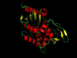
In biology and biochemistry, the active site is the region of an enzyme where substrate molecules bind and undergo a chemical reaction. The active site consists of amino acid residues that form temporary bonds with the substrate, the binding site, and residues that catalyse a reaction of that substrate, the catalytic site. Although the active site occupies only ~10–20% of the volume of an enzyme, it is the most important part as it directly catalyzes the chemical reaction. It usually consists of three to four amino acids, while other amino acids within the protein are required to maintain the tertiary structure of the enzymes.

<span class="mw-page-title-main">Histone acetyltransferase</span> Enzymes that catalyze acyl group transfer from acetyl-CoA to histones

Histone acetyltransferases (HATs) are enzymes that acetylate conserved lysine amino acids on histone proteins by transferring an acetyl group from acetyl-CoA to form ε-N-acetyllysine. DNA is wrapped around histones, and, by transferring an acetyl group to the histones, genes can be turned on and off. In general, histone acetylation increases gene expression.

<span class="mw-page-title-main">Ribonucleotide reductase</span> Class of enzymes

Ribonucleotide reductase (RNR), also known as ribonucleoside diphosphate reductase (rNDP), is an enzyme that catalyzes the formation of deoxyribonucleotides from ribonucleotides. It catalyzes this formation by removing the 2'-hydroxyl group of the ribose ring of nucleoside diphosphates. This reduction produces deoxyribonucleotides. Deoxyribonucleotides in turn are used in the synthesis of DNA. The reaction catalyzed by RNR is strictly conserved in all living organisms. Furthermore, RNR plays a critical role in regulating the total rate of DNA synthesis so that DNA to cell mass is maintained at a constant ratio during cell division and DNA repair. A somewhat unusual feature of the RNR enzyme is that it catalyzes a reaction that proceeds via a free radical mechanism of action. The substrates for RNR are ADP, GDP, CDP and UDP. dTDP is synthesized by another enzyme from dTMP.

<span class="mw-page-title-main">Pyridoxal phosphate</span> Active form of vitamin B6

Pyridoxal phosphate (PLP, pyridoxal 5'-phosphate, P5P), the active form of vitamin B6, is a coenzyme in a variety of enzymatic reactions. The International Union of Biochemistry and Molecular Biology has catalogued more than 140 PLP-dependent activities, corresponding to ~4% of all classified activities. The versatility of PLP arises from its ability to covalently bind the substrate, and then to act as an electrophilic catalyst, thereby stabilizing different types of carbanionic reaction intermediates.

DNA gyrase, or simply gyrase, is an enzyme within the class of topoisomerase and is a subclass of Type II topoisomerases that reduces topological strain in an ATP dependent manner while double-stranded DNA is being unwound by elongating RNA-polymerase or by helicase in front of the progressing replication fork. It is the only known enzyme to actively contribute negative supercoiling to DNA, while it also is capable of relaxing positive supercoils. It does so by looping the template to form a crossing, then cutting one of the double helices and passing the other through it before releasing the break, changing the linking number by two in each enzymatic step. This process occurs in bacteria, whose single circular DNA is cut by DNA gyrase and the two ends are then twisted around each other to form supercoils. Gyrase is also found in eukaryotic plastids: it has been found in the apicoplast of the malarial parasite Plasmodium falciparum and in chloroplasts of several plants. Bacterial DNA gyrase is the target of many antibiotics, including nalidixic acid, novobiocin, albicidin, and ciprofloxacin.

<span class="mw-page-title-main">DD-transpeptidase</span>

DD-transpeptidase is a bacterial enzyme that catalyzes the transfer of the R-L-αα-D-alanyl moiety of R-L-αα-D-alanyl-D-alanine carbonyl donors to the γ-OH of their active-site serine and from this to a final acceptor. It is involved in bacterial cell wall biosynthesis, namely, the transpeptidation that crosslinks the peptide side chains of peptidoglycan strands.

<span class="mw-page-title-main">Glutamine synthetase</span> Class of enzymes

Glutamine synthetase (GS) is an enzyme that plays an essential role in the metabolism of nitrogen by catalyzing the condensation of glutamate and ammonia to form glutamine:

<span class="mw-page-title-main">Novobiocin</span> Chemical compound

Novobiocin, also known as albamycin or cathomycin, is an aminocoumarin antibiotic that is produced by the actinomycete Streptomyces niveus, which has recently been identified as a subjective synonym for S. spheroides a member of the class Actinomycetia. Other aminocoumarin antibiotics include clorobiocin and coumermycin A1. Novobiocin was first reported in the mid-1950s.

<span class="mw-page-title-main">Enzyme inhibitor</span> Molecule that blocks enzyme activity

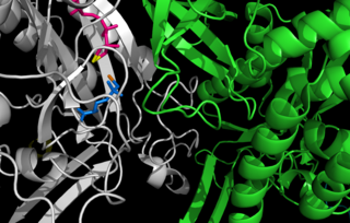
An enzyme inhibitor is a molecule that binds to an enzyme and blocks its activity. Enzymes are proteins that speed up chemical reactions necessary for life, in which substrate molecules are converted into products. An enzyme facilitates a specific chemical reaction by binding the substrate to its active site, a specialized area on the enzyme that accelerates the most difficult step of the reaction.

<span class="mw-page-title-main">Amino acid synthesis</span> The set of biochemical processes by which amino acids are produced

Amino acid synthesis is the set of biochemical processes by which the amino acids are produced. The substrates for these processes are various compounds in the organism's diet or growth media. Not all organisms are able to synthesize all amino acids. For example, humans can synthesize 11 of the 20 standard amino acids. These 11 are called the non-essential amino acids).

<span class="mw-page-title-main">Type II topoisomerase</span>

Type II topoisomerases are topoisomerases that cut both strands of the DNA helix simultaneously in order to manage DNA tangles and supercoils. They use the hydrolysis of ATP, unlike Type I topoisomerase. In this process, these enzymes change the linking number of circular DNA by ±2. Topoisomerases are ubiquitous enzymes, found in all living organisms.

<span class="mw-page-title-main">Acetolactate synthase</span> Class of enzymes

The acetolactate synthase (ALS) enzyme is a protein found in plants and micro-organisms. ALS catalyzes the first step in the synthesis of the branched-chain amino acids.

<span class="mw-page-title-main">Amidophosphoribosyltransferase</span> Mammalian protein found in Homo sapiens

Amidophosphoribosyltransferase (ATase), also known as glutamine phosphoribosylpyrophosphate amidotransferase (GPAT), is an enzyme responsible for catalyzing the conversion of 5-phosphoribosyl-1-pyrophosphate (PRPP) into 5-phosphoribosyl-1-amine (PRA), using the amine group from a glutamine side-chain. This is the committing step in de novo purine synthesis. In humans it is encoded by the PPAT gene. ATase is a member of the purine/pyrimidine phosphoribosyltransferase family.

In enzymology, an aspartate racemase is an enzyme that catalyzes the following chemical reaction:

<span class="mw-page-title-main">Diaminopimelate epimerase</span>

In enzymology, a diaminopimelate epimerase is an enzyme that catalyzes the chemical reaction

<span class="mw-page-title-main">Cystathionine beta-lyase</span> Enzyme

Cystathionine beta-lyase, also commonly referred to as CBL or β-cystathionase, is an enzyme that primarily catalyzes the following α,β-elimination reaction

<span class="mw-page-title-main">Diaminopimelate decarboxylase</span>

The enzyme diaminopimelate decarboxylase (EC 4.1.1.20) catalyzes the cleavage of carbon-carbon bonds in meso 2,6 diaminoheptanedioate to produce CO2 and L-lysine, the essential amino acid. It employs the cofactor pyridoxal phosphate, also known as PLP, which participates in numerous enzymatic transamination, decarboxylation and deamination reactions.

<span class="mw-page-title-main">Phospholipase C</span> Class of enzymes

Phospholipase C (PLC) is a class of membrane-associated enzymes that cleave phospholipids just before the phosphate group (see figure). It is most commonly taken to be synonymous with the human forms of this enzyme, which play an important role in eukaryotic cell physiology, in particular signal transduction pathways. Phospholipase C's role in signal transduction is its cleavage of phosphatidylinositol 4,5-bisphosphate (PIP2) into diacyl glycerol (DAG) and inositol 1,4,5-trisphosphate (IP3), which serve as second messengers. Activators of each PLC vary, but typically include heterotrimeric G protein subunits, protein tyrosine kinases, small G proteins, Ca2+, and phospholipids.

<span class="mw-page-title-main">Morpheein</span> Model of protein allosteric regulation

Morpheeins are proteins that can form two or more different homo-oligomers, but must come apart and change shape to convert between forms. The alternate shape may reassemble to a different oligomer. The shape of the subunit dictates which oligomer is formed. Each oligomer has a finite number of subunits (stoichiometry). Morpheeins can interconvert between forms under physiological conditions and can exist as an equilibrium of different oligomers. These oligomers are physiologically relevant and are not misfolded protein; this distinguishes morpheeins from prions and amyloid. The different oligomers have distinct functionality. Interconversion of morpheein forms can be a structural basis for allosteric regulation, an idea noted many years ago, and later revived. A mutation that shifts the normal equilibrium of morpheein forms can serve as the basis for a conformational disease. Features of morpheeins can be exploited for drug discovery. The dice image represents a morpheein equilibrium containing two different monomeric shapes that dictate assembly to a tetramer or a pentamer. The one protein that is established to function as a morpheein is porphobilinogen synthase, though there are suggestions throughout the literature that other proteins may function as morpheeins.

The bacterial cell wall provides strength and rigidity to counteract internal osmotic pressure, and protection against the environment. The peptidoglycan layer gives the cell wall its strength, and helps maintain the overall shape of the cell. The basic peptidoglycan structure of both Gram-positive and Gram-negative bacteria comprises a sheet of glycan chains connected by short cross-linking polypeptides. Biosynthesis of peptidoglycan is a multi-step process comprising three main stages:

  1. formation of UDP-N-acetylmuramic acid (UDPMurNAc) from N-acetylglucosamine (GlcNAc).
  2. addition of a short polypeptide chain to the UDPMurNAc.
  3. addition of a second GlcNAc to the disaccharide-pentapeptide building block and transport of this unit through the cytoplasmic membrane and incorporation into the growing peptidoglycan layer.

References

  1. 1 2 3 4 5 Kim KH, Bong YJ, Park JK, Shin KJ, Hwang KY, Kim EE (September 2007). "Structural basis for glutamate racemase inhibition". J. Mol. Biol. 372 (2): 434–43. doi:10.1016/j.jmb.2007.05.003. PMID   17658548.
  2. 1 2 3 4 5 6 7 8 9 10 11 12 13 14 Sengupta S, Ghosh S, Nagaraja V (September 2008). "Moonlighting function of glutamate racemase from Mycobacterium tuberculosis: racemization and DNA gyrase inhibition are two independent activities of the enzyme". Microbiology. 154 (Pt 9): 2796–803. doi: 10.1099/mic.0.2008/020933-0 . PMID   18757813.
  3. 1 2 Reece RJ, Maxwell A (1991). "DNA gyrase: structure and function". Crit. Rev. Biochem. Mol. Biol. 26 (3–4): 335–75. doi:10.3109/10409239109114072. PMID   1657531.
  4. 1 2 Hwang KY, Cho CS, Kim SS, Sung HC, Yu YG, Cho Y (May 1999). "Structure and mechanism of glutamate racemase from Aquifex pyrophilus". Nat. Struct. Biol. 6 (5): 422–6. doi:10.1038/8223. PMID   10331867. S2CID   10925922.
  5. 1 2 3 4 5 Ruzheinikov SN, Taal MA, Sedelnikova SE, Baker PJ, Rice DW (November 2005). "Substrate-induced conformational changes in Bacillus subtilis glutamate racemase and their implications for drug discovery". Structure. 13 (11): 1707–13. doi: 10.1016/j.str.2005.07.024 . PMID   16271894.
  6. Schleifer KH, Kandler O (December 1972). "Peptidoglycan types of bacterial cell walls and their taxonomic implications". Bacteriol Rev. 36 (4): 407–77. doi:10.1128/MMBR.36.4.407-477.1972. PMC   408328 . PMID   4568761.
  7. 1 2 3 4 Glavas S, Tanner ME (May 2001). "Active site residues of glutamate racemase". Biochemistry. 40 (21): 6199–204. doi:10.1021/bi002703z. PMID   11371180.
  8. Lundqvist T, Fisher SL, Kern G, Folmer RH, Xue Y, Newton DT, Keating TA, Alm RA, de Jonge BL (June 2007). "Exploitation of structural and regulatory diversity in glutamate racemases". Nature. 447 (7146): 817–22. Bibcode:2007Natur.447..817L. doi:10.1038/nature05689. PMID   17568739. S2CID   4408683.
  9. Sengupta S, Shah M, Nagaraja V (2006). "Glutamate racemase from Mycobacterium tuberculosis inhibits DNA gyrase by affecting its DNA-binding". Nucleic Acids Res. 34 (19): 5567–76. doi:10.1093/nar/gkl704. PMC   1635304 . PMID   17020913.
  10. 1 2 3 4 Sengupta S, Nagaraja V (February 2008). "Inhibition of DNA gyrase activity by Mycobacterium smegmatis MurI". FEMS Microbiol. Lett. 279 (1): 40–7. doi: 10.1111/j.1574-6968.2007.01005.x . PMID   18177305.
  11. PDB: 2OHV ; Kim KH, Bong YJ, Park JK, Shin KJ, Hwang KY, Kim EE (September 2007). "Structural basis for glutamate racemase inhibition". J. Mol. Biol. 372 (2): 434–43. doi:10.1016/j.jmb.2007.05.003. PMID   17658548.
  12. Ashiuchi M, Kuwana E, Komatsu K, Soda K, Misono H (June 2003). "Differences in effects on DNA gyrase activity between two glutamate racemases of Bacillus subtilis, the poly-gamma-glutamate synthesis-linking Glr enzyme and the YrpC (MurI) isozyme". FEMS Microbiol. Lett. 223 (2): 221–5. doi: 10.1016/s0378-1097(03)00381-1 . PMID   12829290.
  13. T. Selwood; E. K. Jaffe. (2011). "Dynamic dissociating homo-oligomers and the control of protein function". Arch. Biochem. Biophys. 519 (2): 131–43. doi:10.1016/j.abb.2011.11.020. PMC   3298769 . PMID   22182754.

Further reading