Glutamine synthetase

Last updated
glutamate—ammonia ligase
MN MN ADP PPQ.png
Active site between two monomers of glutamine synthetase from Salmonella typhimurium . Cation binding sites are yellow and orange; ADP is pink; phosphinothricin is blue. [1]
Identifiers
EC no. 6.3.1.2
CAS no. 9023-70-5
Databases
IntEnz IntEnz view
BRENDA BRENDA entry
ExPASy NiceZyme view
KEGG KEGG entry
MetaCyc metabolic pathway
PRIAM profile
PDB structures RCSB PDB PDBe PDBsum
Gene Ontology AmiGO / QuickGO
Search
PMC articles
PubMed articles
NCBI proteins
Glutamine synthetase,
beta-Grasp domain
Identifiers
SymbolGln-synt_N
Pfam PF03951
InterPro IPR008147
PROSITE PDOC00162
SCOP2 2gls / SCOPe / SUPFAM
Available protein structures:
Pfam   structures / ECOD  
PDB RCSB PDB; PDBe; PDBj
PDBsum structure summary
PDB 1f1h , 1f52 , 1fpy , 1hto , 1lgr , 2bvc , 2gls , 2qc8 , 2ojw
Glutamine synthetase,
catalytic domain
PDB 2gls EBI.jpg
12-subunit enzyme glutamine synthetase from Salmonella typhimurium . [2]
Identifiers
SymbolGln-synt_C
Pfam PF00120
Pfam clan CL0286
InterPro IPR008146
PROSITE PDOC00162
SCOP2 2gls / SCOPe / SUPFAM
Available protein structures:
Pfam   structures / ECOD  
PDB RCSB PDB; PDBe; PDBj
PDBsum structure summary
PDB 1f1h , 1f52 , 1fpy , 1hto , 1lgr , 2bvc , 2gls , 2qc8 , 2ojw
glutamate-ammonia ligase (glutamine synthetase)
Identifiers
SymbolGLUL
Alt. symbolsGLNS
NCBI gene 2752
HGNC 4341
OMIM 138290
PDB 2qc8
RefSeq NM_002065
UniProt P15104
Other data
EC number 6.3.1.2
Locus Chr. 1 q31
Search for
Structures Swiss-model
Domains InterPro

Glutamine synthetase (GS) (EC 6.3.1.2) [3] is an enzyme that plays an essential role in the metabolism of nitrogen by catalyzing the condensation of glutamate and ammonia to form glutamine:

Contents

Glutamate + ATP + NH3 → Glutamine + ADP + phosphate

Glutamine synthetase catalyzed reaction Glutamine synthetase reaction.svg
Glutamine synthetase catalyzed reaction

Glutamine synthetase uses ammonia produced by nitrate reduction, amino acid degradation, and photorespiration. [4] The amide group of glutamate is a nitrogen source for the synthesis of glutamine pathway metabolites. [5]

Other reactions may take place via GS. Competition between ammonium ion and water, their binding affinities, and the concentration of ammonium ion, influences glutamine synthesis and glutamine hydrolysis. Glutamine is formed if an ammonium ion attacks the acyl-phosphate intermediate, while glutamate is remade if water attacks the intermediate. [6] [7] Ammonium ion binds more strongly than water to GS due to electrostatic forces between a cation and a negatively charged pocket. [4] Another possible reaction is upon NH2OH binding to GS, rather than NH4+, yields γ-glutamylhydroxamate. [6] [7]

Structure

Glutamine synthetase, 12 subunits Gs1.png
Glutamine synthetase, 12 subunits

Glutamine synthetase can be composed of 8, 10, or 12 identical subunits separated into two face-to-face rings. [6] [8] [9] [10] Bacterial GS are dodecamers with 12 active sites between each monomer. [6] Each active site creates a ‘tunnel’ which is the site of three distinct substrate binding sites: nucleotide, ammonium ion, and amino acid. [4] [6] [10] [11] ATP binds to the top of the bifunnel that opens to the external surface of GS. [4] Glutamate binds at the bottom of the active site. [7] The middle of the bifunnel contains two sites in which divalent cations bind (Mn+2 or Mg+2). One cation binding site is involved in phosphoryl transfer of ATP to glutamate, while the second stabilizes active GS and helps with the binding of glutamate. [6]

Hydrogen bonding and hydrophobic interactions hold the two rings of GS together. Each subunit possesses a C-terminus and an N-terminus in its sequence. The C-terminus (helical thong) stabilizes the GS structure by inserting into the hydrophobic region of the subunit across in the other ring. The N-terminus is exposed to the solvent. In addition, the central channel is formed via six four-stranded β-sheets composed of anti-parallel loops from the twelve subunits. [6]

Mechanism

GS catalyzes the ATP-dependent condensation of glutamate with ammonia to yield glutamine. [4] The hydrolysis of ATP drives [8] the first step of a two-part, concerted mechanism. [4] [6] ATP phosphorylates glutamate to form ADP and an acyl-phosphate intermediate, γ-glutamyl phosphate, which reacts with ammonia, forming glutamine and inorganic phosphate. ADP and Pi do not dissociate until ammonia binds and glutamine is released. [6]

ATP binds first to the top of the active site near a cation binding site, while glutamate binds near the second cation binding site at the bottom of the active site. [5] [7] The presence of ADP causes a conformational shift in GS that stabilizes the γ-glutamyl phosphate moiety. Ammonium binds strongly to GS only if the acyl-phosphate intermediate is present. Ammonium, rather than ammonia, binds to GS because the binding site is polar and exposed to solvent. [7] In the second step, deprotonation of ammonium allows ammonia to attack the intermediate from its nearby site to form glutamine. [12] Phosphate leaves through the top of the active site, while glutamine leaves through the bottom (between two rings). [13] [7]

Two views of glutamine synthetase PDB ID: 1FPY 030-GlutamineSynthetase-1fpy-twoviews.tiff
Two views of glutamine synthetase PDB ID: 1FPY

Biological function

GS is present predominantly in the brain, kidneys, and liver. [4] [10] GS in the brain participates in the metabolic regulation of glutamate, the detoxification of brain ammonia, the assimilation of ammonia, recyclization of neurotransmitters, and termination of neurotransmitter signals. [4] [14] GS, in the brain, is found primarily in astrocytes. [15] Astrocytes protect neurons against excitotoxicity by taking up excess ammonia and glutamate. [14] In hyperammonemic environments (high levels of ammonia), astroglial swelling occurs. [14] [16] [17] Different perspectives have approached the problem of astroglial swelling. One study shows that morphological changes occur that increase GS expression in glutamatergic areas or other adaptations that alleviates high levels of glutamate and ammonia. [14] Another perspective is that astrocyte swelling is due to glutamine accumulation. To prevent increased levels of cortical glutamate and cortical water content, a study has been conducted to prevent GS activity in rats by the use of MSO. [16]

Classes

There seem to be three different classes of GS: [18] [19] [20]

Plants have two or more isozymes of GSII, one of the isozymes is translocated into the chloroplast. Another form is cytosolic. The cytosolic GS gene translation is regulated by its 5' untranslated region (UTR), while its 3' UTR plays role in transcript turnover. [23]

While the three classes of GSs are clearly structurally related, the sequence similarities are not so extensive.

Regulation and inhibition

GS is subject to reversible covalent modification. Tyr397 of all 12 subunits can undergo adenylylation or deadenylylation by adenylyl transferase (AT), a bifunctional regulatory enzyme. [25] Adenylylation is a post-translational modification involving the covalent attachment of AMP to a protein side chain. Each adenylylation requires an ATP and complete inhibition of GS requires 12 ATP. Deadenylylation by AT involves phosphorolytic removal of the Tyr-linked adenylyl groups as ADP. AT activity is influenced by the regulatory protein that is associated with it: PII, a 44-kD trimer. [25] PII also undergoes post-translational modification by uridylyl transferase, thus PII has two forms. The state of PII dictates the activity of adenylyl transferase. If PII is not uridylylated, then it will take on the PIIA form. The AT:PIIA complex will deactivate GS by adenylylation. If PII is uridylylated, then it will take on the PIID form. The AT:PIID complex will activate GS by deadenylylation. [25] The AT:PIIA and AT:PIID complexes are allosterically regulated in a reciprocal fashion by α-ketoglutarate (α-KG) and glutamine (Gln). Gln will activate AT:PIIA activity and inhibits AT:PIID, leading to adenylylation and subsequent deactivation of GS. Furthermore, Gln favors the conversion of PIID to PIIA. The effects of α-KG on the complexes are opposite. [25] In the majority of gram-negative bacteria, GS can be modified by adenylylation (some cyanobacteria and green algae or exceptions). [26]

Inhibition of GS has largely focused on amino site ligands. [6] Other inhibitors are the result of glutamine metabolism: tryptophan, histidine, carbamoyl phosphate, glucosamine-6-phosphate, cytidine triphosphate (CTP), and adenosine monophosphate (AMP). [5] [8] [27] Other inhibitors/regulators are glycine and alanine. Alanine, glycine, and serine bind to the glutamate substrate site. GDP, AMP, ADP bind to the ATP site. [6] L-serine, L-alanine, and glycine bind to the site for L-glutamate in unadenylated GS. The four amino acids bind to the site by their common atoms, “the main chain” of amino acids. [5] Glutamate is another product of glutamine metabolism; however, glutamate is a substrate for GS inhibiting it to act as a regulator to GS.2 Each inhibitor can reduce the activity of the enzyme; once all final glutamine metabolites are bound to GS, the activity of GS is almost completely inhibited. [8] Many inhibitory input signals allows for fine tuning of GS by reflecting nitrogen levels in the organism.

Feedback regulation distinguishes the difference between two eukaryotic types of GS: brain and non-brain tissues. Non-brain GS responds to end-product feedback inhibition, while brain GS does not. [6] High concentrations of glutamine-dependent metabolites should inhibit GS activity, while low concentrations should activate GS activity. [6]

Methionine sulfoximine acting as an inhibitor to the glutamate binding site Methionine Sulfoximine.PNG
Methionine sulfoximine acting as an inhibitor to the glutamate binding site

Inhibitors:

Research on E. coli revealed that GS is regulated through gene expression. The gene that encodes the GS subunit is designated glnA. Transcription of glnA is dependent on NRI (a specific transcriptional enhancer). Active transcription occurs if NRI is in its phosphorylated form, designated NRI-P. Phosphorylation of NRI is catalyzed by NRII, a protein kinase. If NRII is complexed with PIIA then it will function as a phosphatase and NRI-P is converted back to NRI. In this case, transcription of glnA ceases. [25]

GS is subject to completely different regulatory mechanisms in cyanobacteria. [28] Instead of the common NtrC-NtrB two component system, [29] [30] cyanobacteria harbour the transcriptional regulator NtcA which is restricted to this clade and controls expression of GS and a multitude of genes involved in nitrogen metabolism. [31] [32] Moreover, GS in cyanobacteria is not covalently modified to raise sensitivity for feedback inhibition. [30] Instead, GS in Cyanobacteria is inhibited by small proteins, termed GS inactivating factors (IFs) whose transcription is negatively regulated by NtcA. [33] [34] These inactivating factors are furthermore regulated by different Non-coding RNAs: The sRNA NsiR4 interacts with the 5'UTR of the mRNA of the GS inactivating factor IF7 (gifA mRNA) and reduces its expression. NsiR4 expression is under positive control of the nitrogen control transcription factor NtcA. [35] In addition, expression of the GS inactivating factor IF17 is controlled by a glutamine-binding riboswitch. [36]

Related Research Articles

<span class="mw-page-title-main">Nitrogenase</span> Class of enzymes

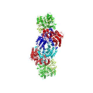
Nitrogenases are enzymes (EC 1.18.6.1EC 1.19.6.1) that are produced by certain bacteria, such as cyanobacteria (blue-green bacteria) and rhizobacteria. These enzymes are responsible for the reduction of nitrogen (N2) to ammonia (NH3). Nitrogenases are the only family of enzymes known to catalyze this reaction, which is a step in the process of nitrogen fixation. Nitrogen fixation is required for all forms of life, with nitrogen being essential for the biosynthesis of molecules (nucleotides, amino acids) that create plants, animals and other organisms. They are encoded by the Nif genes or homologs. They are related to protochlorophyllide reductase.

<span class="mw-page-title-main">Glutamate dehydrogenase</span> Hexameric enzyme

Glutamate dehydrogenase is an enzyme observed in both prokaryotes and eukaryotic mitochondria. The aforementioned reaction also yields ammonia, which in eukaryotes is canonically processed as a substrate in the urea cycle. Typically, the α-ketoglutarate to glutamate reaction does not occur in mammals, as glutamate dehydrogenase equilibrium favours the production of ammonia and α-ketoglutarate. Glutamate dehydrogenase also has a very low affinity for ammonia, and therefore toxic levels of ammonia would have to be present in the body for the reverse reaction to proceed. However, in brain, the NAD+/NADH ratio in brain mitochondria encourages oxidative deamination. In bacteria, the ammonia is assimilated to amino acids via glutamate and aminotransferases. In plants, the enzyme can work in either direction depending on environment and stress. Transgenic plants expressing microbial GLDHs are improved in tolerance to herbicide, water deficit, and pathogen infections. They are more nutritionally valuable.

In molecular biology, biosynthesis is a multi-step, enzyme-catalyzed process where substrates are converted into more complex products in living organisms. In biosynthesis, simple compounds are modified, converted into other compounds, or joined to form macromolecules. This process often consists of metabolic pathways. Some of these biosynthetic pathways are located within a single cellular organelle, while others involve enzymes that are located within multiple cellular organelles. Examples of these biosynthetic pathways include the production of lipid membrane components and nucleotides. Biosynthesis is usually synonymous with anabolism.

<span class="mw-page-title-main">Glutamate dehydrogenase 1</span> Enzyme

GLUD1 is a mitochondrial matrix enzyme, one of the family of glutamate dehydrogenases that are ubiquitous in life, with a key role in nitrogen and glutamate (Glu) metabolism and energy homeostasis. This dehydrogenase is expressed at high levels in liver, brain, pancreas and kidney, but not in muscle. In the pancreatic cells, GLUD1 is thought to be involved in insulin secretion mechanisms. In nervous tissue, where glutamate is present in concentrations higher than in the other tissues, GLUD1 appears to function in both the synthesis and the catabolism of glutamate and perhaps in ammonia detoxification.

<span class="mw-page-title-main">Argininosuccinate synthase</span> Enzyme

Argininosuccinate synthase or synthetase is an enzyme that catalyzes the synthesis of argininosuccinate from citrulline and aspartate. In humans, argininosuccinate synthase is encoded by the ASS gene located on chromosome 9.

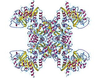
Carbamoyl phosphate synthetase I (CPS I) is a ligase enzyme located in the mitochondria involved in the production of urea. Carbamoyl phosphate synthetase I (CPS1 or CPSI) transfers an ammonia molecule to a molecule of bicarbonate that has been phosphorylated by a molecule of ATP. The resulting carbamate is then phosphorylated with another molecule of ATP. The resulting molecule of carbamoyl phosphate leaves the enzyme.

<span class="mw-page-title-main">Amino acid synthesis</span> The set of biochemical processes by which amino acids are produced

Amino acid synthesis is the set of biochemical processes by which the amino acids are produced. The substrates for these processes are various compounds in the organism's diet or growth media. Not all organisms are able to synthesize all amino acids. For example, humans can synthesize 11 of the 20 standard amino acids. These 11 are called the non-essential amino acids).

<span class="mw-page-title-main">CTP synthetase</span> Enzyme

CTP synthase is an enzyme involved in pyrimidine biosynthesis that interconverts UTP and CTP.

<span class="mw-page-title-main">Glutaminase</span>

Glutaminase is an amidohydrolase enzyme that generates glutamate from glutamine. Glutaminase has tissue-specific isoenzymes. Glutaminase has an important role in glial cells.

<span class="mw-page-title-main">Carbamoyl phosphate synthetase</span> Class of enzymes

Carbamoyl phosphate synthetase catalyzes the ATP-dependent synthesis of carbamoyl phosphate from glutamine or ammonia and bicarbonate. This enzyme catalyzes the reaction of ATP and bicarbonate to produce carboxy phosphate and ADP. Carboxy phosphate reacts with ammonia to give carbamic acid. In turn, carbamic acid reacts with a second ATP to give carbamoyl phosphate plus ADP.

<span class="mw-page-title-main">GMP synthase</span>

Guanosine monophosphate synthetase, also known as GMPS is an enzyme that converts xanthosine monophosphate to guanosine monophosphate.

<span class="mw-page-title-main">Amidophosphoribosyltransferase</span> Mammalian protein found in Homo sapiens

Amidophosphoribosyltransferase (ATase), also known as glutamine phosphoribosylpyrophosphate amidotransferase (GPAT), is an enzyme responsible for catalyzing the conversion of 5-phosphoribosyl-1-pyrophosphate (PRPP) into 5-phosphoribosyl-1-amine (PRA), using the amine group from a glutamine side-chain. This is the committing step in de novo purine synthesis. In humans it is encoded by the PPAT gene. ATase is a member of the purine/pyrimidine phosphoribosyltransferase family.

<span class="mw-page-title-main">Ribose-phosphate diphosphokinase</span> Class of enzymes

Ribose-phosphate diphosphokinase is an enzyme that converts ribose 5-phosphate into phosphoribosyl pyrophosphate (PRPP). It is classified under EC 2.7.6.1.

<span class="mw-page-title-main">Adenylylation</span> Biological process

Adenylylation, more commonly known as AMPylation, is a process in which an adenosine monophosphate (AMP) molecule is covalently attached to the amino acid side chain of a protein. This covalent addition of AMP to a hydroxyl side chain of the protein is a post-translational modification. Adenylylation involves a phosphodiester bond between a hydroxyl group of the molecule undergoing adenylylation, and the phosphate group of the adenosine monophosphate nucleotide. Enzymes that are capable of catalyzing this process are called AMPylators.

<span class="mw-page-title-main">Glutamine riboswitch</span> Glutamine-binding RNA structure

The glutamine riboswitch is a conserved RNA structure that was predicted by bioinformatics. It is present in a variety of lineages of cyanobacteria, as well as some phages that infect cyanobacteria. It is also found in DNA extracted from uncultivated bacteria living in the ocean that are presumably species of cyanobacteria.

<span class="mw-page-title-main">Ammonia transporter</span>

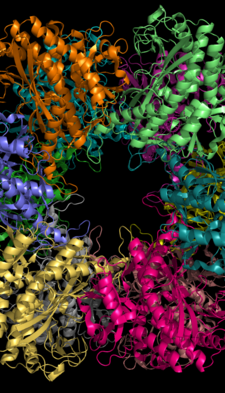
Ammonia transporters are structurally related membrane transport proteins called Amt proteins in bacteria and plants, methylammonium/ammonium permeases (MEPs) in yeast, or Rhesus (Rh) proteins in chordates. In humans, the RhAG, RhBG, and RhCG Rhesus proteins constitute solute carrier family 42 whilst RhD and RhCE form the Rh blood group system. The three-dimensional structure of the ammonia transport protein AmtB from Escherichia coli has been determined by x-ray crystallography revealing a hydrophobic ammonia channel. The human RhCG ammonia transporter was found to have a similar ammonia-conducting channel structure. It was proposed that the erythrocyte Rh complex is a heterotrimer of RhAG, RhD, and RhCE subunits in which RhD and RhCE might play roles in anchoring the ammonia-conducting RhAG subunit to the cytoskeleton. Based on reconstitution experiments, purified RhCG subunits alone can function to transport ammonia. RhCG is required for normal acid excretion by the mouse kidney and epididymis.

<span class="mw-page-title-main">Nif regulon</span>

The Nif regulon is a set of seven operons used to regulate nitrogen fixation in the coliform bacterium Klebsiella pneumoniae under anaerobic and microaerophilic conditions. It includes 17 nif genes, and is situated between the his and the Shi-A operon of the bacterium.

<span class="mw-page-title-main">Pii nitrogen regulatory proteins</span>

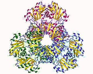
The PII family comprises a group of widely distributed signal transduction proteins found in nearly all Bacteria and also present in Archaea and in the chloroplasts of Algae and plants. PII form barrel-like homotrimers with a flexible loop, namely T-loop, emerging from each subunit. PII proteins have extraordinary sensory properties; they can exist in a vast range of structural status accordingly to the levels of ATP, ADP and 2-oxogluratate. These metabolites interact allosterically with PII in three conserved binding sites located in the lateral cavity between each PII subunit. ATP and ADP bind competitively to the nucleotide binding whereas the 2-oxoglutarate only interacts with PII in the presence of MgATP.

The glnALG operon is an operon that regulates the nitrogen content of a cell. It codes for the structural gene glnA the two regulatory genes glnL and glnG. glnA encodes glutamine synthetase, an enzyme which catalyzes the conversion of glutamate and ammonia to glutamine, thereby controlling the nitrogen level in the cell. glnG encodes NRI which regulates the expression of the glnALG operon at three promoters, which are glnAp1, glnAp2 located upstream of glnA) and glnLp. glnL encodes NRII which regulates the activity of NRI. No significant homology is found in Eukaryotes.

Carbamoyl phosphate synthetase III is one of the three isoforms of the carbamoyl phosphate synthetase, an enzyme that catalyzes the active production of carbamoyl phosphate in many organisms.

References

  1. 1 2 3 PDB: 1FPY ; Gill HS, Eisenberg D (February 2001). "The crystal structure of phosphinothricin in the active site of glutamine synthetase illuminates the mechanism of enzymatic inhibition". Biochemistry. 40 (7): 1903–12. doi:10.1021/bi002438h. PMID   11329256.
  2. PDB: 2GLS ; Yamashita MM, Almassy RJ, Janson CA, Cascio D, Eisenberg D (October 1989). "Refined atomic model of glutamine synthetase at 3.5 A resolution". J. Biol. Chem. 264 (30): 17681–90. doi:10.2210/pdb2gls/pdb. PMID   2572586.
  3. Eisenberg D, Almassy RJ, Janson CA, Chapman MS, Suh SW, Cascio D, Smith WW (1987). "Some evolutionary relationships of the primary biological catalysts glutamine synthetase and RuBisCO". Cold Spring Harb. Symp. Quant. Biol. 52: 483–90. doi:10.1101/sqb.1987.052.01.055. PMID   2900091.
  4. 1 2 3 4 5 6 7 8 Liaw SH, Kuo I, Eisenberg D (Nov 1995). "Discovery of the ammonium substrate site on glutamine synthetase, a third cation binding site". Protein Sci. 4 (11): 2358–65. doi:10.1002/pro.5560041114. PMC   2143006 . PMID   8563633.
  5. 1 2 3 4 Liaw SH, Pan C, Eisenberg D (Jun 1993). "Feedback inhibition of fully unadenylylated glutamine synthetase from Salmonella typhimurium by glycine, alanine, and serine". Proc. Natl. Acad. Sci. USA. 90 (11): 4996–5000. Bibcode:1993PNAS...90.4996L. doi: 10.1073/pnas.90.11.4996 . PMC   46640 . PMID   8099447.
  6. 1 2 3 4 5 6 7 8 9 10 11 12 13 14 15 Eisenberg D, Gill HS, Pfluegl GM, Rotstein SH (Mar 2000). "Structure-function relationships of glutamine synthetases". Biochim Biophys Acta. 1477 (1–2): 122–45. doi:10.1016/S0167-4838(99)00270-8. PMID   10708854.
  7. 1 2 3 4 5 6 7 Liaw SH, Eisenberg D (Jan 1994). "Structural model for the reaction mechanism of glutamine synthetase, based on five crystal structures of enzyme-substrate complexes". Biochemistry. 33 (3): 675–81. doi:10.1021/bi00169a007. PMID   7904828.
  8. 1 2 3 4 Stryer L, Berg JM, Tymoczko JL (2007). Biochemistry (6th ed.). San Francisco: W.H. Freeman. pp.  679–706. ISBN   978-0-7167-8724-2.
  9. Goodsell DS (June 2002). "Glutamine Synthetase". Molecule of the month. RCSB Protein Data Bank. Archived from the original on 2008-05-31. Retrieved 2010-05-08.
  10. 1 2 3 4 5 Krajewski WW, Collins R, Holmberg-Schiavone L, Jones TA, Karlberg T, Mowbray SL (Jan 2008). "Crystal structures of mammalian glutamine synthetases illustrate substrate-induced conformational changes and provide opportunities for drug and herbicide design". J Mol Biol. 375 (1): 317–28. doi:10.1016/j.jmb.2007.10.029. PMID   18005987.
  11. Ginsburg A, Yeh J, Hennig SB, Denton MD (Feb 1970). "Some effects of adenylylation on the biosynthetic properties of the glutamine synthetase from Escherichia coli". Biochemistry. 9 (3): 633–49. doi:10.1021/bi00805a025. PMID   4906326.
  12. Hunt JB, Smyrniotis PZ, Ginsburg A, Stadtman ER (Jan 1975). "Metal ion requirement by glutamine synthetase of Escherichia coli in catalysis of gamma-glutamyl transfer". Arch Biochem Biophys. 166 (1): 102–24. doi:10.1016/0003-9861(75)90370-7. PMID   235885.
  13. Goodsell, DS (June 2002). "Glutamine Synthetase". RCSB Protein Data Bank. Archived from the original on 31 May 2008. Retrieved 8 May 2010.
  14. 1 2 3 4 Suarez I, Bodega G, Fernandez B (Aug–Sep 2002). "Glutamine synthetase in brain: effect of ammonia". Neurochem. Int. 41 (2–3): 123–42. doi:10.1016/S0197-0186(02)00033-5. PMID   12020613. S2CID   24661063.
  15. Venkatesh K, Srikanth L, Vengamma B, Chandrasekhar C, Sanjeevkumar A, Mouleshwara Prasad BC, Sarma PV (2013). "In vitro differentiation of cultured human CD34+ cells into astrocytes". Neurol India. 61 (4): 383–8. doi: 10.4103/0028-3886.117615 . PMID   24005729.
  16. 1 2 Willard-Mack CL, Koehler RC, Hirata T, et al. (March 1996). "Inhibition of glutamine synthetase reduces ammonia-induced astrocyte swelling in rat". Neuroscience. 71 (2): 589–99. doi:10.1016/0306-4522(95)00462-9. PMID   9053810. S2CID   31674240.
  17. Tanigami H, Rebel A, Martin LJ, Chen TY, Brusilow SW, Traystman RJ, Koehler RC (2005). "Effect of glutamine synthetase inhibition on astrocyte swelling and altered astroglial protein expression during hyperammonemia in rats". Neuroscience. 131 (2): 437–49. doi:10.1016/j.neuroscience.2004.10.045. PMC   1819407 . PMID   15708485.
  18. Kumada Y, Benson DR, Hillemann D, Hosted TJ, Rochefort DA, Thompson CJ, Wohlleben W, Tateno Y (April 1993). "Evolution of the glutamine synthetase gene, one of the oldest existing and functioning genes". Proc. Natl. Acad. Sci. U.S.A. 90 (7): 3009–13. Bibcode:1993PNAS...90.3009K. doi: 10.1073/pnas.90.7.3009 . PMC   46226 . PMID   8096645.
  19. Shatters RG, Kahn ML (November 1989). "Glutamine synthetase II in Rhizobium: reexamination of the proposed horizontal transfer of DNA from eukaryotes to prokaryotes". J. Mol. Evol. 29 (5): 422–8. Bibcode:1989JMolE..29..422S. doi:10.1007/BF02602912. PMID   2575672. S2CID   36704558.
  20. Brown JR, Masuchi Y, Robb FT, Doolittle WF (June 1994). "Evolutionary relationships of bacterial and archaeal glutamine synthetase genes". J. Mol. Evol. 38 (6): 566–76. Bibcode:1994JMolE..38..566B. doi:10.1007/BF00175876. PMID   7916055. S2CID   21493521.
  21. "GSI structure". Archived from the original on 2008-12-17. Retrieved 2009-03-31.
  22. InterPro:IPR001637 Glutamine synthetase class-I, adenylation site
  23. Ortega JL, Wilson OL, Sengupta-Gopalan C (December 2012). "The 5' untranslated region of the soybean cytosolic glutamine synthetase β(1) gene contains prokaryotic translation initiation signals and acts as a translational enhancer in plants". Molecular Genetics and Genomics. 287 (11–12): 881–93. doi:10.1007/s00438-012-0724-6. PMC   3881598 . PMID   23080263.
  24. van Rooyen JM, Abratt VR, Sewell BT (August 2006). "Three-dimensional structure of a type III glutamine synthetase by single-particle reconstruction". J. Mol. Biol. 361 (4): 796–810. doi:10.1016/j.jmb.2006.06.026. hdl: 11394/1617 . PMID   16879836.
  25. 1 2 3 4 5 Garrett, Grisham (2017). Biochemistry (6th ed.). United States of America: Cengage Learning. pp. 886–889. ISBN   978-1-305-57720-6.
  26. Ivanovsky RN, Khatipov EA (1994). "Evidence of covalent modification of glutamine synthetase in the purple sulfur bacterium". FEMS Microbiology Letters. 122 (1–2): 115–119. doi: 10.1111/j.1574-6968.1994.tb07153.x .
  27. Krishnan IS, Singhal RK, Dua RD (Apr 1986). "Purification and characterization of glutamine synthetase from Clostridium pasteurianum". Biochemistry. 25 (7): 1589–99. doi:10.1021/bi00355a021. PMID   2871863.
  28. Bolay, Paul; Muro-Pastor, M.; Florencio, Francisco; Klähn, Stephan (27 October 2018). "The Distinctive Regulation of Cyanobacterial Glutamine Synthetase". Life. 8 (4): 52. doi: 10.3390/life8040052 . PMC   6316151 . PMID   30373240.
  29. Merrick MJ, Edwards RA (December 1995). "Nitrogen control in bacteria". Microbiological Reviews. 59 (4): 604–22. doi:10.1128/MR.59.4.604-622.1995. PMC   239390 . PMID   8531888.
  30. 1 2 Fisher R, Tuli R, Haselkorn R (June 1981). "A cloned cyanobacterial gene for glutamine synthetase functions in Escherichia coli, but the enzyme is not adenylylated". Proceedings of the National Academy of Sciences of the United States of America. 78 (6): 3393–7. Bibcode:1981PNAS...78.3393F. doi: 10.1073/pnas.78.6.3393 . PMC   319574 . PMID   6115380.
  31. Vega-Palas MA, Flores E, Herrero A (July 1992). "NtcA, a global nitrogen regulator from the cyanobacterium Synechococcus that belongs to the Crp family of bacterial regulators". Molecular Microbiology. 6 (13): 1853–9. doi:10.1111/j.1365-2958.1992.tb01357.x. PMID   1630321. S2CID   32757978.
  32. Reyes JC, Muro-Pastor MI, Florencio FJ (April 1997). "Transcription of glutamine synthetase genes (glnA and glnN) from the cyanobacterium Synechocystis sp. strain PCC 6803 is differently regulated in response to nitrogen availability". Journal of Bacteriology. 179 (8): 2678–89. doi:10.1128/jb.179.8.2678-2689.1997. PMC   179018 . PMID   9098067.
  33. García-Domínguez M, Reyes JC, Florencio FJ (June 1999). "Glutamine synthetase inactivation by protein-protein interaction". Proceedings of the National Academy of Sciences of the United States of America. 96 (13): 7161–6. Bibcode:1999PNAS...96.7161G. doi: 10.1073/pnas.96.13.7161 . PMC   22038 . PMID   10377385.
  34. García-Domínguez M, Reyes JC, Florencio FJ (March 2000). "NtcA represses transcription of gifA and gifB, genes that encode inhibitors of glutamine synthetase type I from Synechocystis sp. PCC 6803". Molecular Microbiology. 35 (5): 1192–201. doi:10.1046/j.1365-2958.2000.01789.x. PMID   10712699. S2CID   23804565.
  35. Klähn S, Schaal C, Georg J, Baumgartner D, Knippen G, Hagemann M, Muro-Pastor AM, Hess WR (November 2015). "The sRNA NsiR4 is involved in nitrogen assimilation control in cyanobacteria by targeting glutamine synthetase inactivating factor IF7". Proceedings of the National Academy of Sciences of the United States of America. 112 (45): E6243-52. Bibcode:2015PNAS..112E6243K. doi: 10.1073/pnas.1508412112 . PMC   4653137 . PMID   26494284.
  36. Klähn S, Bolay P, Wright PR, Atilho RM, Brewer KI, Hagemann M, Breaker RR, Hess WR (August 2018). "A glutamine riboswitch is a key element for the regulation of glutamine synthetase in cyanobacteria". Nucleic Acids Research. 46 (19): 10082–10094. doi:10.1093/nar/gky709. PMC   6212724 . PMID   30085248.