Related Research Articles
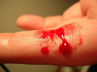
Bleeding, hemorrhage, haemorrhage or blood loss is blood escaping from the circulatory system from damaged blood vessels. Bleeding can occur internally, or externally either through a natural opening such as the mouth, nose, ear, urethra, vagina or anus, or through a puncture in the skin. Hypovolemia is a massive decrease in blood volume, and death by excessive loss of blood is referred to as exsanguination. Typically, a healthy person can endure a loss of 10–15% of the total blood volume without serious medical difficulties. The stopping or controlling of bleeding is called hemostasis and is an important part of both first aid and surgery.

Heart murmurs are unique heart sounds produced when blood flows across a heart valve or blood vessel. This occurs when turbulent blood flow creates a sound loud enough to hear with a stethoscope. The sound differs from normal heart sounds by their characteristics. For example, heart murmurs may have a distinct pitch, duration and timing. The major way health care providers examine the heart on physical exam is heart auscultation; another clinical technique is palpation, which can detect by touch when such turbulence causes the vibrations called cardiac thrill. A murmur is a sign found during the cardiac exam. Murmurs are of various types and are important in the detection of cardiac and valvular pathologies.

Shaken baby syndrome (SBS), also known as abusive head trauma (AHT), is a medical condition in children younger than five years old, generally caused by blunt trauma, vigorous shaking, or a combination of both. SBS is the leading cause of fatal head injuries in children under two, with a risk of death of about 25%. The most common symptoms include retinal bleeds, multiple fractures of the long bones, and subdural hematomas.

Pulmonary edema, also known as pulmonary congestion, is excessive fluid accumulation in the tissue or air spaces of the lungs. This leads to impaired gas exchange, most often leading to shortness of breath (dyspnea) which can progress to hypoxemia and respiratory failure. Pulmonary edema has multiple causes and is traditionally classified as cardiogenic or noncardiogenic.

An injury is any physiological damage to living tissue caused by immediate physical stress. Injuries to humans can occur intentionally or unintentionally and may be caused by blunt trauma, penetrating trauma, burning, toxic exposure, asphyxiation, or overexertion. Injuries can occur in any part of the body, and different symptoms are associated with different injuries.
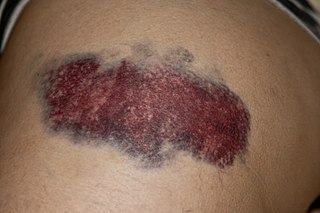
A bruise, also known as a contusion, is a type of hematoma of tissue, the most common cause being capillaries damaged by trauma, causing localized bleeding that extravasates into the surrounding interstitial tissues. Most bruises occur close enough to the epidermis such that the bleeding causes a visible discoloration. The bruise then remains visible until the blood is either absorbed by tissues or cleared by immune system action. Bruises which do not blanch under pressure can involve capillaries at the level of skin, subcutaneous tissue, muscle, or bone.

The mediastinum is the central compartment of the thoracic cavity. Surrounded by loose connective tissue, it is an undelineated region that contains a group of structures within the thorax, namely the heart and its vessels, the esophagus, the trachea, the phrenic and cardiac nerves, the thoracic duct, the thymus and the lymph nodes of the central chest.

Pneumomediastinum is pneumatosis in the mediastinum, the central part of the chest cavity. First described in 1819 by René Laennec, the condition can result from physical trauma or other situations that lead to air escaping from the lungs, airways, or bowel into the chest cavity. In underwater divers it is usually the result of pulmonary barotrauma.

A chest injury, also known as chest trauma, is any form of physical injury to the chest including the ribs, heart and lungs. Chest injuries account for 25% of all deaths from traumatic injury. Typically chest injuries are caused by blunt mechanisms such as direct, indirect, compression, contusion, deceleration, or blasts caused by motor vehicle collisions or penetrating mechanisms such as stabbings.
Hamman's syndrome, also known as Macklin's syndrome, is a syndrome of spontaneous subcutaneous emphysema and pneumomediastinum, sometimes associated with pain and, less commonly, dyspnea, dysphonia, and a low-grade fever.

The carina of trachea is a ridge of cartilage at the base of the trachea separating the openings of the left and right main bronchi.

Athletic heart syndrome (AHS) is a non-pathological condition commonly seen in sports medicine in which the human heart is enlarged, and the resting heart rate is lower than normal.
A gunshot wound (GSW) is a penetrating injury caused by a projectile from a gun. Damage may include bleeding, bone fractures, organ damage, wound infection, loss of the ability to move part of the body, and in severe cases, death. Damage depends on the part of the body hit, the path the bullet follows through the body, and the type and speed of the bullet. Long-term complications can include bowel obstruction, failure to thrive, neurogenic bladder and paralysis, recurrent cardiorespiratory distress and pneumothorax, hypoxic brain injury leading to early dementia, amputations, chronic pain and pain with light touch (hyperalgesia), deep venous thrombosis with pulmonary embolus, limb swelling and debility, and lead poisoning.
Pneumopericardium is a medical condition where air enters the pericardial cavity. This condition has been recognized in preterm neonates, in which it is associated with severe lung pathology, after vigorous resuscitation, or in the presence of assisted ventilation. This is a serious complication, which if untreated may lead to cardiac tamponade and death. Pneumomediastinum, which is the presence of air in the mediastinum, may mimic and also coexist with pneumopericardium.
A pulmonary contusion, also known as lung contusion, is a bruise of the lung, caused by chest trauma. As a result of damage to capillaries, blood and other fluids accumulate in the lung tissue. The excess fluid interferes with gas exchange, potentially leading to inadequate oxygen levels (hypoxia). Unlike pulmonary laceration, another type of lung injury, pulmonary contusion does not involve a cut or tear of the lung tissue.

Subcutaneous emphysema occurs when gas or air accumulates and seeps under the skin, where normally no gas should be present. Subcutaneous refers to the subcutaneous tissue, and emphysema refers to trapped air pockets. Since the air generally comes from the chest cavity, subcutaneous emphysema usually occurs around the upper torso, such as on the chest, neck, face, axillae and arms, where it is able to travel with little resistance along the loose connective tissue within the superficial fascia. Subcutaneous emphysema has a characteristic crackling-feel to the touch, a sensation that has been described as similar to touching warm Rice Krispies. This sensation of air under the skin is known as subcutaneous crepitation, a form of crepitus.

A sternal fracture is a fracture of the sternum, located in the center of the chest. The injury, which occurs in 5–8% of people who experience significant blunt chest trauma, may occur in vehicle accidents, when the still-moving chest strikes a steering wheel or dashboard or is injured by a seatbelt. Cardiopulmonary resuscitation (CPR), has also been known to cause thoracic injury, including sternum and rib fractures. Sternal fractures may also occur as a pathological fracture, in people who have weakened bone in their sternum, due to another disease process. Sternal fracture can interfere with breathing by making it more painful; however, its primary significance is that it can indicate the presence of serious associated internal injuries, especially to the heart and lungs.

Tracheobronchial injury is damage to the tracheobronchial tree. It can result from blunt or penetrating trauma to the neck or chest, inhalation of harmful fumes or smoke, or aspiration of liquids or objects.

Diaphragmatic rupture is a tear of the diaphragm, the muscle across the bottom of the ribcage that plays a crucial role in breathing. Most commonly, acquired diaphragmatic tears result from physical trauma. Diaphragmatic rupture can result from blunt or penetrating trauma and occurs in about 0.5% of all people with trauma.
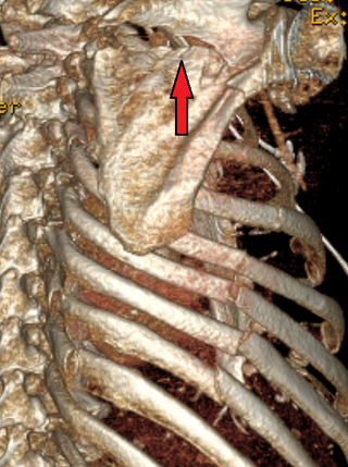
A scapular fracture is a fracture of the scapula, the shoulder blade. The scapula is sturdy and located in a protected place, so it rarely breaks. When it does, it is an indication that the individual was subjected to a considerable amount of force and that severe chest trauma may be present. High-speed vehicle accidents are the most common cause. This could be anywhere from a car accident, motorcycle crash, or high speed bicycle crash but falls and blows to the area can also be responsible for the injury. Signs and symptoms are similar to those of other fractures: they include pain, tenderness, and reduced motion of the affected area although symptoms can take a couple of days to appear. Imaging techniques such as X-ray are used to diagnose scapular fracture, but the injury may not be noticed in part because it is so frequently accompanied by other, severe injuries that demand attention. The injuries that usually accompany scapular fracture generally have the greatest impact on the patient's outcome. However, the injury can also occur by itself; when it does, it does not present a significant threat to life. Treatment involves pain control and immobilizing the affected area, and, later, physical therapy.
References
- ↑ Shabetai, Ralph (2003-10-31). The Pericardium - Google Book Search. Springer. ISBN 9781402076398 . Retrieved 2008-11-26.
- ↑ Hadjis T, Palisaitis D, Dontigny L, Allard M (March 1995). "Benign pneumopericardium and tamponade". Can J Cardiol. 11 (3): 232–4. PMID 7889442.
- ↑ "Hamman sign" at Dorland's Medical Dictionary
- ↑ synd/3001 at Who Named It?
- ↑ McCann], [executive publisher, Judith A. Shilling (2008). Nursing. Ambler, PA: Lippincott Williams & Wilkins. p. 171. ISBN 9781582556680.
{{cite book}}: CS1 maint: multiple names: authors list (link) - ↑ . Chu CP, Chen PP (April 2002). "Tracheobronchial injury secondary to blunt chest trauma: Diagnosis and management". Anaesth Intensive Care. 30 (2): 145–52. doi: 10.1177/0310057X0203000204 . PMID 12002920.