
Anotia describes a rare congenital deformity that involves the complete absence of the auricle, the outer projected portion of the ear, and narrowing or absence of the ear canal. This contrasts with microtia, in which a small part of the auricle is present. Anotia and microtia may occur unilaterally or bilaterally. This deformity results in conductive hearing loss, deafness.

The facial nerve, also known as the seventh cranial nerve, cranial nerve VII, or simply CN VII, is a cranial nerve that emerges from the pons of the brainstem, controls the muscles of facial expression, and functions in the conveyance of taste sensations from the anterior two-thirds of the tongue. The nerve typically travels from the pons through the facial canal in the temporal bone and exits the skull at the stylomastoid foramen. It arises from the brainstem from an area posterior to the cranial nerve VI and anterior to cranial nerve VIII.

The vestibulocochlear nerve or auditory vestibular nerve, also known as the eighth cranial nerve, cranial nerve VIII, or simply CN VIII, is a cranial nerve that transmits sound and equilibrium (balance) information from the inner ear to the brain. Through olivocochlear fibers, it also transmits motor and modulatory information from the superior olivary complex in the brainstem to the cochlea.

The temporal bones are situated at the sides and base of the skull, and lateral to the temporal lobes of the cerebral cortex.

An ear is the organ that enables hearing and body balance using the vestibular system. In mammals, the ear is usually described as having three parts: the outer ear, the middle ear and the inner ear. The outer ear consists of the pinna and the ear canal. Since the outer ear is the only visible portion of the ear in most animals, the word "ear" often refers to the external part alone. The middle ear includes the tympanic cavity and the three ossicles. The inner ear sits in the bony labyrinth, and contains structures which are key to several senses: the semicircular canals, which enable balance and eye tracking when moving; the utricle and saccule, which enable balance when stationary; and the cochlea, which enables hearing. The ear canal is cleaned via earwax, which naturally migrates to the auricle. The ears of vertebrates are placed somewhat symmetrically on either side of the head, an arrangement that aids sound localization.

Elastic cartilage, fibroelastic cartilage or yellow fibrocartilage is a type of cartilage present in the pinnae (auricles) of the ear giving it shape, provides shape for the lateral region of the external auditory meatus, medial part of the auditory canal Eustachian tube, corniculate and cuneiform laryneal cartilages, and the epiglottis. It contains elastic fiber networks and collagen type II fibers. The principal protein is elastin.

The otic ganglion is a small parasympathetic ganglion located immediately below the foramen ovale in the infratemporal fossa and on the medial surface of the mandibular nerve. It is functionally associated with the glossopharyngeal nerve and innervates the parotid gland for salivation.

The tympanic cavity is a small cavity surrounding the bones of the middle ear. Within it sit the ossicles, three small bones that transmit vibrations used in the detection of sound.
The vestibular ganglion is a collection of cell bodies belonging to first order sensory neurons of the vestibular nerve. It is located within the internal auditory canal.

The bony labyrinth is the rigid, bony outer wall of the inner ear in the temporal bone. It consists of three parts: the vestibule, semicircular canals, and cochlea. These are cavities hollowed out of the substance of the bone, and lined by periosteum. They contain a clear fluid, the perilymph, in which the membranous labyrinth is situated.

Otic vesicle, or auditory vesicle, consists of either of the two sac-like invaginations formed and subsequently closed off during embryonic development. It is part of the neural ectoderm, which will develop into the membranous labyrinth of the inner ear. This labyrinth is a continuous epithelium, giving rise to the vestibular system and auditory components of the inner ear. During the earlier stages of embryogenesis, the otic placode invaginates to produce the otic cup. Thereafter, the otic cup closes off, creating the otic vesicle. Once formed, the otic vesicle will reside next to the neural tube medially, and on the lateral side will be paraxial mesoderm. Neural crest cells will migrate rostral and caudal to the placode.
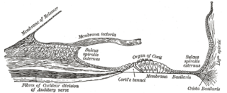
The spiral ligament is a fibrous cushion located between the stria vascularis and the bony otic capsule.

Human embryonic development or human embryogenesis is the development and formation of the human embryo. It is characterised by the processes of cell division and cellular differentiation of the embryo that occurs during the early stages of development. In biological terms, the development of the human body entails growth from a one-celled zygote to an adult human being. Fertilization occurs when the sperm cell successfully enters and fuses with an egg cell (ovum). The genetic material of the sperm and egg then combine to form the single cell zygote and the germinal stage of development commences. Human embryonic development covers the first eight weeks of development, which have 23 stages, called Carnegie stages. At the beginning of the ninth week, the embryo is termed a fetus. In comparison to the embryo, the fetus has more recognizable external features and a more complete set of developing organs.

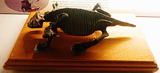
Dissorophus (DI-soh-ROH-fus) is an extinct genus of temnospondyl amphibian that lived during the Early Permian Period about 273 million years ago. Its fossils have been found in Texas and in Oklahoma in North America. Its heavy armor and robust build indicate Dissorophus was active on land, similar to other members of the clade Dissorophidae that are known from the Late Carboniferous to the Early Permian periods. Dissorphus is distinguished by its small body size, disproportionately large head and short trunk.
Quasicyclotosaurus is an extinct genus of mastodonsauroid temnospondyl. It had a closed otic notch.
Kawingasaurus is an extinct genus of dicynodont therapsid from the Late Permian Usili Formation of Tanzania. It is a member of the family Cistecephalidae, and like other cistecephalids it is thought to have been fossorial. It is a member of the family Cistecephalidae. Cistephalidae includes genera Cisteceohalus, Cistecephaloides and Kawingasaurus. Greek for Saurus meaning “lizard” appears as a suffix denoting a reptilian origin. Living between 254.17 and 259.9 million years ago in the late Permian and believed to have the first and last recorded appearance in this time period. It lived in deep burrows as a suggested by most burrowing dicynodonts from evaluation of cranial sutures, vestibule inflation and enlarged stapes foot plates which are thought to be functionally correlated with bone-conduction hearing; all observed in fossorial vertebrates which use seismic signals as communication.
In embryology, the otic placode is a thickening of the ectoderm on the outer surface of a developing embryo from which the ear develops. The ear, including both the vestibular system and the auditory system, develops from the otic placode beginning the third week of development. During the fourth week, the otic placode invaginates into the mesenchyme adjacent to the rhombencephalon to form the otic pit, which then pinches off from the surface ectoderm to form the otic vesicle.
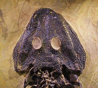
Otic notches are invaginations in the posterior margin of the skull roof, one behind each orbit. Otic notches are one of the features lost in the evolution of amniotes from their tetrapod ancestors.

Nycteroleteridae is a family of procolophonian parareptilians from the Middle to Late Permian of Russia and North America. They are sometimes classified as a sister group to pareiasaurids. The group includes the genera Macroleter, Bashkyroleter, "Bashkyroleter" mesensis, Nycteroleter, Emeroleter, and probably Rhipaeosaurus. They were carnivorous, and occasionally ate insects. The group was most common in European Russia, with only a few fossils in North America. One fossil has also been found in Africa, but this is the only one from Gondwana.

The following diagram is provided as an overview of and topical guide to the human nervous system:
