Eye development

Last updated
Transverse section of head of chick embryo of forty-eight hours' incubation Gray863.png
Transverse section of head of chick embryo of forty-eight hours’ incubation
Transverse section of head of chick embryo of fifty-two hours' incubation, showing the lens and the optic cup Gray864.png
Transverse section of head of chick embryo of fifty-two hours’ incubation, showing the lens and the optic cup

Eye formation in the human embryo begins at approximately three weeks into embryonic development and continues through the tenth week. [1] Cells from both the mesodermal and the ectodermal tissues contribute to the formation of the eye. Specifically, the eye is derived from the neuroepithelium, surface ectoderm, and the extracellular mesenchyme which consists of both the neural crest and mesoderm. [2] [3] [4]

Contents

Neuroepithelium forms the retina, ciliary body, iris, and optic nerves. Surface ectoderm forms the lens, corneal epithelium and eyelid. The extracellular mesenchyme forms the sclera, the corneal endothelium and stroma, blood vessels, muscles, and vitreous.

The eye begins to develop as a pair of optic vesicles on each side of the forebrain at the end of the 4th week of pregnancy. Optic vesicles are outgrowings of the brain which make contact with the surface ectoderm and this contact induces changes necessary for further development of the eye. Through a groove at the bottom of the optic vesicle known as choroid fissure the blood vessels enter the eye. Several layers such as the neural tube, neural crest, surface ectoderm, and mesoderm contribute to the development of the eye. [2] [3] [4]

Eye development is initiated by the master control gene PAX6 , a homeobox gene with known homologues in humans (aniridia), mice (small eye), and Drosophila (eyeless). The PAX6 gene locus is a transcription factor for the various genes and growth factors involved in eye formation. [1] [5] Eye morphogenesis begins with the evagination, or outgrowth, of the optic grooves or sulci. These two grooves in the neural folds transform into optic vesicles with the closure of the neural tube. [6] The optic vesicles then develop into the optic cup with the inner layer forming the retina and the outer portion forming the retinal pigment epithelium. The middle portion of the optic cup develops into the ciliary body and iris. [7] During the invagination of the optic cup, the ectoderm begins to thicken and form the lens placode, which eventually separates from the ectoderm to form the lens vesicle at the open end of the optic cup. [1] [3] [4]

Further differentiation and mechanical rearrangement of cells in and around the optic cup gives rise to the fully developed eye.

Sequential inductions

This development is an example of sequential inductions where the organ is formed from three different tissues:

Neural tube ectoderm (neuroectoderm)

First, there is an outpocketing of the neural tube called optic vesicles. Development of the optic vesicles starts in the 3-week embryo, from a progressively deepening groove in the neural plate called the optic sulcus. Some studies suggest this mechanism is regulated by RX/RAX transcription factor. [8] The proteins Wnt and FGF (fibroblast growth factor) play a part in this early stage and are regulated by another protein called Shisa. [6] As this expands, the rostral neuropore (the exit of the brain cavity out of the embryo) closes and the optic sulcus and the neural plate becomes the optic vesicle. [9] Optic nerves arise from connections of the vesicles to the forebrain. [1]

Neuroectoderm gives rise to the following compartments of the eye:

Surface ectoderm

Lens development is closely related to optic vesicle development. The interaction between the growing vesicle and the ectoderm causes the ectoderm to thicken at that point. This thickened portion of the ectoderm is called the lens placode. Next, the placode invaginates and forms a pouch referred to as the lens pit. [1] [3] [4] Scientists are studying the tension forces necessary for invagination of the lens placode and current research suggests that microfilaments might be present in early retinal cells to allow for invagination behavior. Research has also shown that Rho GTPase dependent filopodia from the precursor lens ectoderm play an important role in the formation of the lens pit. [10] [3] [4] Eventually, the pit becomes completely enclosed. This enclosed structure is the lens vesicle. [1] Studies have shown that lens development requires the presence of the Pax6 gene, which is the master regulatory gene for eye morphogenesis. [11] This master regulatory gene is not necessary for the closely associated optic vesicle development. [12] Additionally, Ras activation has been shown to be sufficient for starting lens differentiation, but not enough for its completion. [11]

The optic vesicles then begin to form the optic cup. [3] [4] Optic cup morphogenesis is the invagination process occurring after neuroectoderm movement forms the spherical optic vesicle (Phase 1). Invagination is when a tissue folds back on itself. Over the course of approximately 12 hours, the distal end of the optic vesicle inner layer begins to flatten (Phase 2). Over the following 18 hours, both the inner and outer layers begin to flex inward at sharp angles, beginning the formation of a C-shaped edge (Phase 3). The final 18 hours involve continuing this apically convex invagination to form the optic cup [3] [4] . At this point, morphologies such as columnar epithelial cells, pseudo-stratified cells, and apically narrow wedge-shaped cells can be observed. [13]

The inner layer of the optic cup is made of neuroepithelium (neural retina), while the outer layer is composed of retinal pigment epithelium (RPE). Experiments have determined that RPE cell differentiation and maintenance requires interaction with neighboring tissues, most likely canonical Wnt signaling, while neural retina differentiation is driven by tissue-autonomous factors. [13]

Bone morphogenic proteins (BMPs) are important regulators of optic cup development. In fact, research studies have shown that BMP agonists and antagonists are necessary for precision of optic cup development. [12] Interactions between tissues and signaling pathways also play a major role in morphogenesis of the optic cup. [8]

It is of interest to note that research has shown isolating the optic cup from neighboring tissue after completed invagination in tissue culture medium can lead to the development of most major parts of the eye, including photoreceptors, ganglion cells, bipolar cells, horizontal cells, amacrine cells and Muller glia. This indicates that morphogenesis of the optic cup occurs independently of external cues from its environment, including presence of lens. [13] However, the lens is necessary to act as an inducer for the ectoderm to transform it into the cornea.

Surface ectoderm produces the following parts:

Neural crest

Neural crest cells are themselves derived from the ectoderm and lie close to the neural tube:

Mesoderm

Mesoderm contributes to the following structures:

Developmental cascade

According to Liem et al., the organogenesis of the eye is pointed out as an example of a developmental cascade of inductions. The eye is essentially a derivative of the ectoderm from the somatic ectoderm and neural tube, with a succession of inductions by the chordamesoderm.

Chordamesoderm induces the anterior portion of the neural tube to form the precursors of the synapomorphic tripartite brain of vertebrates, and it will form a bulge called the diencephalon. Further induction by the chordamesoderm will form a protrusion: the optic vesicle. This vesicle will be subsequently invaginated by means of further inductions from the chordamesoderm. The optic vesicle will then induce the ectoderm that thickens (lens placode) and further invaginates to a point that detaches from the ectoderm and forms a neurogenic placode by itself. The lens placode is affected by the chordamesoderm making it invaginate and forms the optic cup composed by an inner layer of the neural retina and outer layer of the pigmented retina that will unite and form the optic stalk. The pigmented retina is formed by rods and cones and composed of small cilia typical of the ependymal epithelium of the neural tube. Some cells in the lens vesicle will be fated to form the cornea and the lens vesicle will develop completely to form the definitive lens. Iris is formed from the optic cup cells.

Responsivity of head epidermis

Only the epidermis in the head is competent to respond to the signal from the optic vesicles. Both the optic vesicle and the head epidermis are required for eye development. The competence of the head epidermis to respond to the optic vesicle signals comes from the expression of Pax6 in the epidermis. Pax6 is necessary and sufficient for eye induction. This competence is acquired gradually during gastrulation and neurulation from interactions with the endoderm, mesoderm, and neural plate.

Regulation and inhibition

Sonic hedgehog reduces the expression of Pax6. When Shh is inhibited during development, the domain of expression for Pax6 is expanded and the eyes fail to separate causing cyclopia. [14] Overexpression of Shh causes a loss of eye structures.

Retinoic acid generated from vitamin A in the retina plays an essential role in eye development as a secreted paracrine signal which restricts invasion of perioptic mesenchyme around the optic cup. [15] Vitamin A deficiency during embryogenesis results in anterior segment defects (particularly cornea and eyelids) that lead to vision loss or blindness.

There is some evidence that LMX1B plays a role in periocular mesenchymal survival. [16]

Additional images

Related Research Articles

<span class="mw-page-title-main">Mesoderm</span> Middle germ layer of embryonic development

The mesoderm is the middle layer of the three germ layers that develops during gastrulation in the very early development of the embryo of most animals. The outer layer is the ectoderm, and the inner layer is the endoderm.

<span class="mw-page-title-main">Lens (vertebrate anatomy)</span> Eye structure

The lens, or crystalline lens, is a transparent biconvex structure in most land vertebrate eyes. Along with the cornea, aqueous and vitreous humours it refracts light, focusing it onto the retina. In many land animals the shape of the lens can be altered, effectively changing the focal length of the eye, enabling them to focus on objects at various distances. This adjustment of the lens is known as accommodation. In many fully aquatic vertebrates such as fish other methods of accommodation are used such as changing the lens's position relative to the retina rather than changing lens shape. Accommodation is analogous to the focusing of a photographic camera via changing its lenses. In land vertebrates the lens is flatter on its anterior side than on its posterior side, while in fish the lens is often close to spherical.

<span class="mw-page-title-main">Ectoderm</span> Outer germ layer of embryonic development

The ectoderm is one of the three primary germ layers formed in early embryonic development. It is the outermost layer, and is superficial to the mesoderm and endoderm. It emerges and originates from the outer layer of germ cells. The word ectoderm comes from the Greek ektos meaning "outside", and derma meaning "skin".

<span class="mw-page-title-main">Invagination</span> Process in embryonic development

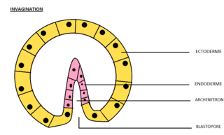
Invagination is the process of a surface folding in on itself to form a cavity, pouch or tube. In developmental biology, invagination is a mechanism that takes place during gastrulation. This mechanism or cell movement happens mostly in the vegetal pole. Invagination consists of the folding of an area of the exterior sheet of cells towards the inside of the blastula. In each organism, the complexity will be different depending on the number of cells. Invagination can be referenced as one of the steps of the establishment of the body plan. The term, originally used in embryology, has been adopted in other disciplines as well.

<span class="mw-page-title-main">Olfactory epithelium</span> Specialised epithelial tissue in the nasal cavity that detects odours

The olfactory epithelium is a specialized epithelial tissue inside the nasal cavity that is involved in smell. In humans, it measures 5 cm2 (0.78 sq in) and lies on the roof of the nasal cavity about 7 cm (2.8 in) above and behind the nostrils. The olfactory epithelium is the part of the olfactory system directly responsible for detecting odors.

<span class="mw-page-title-main">Neurulation</span> Embryological process forming the neural tube

Neurulation refers to the folding process in vertebrate embryos, which includes the transformation of the neural plate into the neural tube. The embryo at this stage is termed the neurula.

A germ layer is a primary layer of cells that forms during embryonic development. The three germ layers in vertebrates are particularly pronounced; however, all eumetazoans produce two or three primary germ layers. Some animals, like cnidarians, produce two germ layers making them diploblastic. Other animals such as bilaterians produce a third layer between these two layers, making them triploblastic. Germ layers eventually give rise to all of an animal's tissues and organs through the process of organogenesis.

Organogenesis is the phase of embryonic development that starts at the end of gastrulation and continues until birth. During organogenesis, the three germ layers formed from gastrulation form the internal organs of the organism.

A neurogenic placode is an area of thickening of the epithelium in the embryonic head ectoderm layer that gives rise to neurons and other structures of the sensory nervous system.

<span class="mw-page-title-main">Optic vesicle</span> Sac that protrudes from the embryonic forebrain to form each eye

The eyes begin to develop as a pair of diverticula (pouches) from the lateral aspects of the forebrain. These diverticula make their appearance before the closure of the anterior end of the neural tube; after the closure of the tube around the 4th week of development, they are known as the optic vesicles. Previous studies of optic vesicles suggest that the surrounding extraocular tissues – the surface ectoderm and extraocular mesenchyme – are necessary for normal eye growth and differentiation.

<span class="mw-page-title-main">Bone morphogenetic protein 4</span> Human protein and coding gene

Bone morphogenetic protein 4 is a protein that in humans is encoded by BMP4 gene. BMP4 is found on chromosome 14q22-q23.

<span class="mw-page-title-main">Mesenchyme</span> Type of animal embryonic connective tissue

Mesenchyme is a type of loosely organized animal embryonic connective tissue of undifferentiated cells that give rise to most tissues, such as skin, blood or bone. The interactions between mesenchyme and epithelium help to form nearly every organ in the developing embryo.

<span class="mw-page-title-main">Otic vesicle</span> Two sac-like invaginations formed and subsequently closed off during embryonic development

Otic vesicle, or auditory vesicle, consists of either of the two sac-like invaginations formed and subsequently closed off during embryonic development. It is part of the neural ectoderm, which will develop into the membranous labyrinth of the inner ear. This labyrinth is a continuous epithelium, giving rise to the vestibular system and auditory components of the inner ear. During the earlier stages of embryogenesis, the otic placode invaginates to produce the otic cup. Thereafter, the otic cup closes off, creating the otic vesicle. Once formed, the otic vesicle will reside next to the neural tube medially, and on the lateral side will be paraxial mesoderm. Neural crest cells will migrate rostral and caudal to the placode.

<span class="mw-page-title-main">Human embryonic development</span> Development and formation of the human embryo

Human embryonic development or human embryogenesis is the development and formation of the human embryo. It is characterised by the processes of cell division and cellular differentiation of the embryo that occurs during the early stages of development. In biological terms, the development of the human body entails growth from a one-celled zygote to an adult human being. Fertilization occurs when the sperm cell successfully enters and fuses with an egg cell (ovum). The genetic material of the sperm and egg then combine to form the single cell zygote and the germinal stage of development commences. Embryonic development in the human, covers the first eight weeks of development; at the beginning of the ninth week the embryo is termed a fetus. The eight weeks has 23 stages.

<span class="mw-page-title-main">Surface ectoderm</span>

The surface ectoderm, AKA external ectoderm, is one of the two early embryonic divisions of the ectoderm. The other early division of the ectoderm is the neuroectoderm.

<span class="mw-page-title-main">Lens placode</span> Thickened portion of ectoderm which serves as the precursor to the lens

The lens placode is a thickened portion of ectoderm that serves as the precursor to the lens.

In embryology, the otic placode is a thickening of the ectoderm on the outer surface of a developing embryo from which the ear develops. The ear, including both the vestibular system and the auditory system, develops from the otic placode beginning the third week of development. During the fourth week, the otic placode invaginates into the mesenchyme adjacent to the rhombencephalon to form the otic pit, which then pinches off from the surface ectoderm to form the otic vesicle.

<span class="mw-page-title-main">Retinal precursor cells</span> Type of cell in the human eye

Retinal precursor cells are biological cells that differentiate into the various cell types of the retina during development. In the vertebrate, these retinal cells differentiate into seven cell types, including retinal ganglion cells, amacrine cells, bipolar cells, horizontal cells, rod photoreceptors, cone photoreceptors, and Müller glia cells. During embryogenesis, retinal cells originate from the anterior portion of the neural plate termed the eye field. Eye field cells with a retinal fate express several transcription factor markers including Rx1, Pax6, and Lhx2. The eye field gives rise to the optic vesicle and then to the optic cup. The retina is generated from the precursor cells within the inner layer of the optic cup, as opposed to the retinal pigment epithelium that originate from the outer layer of the optic cup. In general, the developing retina is organized so that the least-committed precursor cells are located in the periphery of the retina, while the committed cells are located in the center of the retina. The differentiation of retinal precursor cells into the mature cell types found in the retina is coordinated in time and space by factors within the cell as well as factors in the environment of the cell. One example of an intrinsic regulator of this process is the transcription factor Ath5. Ath5 expression in retinal progenitor cells biases their differentiation into a retinal ganglion cell fate. An example of an environmental factor is the morphogen sonic hedge hog (Shh). Shh has been shown to repress the differentiation of precursor cells into retinal ganglion cells.

The face and neck development of the human embryo refers to the development of the structures from the third to eighth week that give rise to the future head and neck. They consist of three layers, the ectoderm, mesoderm and endoderm, which form the mesenchyme, neural crest and neural placodes. The paraxial mesoderm forms structures named somites and somitomeres that contribute to the development of the floor of the brain and voluntary muscles of the craniofacial region. The lateral plate mesoderm consists of the laryngeal cartilages. The three tissue layers give rise to the pharyngeal apparatus, formed by six pairs of pharyngeal arches, a set of pharyngeal pouches and pharyngeal grooves, which are the most typical feature in development of the head and neck. The formation of each region of the face and neck is due to the migration of the neural crest cells which come from the ectoderm. These cells determine the future structure to develop in each pharyngeal arch. Eventually, they also form the neurectoderm, which forms the forebrain, midbrain and hindbrain, cartilage, bone, dentin, tendon, dermis, pia mater and arachnoid mater, sensory neurons, and glandular stroma.

Pax-6, member of the Pax gene class, is responsible for carrying the genetic information that will encode Pax-6 (protein) which dictates the development of the olfactory epithelium, eyes and central nervous system in vertebrates. Pax-6 is expressed as a transcription factor when neural ectoderm receives a combination of weak sonic hedgehog and a strong TGF-Beta signaling gradients. Expression is first seen in the forebrain, hindbrain, head ectoderm and spinal cord followed by later expression in midbrain. Expression in the head ectoderm will give rise to the nasal placodes and to the eye placodes. The nasal placodes will give rise to the olfactory epithelium while eye placodes give rise to the lens and cornea. Expression in the different brain regions is orchestrated with the combinatorial expression of other transcription factors to give rise to the central nervous system. Experiments in mice demonstrate that a deficiency in Pax-6 leads to decrease in brain size, brain structure abnormality leading to Autism, lack of iris formation or a thin cornea. Knockout experiments produced eyeless phenotypes reinforcing the gene’s role in eye development. Advancing research in this area may lead us to better understanding of the complexity seen in neural development and maybe one day be able to grow eye tissue in vitro.

References

  1. 1 2 3 4 5 6 Ort, D., David, H., "Development of the Eye". Retrieved 22 April 2015.
  2. 1 2 Sadler, T.W (1990). Langman's medical embryology (6th ed.). Williams and Wilkins. ISBN   978-0683074932.
  3. 1 2 3 4 5 6 7 Hosseini, Hadi S.; Beebe, David C.; Taber, Larry A. (2014). "Mechanical effects of the surface ectoderm on optic vesicle morphogenesis in the chick embryo". Journal of Biomechanics. 47 (16): 3837–3846. doi:10.1016/j.jbiomech.2014.10.018. PMC   4261019 . PMID   25458577.
  4. 1 2 3 4 5 6 7 Hosseini, Hadi S.; Taber, Larry A. (2018). "How mechanical forces shape the developing eye". Progress in Biophysics and Molecular Biology. 137 (16): 25–36. doi:10.1016/j.pbiomolbio.2018.01.004. PMC   6085168 . PMID   29432780.
  5. Keller, A. M. V., "Embryonic Development of the Eye". Retrieved 22 April 2015.
  6. 1 2 Fuhrmann, S., Levine, E. M. and Reh, T. A. (2000). "Extraocular mesenchyme patterns the optic vesicle during early eye development in the embryonic chick". Development 127, 4599–4609.
  7. LifeMap Science, Inc. "Embryonic and Postnatal Development of the Eye". Retrieved 22 April 2015.
  8. 1 2 Fuhrmann, S., “Eye Morphogenesis and Patterning of the Optic Vesicle” Current Topics in Developmental Biology 93, 61-84 (07 October 2010)
  9. "eye(vertebrate) McGraw-Hil; Encyclopedia of Science and Technology (2007), vol.6, pp.801-802
  10. Fuhrmann, Sabine, “Eye Morphogenesis and Patterning of the Optic Vesicle” Current Topics in Developmental Biology 93, 61-84 (07 October 2010)
  11. 1 2 Hill, M.A. (2015) Embryology Vision - Lens Development. Retrieved April 22, 2015.
  12. 1 2 Adler, R., Canto-Soler, M.V., “Molecular mechanisms of optic vesicle development: Complexities, ambiguities and controversies”, Developmental Biology 305,1,1-13 (1 May 2007)
  13. 1 2 3 Eiraku et al., "Self-organizing optic-cup morphogenesis in three-dimensional cell culture", Nature 472, 51–56 (07 April 2011)
  14. "Development of the Vertebrate Eye -- Developmental Biology -- NCBI Bookshelf" . Retrieved 2010-04-10.
  15. Duester, G (September 2008). "Retinoic acid synthesis and signaling during early organogenesis". Cell. 134 (6): 921–31. doi:10.1016/j.cell.2008.09.002. PMC   2632951 . PMID   18805086.
  16. McMahon C, Gestri G, Wilson SW, Link BA (August 2009). "Lmx1b is essential for survival of periocular mesenchymal cells and influences Fgf-mediated retinal patterning in zebrafish". Dev. Biol. 332 (2): 287–98. doi:10.1016/j.ydbio.2009.05.577. PMC   2716413 . PMID   19500562.

Further reading