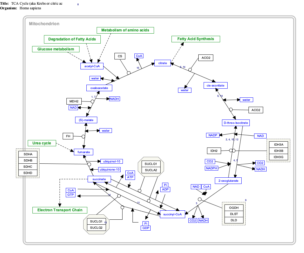
PDK4

Last updated
PDK4
Protein PDK4 PDB 2E0A.png
Available structures
PDB Ortholog search: PDBe RCSB
Identifiers
Aliases PDK4 , pyruvate dehydrogenase kinase 4
External IDs OMIM: 602527 MGI: 1351481 HomoloGene: 129720 GeneCards: PDK4
Orthologs
SpeciesHumanMouse
Entrez
Ensembl
UniProt
RefSeq (mRNA)

NM_002612

NM_013743

RefSeq (protein)

NP_002603

NP_038771

Location (UCSC) Chr 7: 95.58 – 95.6 Mb Chr 6: 5.48 – 5.5 Mb
PubMed search [3] [4]
Wikidata
View/Edit Human View/Edit Mouse

Pyruvate dehydrogenase lipoamide kinase isozyme 4, mitochondrial (PDK4) is an enzyme that in humans is encoded by the PDK4 gene. [5] [6] It codes for an isozyme of pyruvate dehydrogenase kinase.

Contents

This gene is a member of the PDK/BCKDK protein kinase family and encodes a mitochondrial protein with a histidine kinase domain. This protein is located in the matrix of the mitochondria and inhibits the pyruvate dehydrogenase complex by phosphorylating one of its subunits, reducing the conversion of pyruvate, which is produced from the oxidation of glucose and amino acids, to acetyl-CoA and contributing to the regulation of glucose metabolism. Expression of this gene is regulated by glucocorticoids, retinoic acid and insulin. [6] PDK4 is increased in hibernation and helps to decrease metabolism and conserve glucose by decreasing its conversion to acetyl-CoA, which enters the citric acid cycle and is converted to ATP. [7]

Structure

The mature protein encoded by the PDK4 gene contains 294 amino acids in its sequence. To form the active protein, two of the polypeptide chains come together to form an open conformation. [6] Specifically, the two subunits come together to form a nucleotide-binding pocket; this pocket is targeted most often by inhibitors. [8]

Function

The pyruvate dehydrogenase (PDH) complex must be tightly regulated due to its central role in general metabolism. Within the complex, there are three serine residues on the E1 component that are sites for phosphorylation; this phosphorylation inactivates the complex. In humans, there have been four isozymes of pyruvate dehydrogenase kinase that have been shown to phosphorylate these three sites: PDK1, PDK2, PDK3, and PDK4. PDK4 does not incorporate the most phosphate groups per catalytic event, because it can only phosphorylate site 1 and site 2; its rate of phosphorylation is less than PDK1, equal to PDK3, and more than PDK2. When the thiamine pyrophosphate (TPP) coenzyme is bound, the rates of phosphorylation by all four isozymes are drastically affected. Site 1 is the most affected, with the rate being significantly decreased. However, overall activity by PDK4 is not affected. [9]

Regulation

As the primary regulators of a crucial step in the central metabolic pathway, the pyruvate dehydrogenase family is tightly regulated itself by a myriad of factors including transcription factors Sp1 and CCAAT box binding factor (CBF). Retinoic acid enhances PDK4 transcription by enabling retinoic acid receptor family members to recruit transcriptional coactivators to retinoic acid response elements (RAREs) in the PDK4 promoter. Transcription is also increased by inhibiting inhibitory histone deacetylases (HDACs) using trichostatin A (TSA). [10] Rosiglitazone, a thiazolidinedione known to activate the glycerol biogenesis pathway, increases PDK4 mRNA transcription in white adipose tissue, but not in liver or muscle tissue. [11] Farnesoid X receptor, or FXR, suppresses glycolysis and enhances fatty acid oxidation by increasing PDK4 expression and inactivating the PDH complex. [12] Other factors, such as insulin, directly downregulate both PDK2 and PDK4 mRNA transcription. This is done through a proposed phosphatidylinositol 3-kinase (PI3K)-dependent pathway. In fact, even when cells are exposed to dexamethasone to increase mRNA expression, insulin blocks this effect. [13] Peroxisome proliferator-activated receptors also regulate expression; PPAR alpha and delta were found to upregulate PDK4 mRNA, but PPAR gamma activation downregulated expression. [14]

Clinical significance

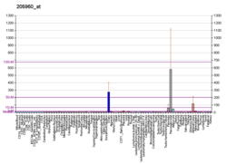
PDK4 is relevant in a variety of clinical conditions. Short-term fasting induces an increase in PDK4 transcription by about 10-fold. [15] Upon refeeding, transcription of PDK4 increased further, a surprising outlook, by about 50-fold over levels before fasting began. [16] This effect can be seen long term as well. PDK4 is overexpressed in skeletal muscle in type 2 diabetes, resulting in impaired glucose utilization. [17] In post-obese patients, there is a significant decrease in PDK4 mRNA expression, in conjunction with increased glucose uptake; this is likely due to the downregulation of PDK4 by insulin. This corroborates the concept that a lowered availability of free fatty acids affects glucose metabolism by PDH complex regulation. [18] In fact, it has been shown that insufficient downregulation of PDK mRNA in insulin-resistant individuals could be a cause of increased PDK expression leading to impaired glucose oxidation followed by increased fatty acid oxidation. [19]

Exercise has been shown to induce changes in this gene as well, and that transient changes can have a cumulative effect across many exercise sessions. The mRNA of PDK4, along with PPARGC1A , increases in both types of muscle tissue after exercise. [20] [21]

These metabolic effects can be seen in other conditions. Hypoxia is shown to induce PDK4 gene expression through the ERR gamma mechanism. [22] Conversely, PDK4 is downregulated in cardiac muscle tissue during heart failure. [23]

Cancer

The ubiquitous role of this gene lends itself to being involved in a variety of disease pathologies, including cancer. One metabolite, butyrate, induces hyperacetylation of the histones around the PDK4 gene. This is associated with a greater transcription level of PDK4 mRNA, thereby reversing the downregulation of PDK4 in colon carcinoma cells. In human colon cancer cells, targeting and inactivating the PDH complex limits the metabolic rate and regulates glutamine metabolism, thereby partially inhibiting cell growth. [24] However, PDK4 has also been shown to promote tumor genesis and proliferation through a different pathway, the CREB-RHEB-mTORC1 signaling cascade. [25]

Interactive pathway map

Click on genes, proteins and metabolites below to link to respective articles. [§ 1]

[[File:
TCACycle WP78.png Go to articleGo to articleGo to articleGo to articleGo to articleGo to articleGo to articleGo to articleGo to articleGo to articleGo to articleGo to articleGo to articleGo to articleGo to articleGo to articleGo to articleGo to articleGo to articleGo to articleGo to articleGo to articleGo to articleGo to articleGo to articleGo to articleGo to articleGo to articleGo to articleGo to articleGo to articleGo to articleGo to articleGo to articleGo to articleGo to articleGo to articleGo to articleGo to articleGo to articleGo to articleGo to HMDBGo to articleGo to articleGo to articleGo to articleGo to articleGo to articleGo to articleGo to articleGo to articleGo to article
[[
]]
[[
]]
[[
]]
[[
]]
[[
]]
[[
]]
[[
]]
[[
]]
[[
]]
[[
]]
[[
]]
[[
]]
[[
]]
[[
]]
[[
]]
[[
]]
[[
]]
[[
]]
[[
]]
[[
]]
[[
]]
[[
]]
[[
]]
[[
]]
[[
]]
[[
]]
[[
]]
[[
]]
[[
]]
[[
]]
[[
]]
[[
]]
[[
]]
[[
]]
[[
]]
[[
]]
[[
]]
[[
]]
[[
]]
[[
]]
[[
]]
[[
]]
[[
]]
[[
]]
[[
]]
[[
]]
[[
]]
[[
]]
[[
]]
[[
]]
[[
]]
[[
]]
[[
]]
[[
]]
[[
]]
[[
]]
[[
]]
[[
]]
[[
]]
[[
]]
[[
]]
[[
]]
[[
]]
TCACycle WP78.png Go to articleGo to articleGo to articleGo to articleGo to articleGo to articleGo to articleGo to articleGo to articleGo to articleGo to articleGo to articleGo to articleGo to articleGo to articleGo to articleGo to articleGo to articleGo to articleGo to articleGo to articleGo to articleGo to articleGo to articleGo to articleGo to articleGo to articleGo to articleGo to articleGo to articleGo to articleGo to articleGo to articleGo to articleGo to articleGo to articleGo to articleGo to articleGo to articleGo to articleGo to articleGo to HMDBGo to articleGo to articleGo to articleGo to articleGo to articleGo to articleGo to articleGo to articleGo to articleGo to article
|alt=TCACycle_WP78 edit]]
TCACycle_WP78 edit
  1. The interactive pathway map can be edited at WikiPathways: "TCACycle_WP78".

Related Research Articles

<span class="mw-page-title-main">Glycolysis</span> Catabolic pathway

Glycolysis is the metabolic pathway that converts glucose into pyruvate. The free energy released in this process is used to form the high-energy molecules adenosine triphosphate (ATP) and reduced nicotinamide adenine dinucleotide (NADH). Glycolysis is a sequence of ten reactions catalyzed by enzymes.

Gluconeogenesis (GNG) is a metabolic pathway that results in the generation of glucose from certain non-carbohydrate carbon substrates. It is a ubiquitous process, present in plants, animals, fungi, bacteria, and other microorganisms. In vertebrates, gluconeogenesis occurs mainly in the liver and, to a lesser extent, in the cortex of the kidneys. It is one of two primary mechanisms – the other being degradation of glycogen (glycogenolysis) – used by humans and many other animals to maintain blood sugar levels, avoiding low levels (hypoglycemia). In ruminants, because dietary carbohydrates tend to be metabolized by rumen organisms, gluconeogenesis occurs regardless of fasting, low-carbohydrate diets, exercise, etc. In many other animals, the process occurs during periods of fasting, starvation, low-carbohydrate diets, or intense exercise.

<span class="mw-page-title-main">Tumor hypoxia</span> Situation where tumor cells have been deprived of oxygen

Tumor hypoxia is the situation where tumor cells have been deprived of oxygen. As a tumor grows, it rapidly outgrows its blood supply, leaving portions of the tumor with regions where the oxygen concentration is significantly lower than in healthy tissues. Hypoxic microenvironements in solid tumors are a result of available oxygen being consumed within 70 to 150 μm of tumour vasculature by rapidly proliferating tumor cells thus limiting the amount of oxygen available to diffuse further into the tumor tissue. In order to support continuous growth and proliferation in challenging hypoxic environments, cancer cells are found to alter their metabolism. Furthermore, hypoxia is known to change cell behavior and is associated with extracellular matrix remodeling and increased migratory and metastatic behavior.

<span class="mw-page-title-main">Pyruvate kinase</span> Class of enzymes

Pyruvate kinase is the enzyme involved in the last step of glycolysis. It catalyzes the transfer of a phosphate group from phosphoenolpyruvate (PEP) to adenosine diphosphate (ADP), yielding one molecule of pyruvate and one molecule of ATP. Pyruvate kinase was inappropriately named before it was recognized that it did not directly catalyze phosphorylation of pyruvate, which does not occur under physiological conditions. Pyruvate kinase is present in four distinct, tissue-specific isozymes in animals, each consisting of particular kinetic properties necessary to accommodate the variations in metabolic requirements of diverse tissues.

<span class="mw-page-title-main">AMP-activated protein kinase</span> Class of enzymes

5' AMP-activated protein kinase or AMPK or 5' adenosine monophosphate-activated protein kinase is an enzyme that plays a role in cellular energy homeostasis, largely to activate glucose and fatty acid uptake and oxidation when cellular energy is low. It belongs to a highly conserved eukaryotic protein family and its orthologues are SNF1 in yeast, and SnRK1 in plants. It consists of three proteins (subunits) that together make a functional enzyme, conserved from yeast to humans. It is expressed in a number of tissues, including the liver, brain, and skeletal muscle. In response to binding AMP and ADP, the net effect of AMPK activation is stimulation of hepatic fatty acid oxidation, ketogenesis, stimulation of skeletal muscle fatty acid oxidation and glucose uptake, inhibition of cholesterol synthesis, lipogenesis, and triglyceride synthesis, inhibition of adipocyte lipogenesis, inhibition of adipocyte lipolysis, and modulation of insulin secretion by pancreatic β-cells.

<span class="mw-page-title-main">Pyruvate dehydrogenase lipoamide kinase isozyme 1</span> Protein-coding gene in the species Homo sapiens

Pyruvate dehydrogenase lipoamide kinase isozyme 1, mitochondrial is an enzyme that in humans is encoded by the PDK1 gene. It codes for an isozyme of pyruvate dehydrogenase kinase (PDK).

In biochemistry, lipogenesis is the conversion of fatty acids and glycerol into fats, or a metabolic process through which acetyl-CoA is converted to triglyceride for storage in fat. Lipogenesis encompasses both fatty acid and triglyceride synthesis, with the latter being the process by which fatty acids are esterified to glycerol before being packaged into very-low-density lipoprotein (VLDL). Fatty acids are produced in the cytoplasm of cells by repeatedly adding two-carbon units to acetyl-CoA. Triacylglycerol synthesis, on the other hand, occurs in the endoplasmic reticulum membrane of cells by bonding three fatty acid molecules to a glycerol molecule. Both processes take place mainly in liver and adipose tissue. Nevertheless, it also occurs to some extent in other tissues such as the gut and kidney. A review on lipogenesis in the brain was published in 2008 by Lopez and Vidal-Puig. After being packaged into VLDL in the liver, the resulting lipoprotein is then secreted directly into the blood for delivery to peripheral tissues.

<span class="mw-page-title-main">Branched-chain amino acid</span> Amino acid with a branched carbon chain

A branched-chain amino acid (BCAA) is an amino acid having an aliphatic side-chain with a branch. Among the proteinogenic amino acids, there are three BCAAs: leucine, isoleucine, and valine. Non-proteinogenic BCAAs include 2-aminoisobutyric acid.

Glucose transporter type 4 (GLUT4), also known as solute carrier family 2, facilitated glucose transporter member 4, is a protein encoded, in humans, by the SLC2A4 gene. GLUT4 is the insulin-regulated glucose transporter found primarily in adipose tissues and striated muscle. The first evidence for this distinct glucose transport protein was provided by David James in 1988. The gene that encodes GLUT4 was cloned and mapped in 1989.

<span class="mw-page-title-main">Tumor metabolome</span>

The study of the tumor metabolism, also known as tumor metabolome describes the different characteristic metabolic changes in tumor cells. The characteristic attributes of the tumor metabolome are high glycolytic enzyme activities, the expression of the pyruvate kinase isoenzyme type M2, increased channeling of glucose carbons into synthetic processes, such as nucleic acid, amino acid and phospholipid synthesis, a high rate of pyrimidine and purine de novo synthesis, a low ratio of Adenosine triphosphate and Guanosine triphosphate to Cytidine triphosphate and Uridine triphosphate, low Adenosine monophosphate levels, high glutaminolytic capacities, release of immunosuppressive substances and dependency on methionine.

<span class="mw-page-title-main">Pyruvate dehydrogenase kinase</span> Class of enzymes

Pyruvate dehydrogenase kinase is a kinase enzyme which acts to inactivate the enzyme pyruvate dehydrogenase by phosphorylating it using ATP.

<span class="mw-page-title-main">E3 binding protein</span> Protein-coding gene in the species Homo sapiens

E3 binding protein also known as pyruvate dehydrogenase protein X component, mitochondrial is a protein that in humans is encoded by the PDHX gene. The E3 binding protein is a component of the pyruvate dehydrogenase complex found only in eukaryotes. Defects in this gene are a cause of pyruvate dehydrogenase deficiency which results in neurological dysfunction and lactic acidosis in infancy and early childhood. This protein is also a minor antigen for antimitochondrial antibodies. These autoantibodies are present in nearly 95% of patients with primary biliary cholangitis, an autoimmune disease of the liver. In primary biliary cholangitis, activated T lymphocytes attack and destroy epithelial cells in the bile duct where this protein is abnormally distributed and overexpressed. Primary biliary cholangitis eventually leads to liver failure.

<span class="mw-page-title-main">Pyruvate dehydrogenase phosphatase</span> Protein-coding gene in the species Homo sapiens

Pyruvate dehydrogenase phosphatase catalytic subunit 1, also known as protein phosphatase 2C, is an enzyme that in humans is encoded by the PDP1 gene. PDPC 1 is an enzyme which serves to reverse the effects of pyruvate dehydrogenase kinase upon pyruvate dehydrogenase, activating pyruvate dehydrogenase.

The Randle cycle, also known as the glucose fatty-acid cycle, is a metabolic process involving the competition of glucose and fatty acids for substrates. It is theorized to play a role in explaining type 2 diabetes and insulin resistance.

<span class="mw-page-title-main">Pyruvate dehydrogenase (lipoamide) alpha 1</span> Protein-coding gene in the species Homo sapiens

Pyruvate dehydrogenase E1 component subunit alpha, somatic form, mitochondrial is an enzyme that in humans is encoded by the PDHA1 gene.The pyruvate dehydrogenase complex is a nuclear-encoded mitochondrial matrix multienzyme complex that provides the primary link between glycolysis and the tricarboxylic acid (TCA) cycle by catalyzing the irreversible conversion of pyruvate into acetyl-CoA. The PDH complex is composed of multiple copies of 3 enzymes: E1 (PDHA1); dihydrolipoyl transacetylase (DLAT) ; and dihydrolipoyl dehydrogenase (DLD). The E1 enzyme is a heterotetramer of 2 alpha and 2 beta subunits. The E1-alpha subunit contains the E1 active site and plays a key role in the function of the PDH complex.

<span class="mw-page-title-main">PDK2</span> Protein-coding gene in the species Homo sapiens

Pyruvate dehydrogenase kinase isoform 2 (PDK2) also known as pyruvate dehydrogenase lipoamide kinase isozyme 2, mitochondrial is an enzyme that in humans is encoded by the PDK2 gene. PDK2 is an isozyme of pyruvate dehydrogenase kinase.

<span class="mw-page-title-main">PDK3</span> Protein-coding gene in the species Homo sapiens

Pyruvate dehydrogenase lipoamide kinase isozyme 3, mitochondrial is an enzyme that in humans is encoded by the PDK3 gene. It codes for an isozyme of pyruvate dehydrogenase kinase.The pyruvate dehydrogenase (PDH) complex is a nuclear-encoded mitochondrial multienzyme complex that catalyzes the overall conversion of pyruvate to acetyl-CoA and CO2. It provides the primary link between glycolysis and the tricarboxylic acid (TCA) cycle, and thus is one of the major enzymes responsible for the regulation of glucose metabolism. The enzymatic activity of PDH is regulated by a phosphorylation/dephosphorylation cycle, and phosphorylation results in inactivation of PDH. The protein encoded by this gene is one of the four pyruvate dehydrogenase kinases that inhibits the PDH complex by phosphorylation of the E1 alpha subunit. This gene is predominantly expressed in the heart and skeletal muscles. Alternatively spliced transcript variants encoding different isoforms have been found for this gene.

<span class="mw-page-title-main">Carbohydrate-responsive element-binding protein</span> Protein-coding gene in the species Homo sapiens

Carbohydrate-responsive element-binding protein (ChREBP) also known as MLX-interacting protein-like (MLXIPL) is a protein that in humans is encoded by the MLXIPL gene. The protein name derives from the protein's interaction with carbohydrate response element sequences of DNA.

<span class="mw-page-title-main">PKM2</span> Protein-coding gene in the species Homo sapiens

Pyruvate kinase isozymes M1/M2 (PKM1/M2), also known as pyruvate kinase muscle isozyme (PKM), pyruvate kinase type K, cytosolic thyroid hormone-binding protein (CTHBP), thyroid hormone-binding protein 1 (THBP1), or opa-interacting protein 3 (OIP3), is an enzyme that in humans is encoded by the PKM2 gene.

Fructolysis refers to the metabolism of fructose from dietary sources. Though the metabolism of glucose through glycolysis uses many of the same enzymes and intermediate structures as those in fructolysis, the two sugars have very different metabolic fates in human metabolism. Unlike glucose, which is directly metabolized widely in the body, fructose is almost entirely metabolized in the liver in humans, where it is directed toward replenishment of liver glycogen and triglyceride synthesis. Under one percent of ingested fructose is directly converted to plasma triglyceride. 29% - 54% of fructose is converted in liver to glucose, and about a quarter of fructose is converted to lactate. 15% - 18% is converted to glycogen. Glucose and lactate are then used normally as energy to fuel cells all over the body.

References

  1. 1 2 3 GRCh38: Ensembl release 89: ENSG00000004799 - Ensembl, May 2017
  2. 1 2 3 GRCm38: Ensembl release 89: ENSMUSG00000019577 - Ensembl, May 2017
  3. "Human PubMed Reference:". National Center for Biotechnology Information, U.S. National Library of Medicine.
  4. "Mouse PubMed Reference:". National Center for Biotechnology Information, U.S. National Library of Medicine.
  5. Gudi R, Bowker-Kinley MM, Kedishvili NY, Zhao Y, Popov KM (Dec 1995). "Diversity of the pyruvate dehydrogenase kinase gene family in humans". The Journal of Biological Chemistry. 270 (48): 28989–94. doi: 10.1074/jbc.270.48.28989 . PMID   7499431.
  6. 1 2 3 "Entrez Gene: PDK4 pyruvate dehydrogenase kinase, isozyme 4".
  7. Andrews MT, Squire TL, Bowen CM, Rollins MB (Jul 1998). "Low-temperature carbon utilization is regulated by novel gene activity in the heart of a hibernating mammal". Proceedings of the National Academy of Sciences of the United States of America. 95 (14): 8392–7. Bibcode:1998PNAS...95.8392A. doi: 10.1073/pnas.95.14.8392 . PMC   20986 . PMID   9653197.
  8. Kukimoto-Niino M, Tokmakov A, Terada T, Ohbayashi N, Fujimoto T, Gomi S, Shiromizu I, Kawamoto M, Matsusue T, Shirouzu M, Yokoyama S (Sep 2011). "Inhibitor-bound structures of human pyruvate dehydrogenase kinase 4". Acta Crystallographica Section D. 67 (Pt 9): 763–73. doi:10.1107/S090744491102405X. PMID   21904029.
  9. Kolobova E, Tuganova A, Boulatnikov I, Popov KM (Aug 2001). "Regulation of pyruvate dehydrogenase activity through phosphorylation at multiple sites". The Biochemical Journal. 358 (Pt 1): 69–77. doi:10.1042/0264-6021:3580069. PMC   1222033 . PMID   11485553.
  10. Kwon HS, Huang B, Ho Jeoung N, Wu P, Steussy CN, Harris RA (2006). "Retinoic acids and trichostatin A (TSA), a histone deacetylase inhibitor, induce human pyruvate dehydrogenase kinase 4 (PDK4) gene expression". Biochimica et Biophysica Acta (BBA) - Gene Structure and Expression. 1759 (3–4): 141–51. doi:10.1016/j.bbaexp.2006.04.005. PMID   16757381.
  11. Cadoudal T, Distel E, Durant S, Fouque F, Blouin JM, Collinet M, Bortoli S, Forest C, Benelli C (Sep 2008). "Pyruvate dehydrogenase kinase 4: regulation by thiazolidinediones and implication in glyceroneogenesis in adipose tissue". Diabetes. 57 (9): 2272–9. doi:10.2337/db08-0477. PMC   2518477 . PMID   18519799.
  12. Savkur RS, Bramlett KS, Michael LF, Burris TP (Apr 2005). "Regulation of pyruvate dehydrogenase kinase expression by the farnesoid X receptor". Biochemical and Biophysical Research Communications. 329 (1): 391–6. doi:10.1016/j.bbrc.2005.01.141. PMID   15721319.
  13. Kwon HS, Huang B, Unterman TG, Harris RA (Apr 2004). "Protein kinase B-alpha inhibits human pyruvate dehydrogenase kinase-4 gene induction by dexamethasone through inactivation of FOXO transcription factors". Diabetes. 53 (4): 899–910. doi: 10.2337/diabetes.53.4.899 . PMID   15047604.
  14. Abbot EL, McCormack JG, Reynet C, Hassall DG, Buchan KW, Yeaman SJ (Jun 2005). "Diverging regulation of pyruvate dehydrogenase kinase isoform gene expression in cultured human muscle cells". The FEBS Journal. 272 (12): 3004–14. doi: 10.1111/j.1742-4658.2005.04713.x . PMID   15955060. S2CID   21366281.
  15. Spriet LL, Tunstall RJ, Watt MJ, Mehan KA, Hargreaves M, Cameron-Smith D (Jun 2004). "Pyruvate dehydrogenase activation and kinase expression in human skeletal muscle during fasting". Journal of Applied Physiology. 96 (6): 2082–7. doi:10.1152/japplphysiol.01318.2003. PMID   14966024. S2CID   13601849.
  16. Pilegaard H, Saltin B, Neufer PD (Mar 2003). "Effect of short-term fasting and refeeding on transcriptional regulation of metabolic genes in human skeletal muscle". Diabetes. 52 (3): 657–62. doi: 10.2337/diabetes.52.3.657 . PMID   12606505.
  17. Wynn RM, Kato M, Chuang JL, Tso SC, Li J, Chuang DT (Sep 2008). "Pyruvate dehydrogenase kinase-4 structures reveal a metastable open conformation fostering robust core-free basal activity". The Journal of Biological Chemistry. 283 (37): 25305–15. doi: 10.1074/jbc.M802249200 . PMC   2533096 . PMID   18658136.
  18. Rosa G, Di Rocco P, Manco M, Greco AV, Castagneto M, Vidal H, Mingrone G (Feb 2003). "Reduced PDK4 expression associates with increased insulin sensitivity in postobese patients". Obesity Research. 11 (2): 176–82. doi: 10.1038/oby.2003.28 . PMID   12582211.
  19. Majer M, Popov KM, Harris RA, Bogardus C, Prochazka M (Oct 1998). "Insulin downregulates pyruvate dehydrogenase kinase (PDK) mRNA: potential mechanism contributing to increased lipid oxidation in insulin-resistant subjects". Molecular Genetics and Metabolism. 65 (2): 181–6. doi:10.1006/mgme.1998.2748. PMID   9787110.
  20. Pilegaard H, Ordway GA, Saltin B, Neufer PD (Oct 2000). "Transcriptional regulation of gene expression in human skeletal muscle during recovery from exercise". American Journal of Physiology. Endocrinology and Metabolism. 279 (4): E806-14. doi:10.1152/ajpendo.2000.279.4.e806. PMID   11001762. S2CID   1008940.
  21. Wang L, Sahlin K (Apr 2012). "The effect of continuous and interval exercise on PGC-1α and PDK4 mRNA in type I and type II fibres of human skeletal muscle". Acta Physiologica. 204 (4): 525–32. doi:10.1111/j.1748-1716.2011.02354.x. PMID   21883960. S2CID   13208033.
  22. Lee JH, Kim EJ, Kim DK, Lee JM, Park SB, Lee IK, Harris RA, Lee MO, Choi HS (2012). "Hypoxia induces PDK4 gene expression through induction of the orphan nuclear receptor ERRγ". PLOS ONE. 7 (9): e46324. Bibcode:2012PLoSO...746324L. doi: 10.1371/journal.pone.0046324 . PMC   3457976 . PMID   23050013.
  23. Razeghi P, Young ME, Ying J, Depre C, Uray IP, Kolesar J, Shipley GL, Moravec CS, Davies PJ, Frazier OH, Taegtmeyer H (2002). "Downregulation of metabolic gene expression in failing human heart before and after mechanical unloading". Cardiology. 97 (4): 203–9. doi:10.1159/000063122. PMID   12145475. S2CID   46861699.
  24. Blouin JM, Penot G, Collinet M, Nacfer M, Forest C, Laurent-Puig P, Coumoul X, Barouki R, Benelli C, Bortoli S (Jun 2011). "Butyrate elicits a metabolic switch in human colon cancer cells by targeting the pyruvate dehydrogenase complex". International Journal of Cancer. 128 (11): 2591–601. doi: 10.1002/ijc.25599 . PMID   20715114. S2CID   27407499.
  25. Liu Z, Chen X, Wang Y, Peng H, Wang Y, Jing Y, Zhang H (Oct 2014). "PDK4 protein promotes tumorigenesis through activation of cAMP-response element-binding protein (CREB)-Ras homolog enriched in brain (RHEB)-mTORC1 signaling cascade". The Journal of Biological Chemistry. 289 (43): 29739–49. doi: 10.1074/jbc.M114.584821 . PMC   4207987 . PMID   25164809.

Further reading