PDK2

Last updated
PDK2
Protein PDK2 PDB 1jm6.png
Available structures
PDB Ortholog search: PDBe RCSB
Identifiers
Aliases PDK2 , PDHK2, PDKII, pyruvate dehydrogenase kinase 2
External IDs OMIM: 602525 MGI: 1343087 HomoloGene: 68265 GeneCards: PDK2
Orthologs
SpeciesHumanMouse
Entrez
Ensembl
UniProt
RefSeq (mRNA)

NM_002611
NM_001199898
NM_001199899
NM_001199900

NM_133667
NM_001361915

RefSeq (protein)

NP_001186827
NP_001186828
NP_001186829
NP_002602

NP_598428
NP_001348844

Location (UCSC) Chr 17: 50.09 – 50.11 Mb Chr 11: 94.92 – 94.93 Mb
PubMed search [3] [4]
Wikidata
View/Edit Human View/Edit Mouse

Pyruvate dehydrogenase kinase isoform 2 (PDK2) also known as pyruvate dehydrogenase lipoamide kinase isozyme 2, mitochondrial is an enzyme that in humans is encoded by the PDK2 gene. [5] [6] PDK2 is an isozyme of pyruvate dehydrogenase kinase.

Structure

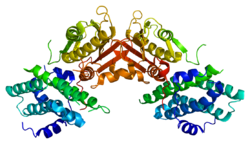
The protein encoded by the PDK2 gene has two sites, an active site and an allosteric site that allow for the activity and regulation of this enzyme. There are many structural motifs that are important to the regulation of this enzyme. Nov3r and AZ12 inhibitors bind at the lipoamide binding site that is located at one end of the R domain. Pfz3 binds in an extended site at the other end of the R domain. One inhibitor, dicholoroacetate (DCA), binds at the center of the R domain. [7] Within the active site, there are three amino acid residues, R250, T302, and Y320, that make the kinase resistant to the inhibitor dichloroacetate, which uncouples the active site from the allosteric site. This supports the theory that R250, T302, and Y320 stabilize the "open" and "closed" conformations of the built-in lid that controls the access of a nucleotide into the nucleotide-binding cavity. This strongly suggests that the mobility of ATP lid is central to the allosteric regulation of PDHK2 activity serving as a conformational switch required for communication between the active site and allosteric sites in the kinase molecule. [8] There is also a DW-motif that is crucial in mediating DCA, nucleotide, and lipoyl domain binding site communication. This network is responsible for rendering PDK2 locked in the closed, or inactive conformation. [9]

Function

The Pyruvate Dehydrogenase (PDH) complex must be tightly regulated due to its central role in general metabolism. Within the complex, there are three serine residues on the E1 component that are sites for phosphorylation; this phosphorylation inactivates the complex. In humans, there have been four isozymes of Pyruvate Dehydrogenase Kinase that have been shown to phosphorylate these three sites: PDK1, PDK2, PDK3, and PDK4. [10] PDK2 has been identified as the most abundant isoform in human tissues. Through many studies, it has been made clear that the activity of this enzyme is essential, even at rest, to regulate glycolysis/carbodydrate oxidation and producing metabolites for oxidative phosphorylation and the electron transport chain. These studies have illustrated that the kinetics of the PDK isoform population, specifically PDK2, is more important in determining PDH activity than measuring PDK activity. [11]

Regulation

As the primary regulators of a crucial step in the central metabolic pathway, the pyruvate dehydrogenase family is tightly regulated itself by a myriad of factors. PDK2 activity is modulated by low levels of hydrogen peroxide; this happens because the compound temporarily oxidizes the cysteine residues 45 and 392 on the enzyme, resulting in an inactive PDK2 and greater PDH activity. These conditions also inactivate the TCA cycle, the next step in aerobic respiration. This alludes to the fact that when there is a high level of O2 production in the mitochondria, which may occur because of nutrient excess, the increase in the products serve as a negative feedback that control mitochondria metabolism. [12] PDK2, in conjunction with PDK3 and PDK4, are primary targets of Peroxisome proliferator-activated receptor delta or beta, with PDK2 having two elements that respond to these receptors. [13]

Clinical significance

All of the pyruvate dehydrogenase isozymes have been associated with various metabolic disorders, including diabetes. This is due to a mechanism by which consistently elevated free fatty acid levels stimulate the PDK enzymes, particularly, PDK2 and PDK4 in the liver. In stimulating this activity, there is less PDH activity, and therefore less glucose uptake. [14]

Cancer

As the PDK enzymes are associated with central metabolism and growth, they are often associated with various mechanisms of cancer progression. Enhanced PDK2 activity leads to increased glycolysis and lactic acid production, known as the Warburg effect. In some studies, the wild-type form of tumor protein p53 prevents manifestation of tumorigenesis by regulating PDK2 activity. [15] Additionally, inhibition of PDK2 subsequently inhibits HIF1A in cancer cells by both a prolyl-hydroxylase (PHD)-dependent mechanism and a PHD-independent mechanism. Therefore, mitochondria-targeting metabolic modulators increase pyruvate dehydrogenase activity, and suppress angiogenesis as well, normalizing the pseudo-hypoxic signals that lead to normoxic HIF1A activation in solid tumors. [16]

Related Research Articles

<span class="mw-page-title-main">Pyruvate kinase</span> Class of enzymes

Pyruvate kinase is the enzyme involved in the last step of glycolysis. It catalyzes the transfer of a phosphate group from phosphoenolpyruvate (PEP) to adenosine diphosphate (ADP), yielding one molecule of pyruvate and one molecule of ATP. Pyruvate kinase was inappropriately named before it was recognized that it did not directly catalyze phosphorylation of pyruvate, which does not occur under physiological conditions. Pyruvate kinase is present in four distinct, tissue-specific isozymes in animals, each consisting of particular kinetic properties necessary to accommodate the variations in metabolic requirements of diverse tissues.

<span class="mw-page-title-main">Pyruvate dehydrogenase complex</span>

Pyruvate dehydrogenase complex (PDC) is a complex of three enzymes that converts pyruvate into acetyl-CoA by a process called pyruvate decarboxylation. Acetyl-CoA may then be used in the citric acid cycle to carry out cellular respiration, and this complex links the glycolysis metabolic pathway to the citric acid cycle. Pyruvate decarboxylation is also known as the "pyruvate dehydrogenase reaction" because it also involves the oxidation of pyruvate.

<span class="mw-page-title-main">AMP-activated protein kinase</span> Class of enzymes

5' AMP-activated protein kinase or AMPK or 5' adenosine monophosphate-activated protein kinase is an enzyme that plays a role in cellular energy homeostasis, largely to activate glucose and fatty acid uptake and oxidation when cellular energy is low. It belongs to a highly conserved eukaryotic protein family and its orthologues are SNF1 in yeast, and SnRK1 in plants. It consists of three proteins (subunits) that together make a functional enzyme, conserved from yeast to humans. It is expressed in a number of tissues, including the liver, brain, and skeletal muscle. In response to binding AMP and ADP, the net effect of AMPK activation is stimulation of hepatic fatty acid oxidation, ketogenesis, stimulation of skeletal muscle fatty acid oxidation and glucose uptake, inhibition of cholesterol synthesis, lipogenesis, and triglyceride synthesis, inhibition of adipocyte lipogenesis, inhibition of adipocyte lipolysis, and modulation of insulin secretion by pancreatic β-cells.

<span class="mw-page-title-main">Pyruvate dehydrogenase lipoamide kinase isozyme 1</span> Protein-coding gene in the species Homo sapiens

Pyruvate dehydrogenase lipoamide kinase isozyme 1, mitochondrial is an enzyme that in humans is encoded by the PDK1 gene. It codes for an isozyme of pyruvate dehydrogenase kinase (PDK).

<span class="mw-page-title-main">Phosphofructokinase 2</span> Class of enzymes

Phosphofructokinase-2 (6-phosphofructo-2-kinase, PFK-2) or fructose bisphosphatase-2 (FBPase-2), is an enzyme indirectly responsible for regulating the rates of glycolysis and gluconeogenesis in cells. It catalyzes formation and degradation of a significant allosteric regulator, fructose-2,6-bisphosphate (Fru-2,6-P2) from substrate fructose-6-phosphate. Fru-2,6-P2 contributes to the rate-determining step of glycolysis as it activates enzyme phosphofructokinase 1 in the glycolysis pathway, and inhibits fructose-1,6-bisphosphatase 1 in gluconeogenesis. Since Fru-2,6-P2 differentially regulates glycolysis and gluconeogenesis, it can act as a key signal to switch between the opposing pathways. Because PFK-2 produces Fru-2,6-P2 in response to hormonal signaling, metabolism can be more sensitively and efficiently controlled to align with the organism's glycolytic needs. This enzyme participates in fructose and mannose metabolism. The enzyme is important in the regulation of hepatic carbohydrate metabolism and is found in greatest quantities in the liver, kidney and heart. In mammals, several genes often encode different isoforms, each of which differs in its tissue distribution and enzymatic activity. The family described here bears a resemblance to the ATP-driven phospho-fructokinases, however, they share little sequence similarity, although a few residues seem key to their interaction with fructose 6-phosphate.

In biochemistry, lipogenesis is the conversion of fatty acids and glycerol into fats, or a metabolic process through which acetyl-CoA is converted to triglyceride for storage in fat. Lipogenesis encompasses both fatty acid and triglyceride synthesis, with the latter being the process by which fatty acids are esterified to glycerol before being packaged into very-low-density lipoprotein (VLDL). Fatty acids are produced in the cytoplasm of cells by repeatedly adding two-carbon units to acetyl-CoA. Triacylglycerol synthesis, on the other hand, occurs in the endoplasmic reticulum membrane of cells by bonding three fatty acid molecules to a glycerol molecule. Both processes take place mainly in liver and adipose tissue. Nevertheless, it also occurs to some extent in other tissues such as the gut and kidney. A review on lipogenes in the brain was published in 2008 by Lopez and Vidal-Puig. After being packaged into VLDL in the liver, the resulting lipoprotein is then secreted directly into the blood for delivery to peripheral tissues.

<span class="mw-page-title-main">Pyruvate dehydrogenase</span> Class of enzymes

Pyruvate dehydrogenase is an enzyme that catalyzes the reaction of pyruvate and a lipoamide to give the acetylated dihydrolipoamide and carbon dioxide. The conversion requires the coenzyme thiamine pyrophosphate.

<span class="mw-page-title-main">Pyruvate dehydrogenase kinase</span>

Pyruvate dehydrogenase kinase is a kinase enzyme which acts to inactivate the enzyme pyruvate dehydrogenase by phosphorylating it using ATP.

<span class="mw-page-title-main">E3 binding protein</span>

E3 binding protein also known as pyruvate dehydrogenase protein X component, mitochondrial is a protein that in humans is encoded by the PDHX gene. The E3 binding protein is a component of the pyruvate dehydrogenase complex found only in eukaryotes. Defects in this gene are a cause of pyruvate dehydrogenase deficiency which results in neurological dysfunction and lactic acidosis in infancy and early childhood. This protein is also a minor antigen for antimitochondrial antibodies. These autoantibodies are present in nearly 95% of patients with primary biliary cholangitis, an autoimmune disease of the liver. In primary biliary cholangitis, activated T lymphocytes attack and destroy epithelial cells in the bile duct where this protein is abnormally distributed and overexpressed. Primary biliary cholangitis eventually leads to liver failure.

<span class="mw-page-title-main">Pyruvate dehydrogenase phosphatase</span> Protein-coding gene in the species Homo sapiens

Pyruvate dehydrogenase phosphatase catalytic subunit 1, also known as protein phosphatase 2C, is an enzyme that in humans is encoded by the PDP1 gene. PDPC 1 is an enzyme which serves to reverse the effects of pyruvate dehydrogenase kinase upon pyruvate dehydrogenase, activating pyruvate dehydrogenase.

<span class="mw-page-title-main">Pyruvate dehydrogenase (lipoamide) alpha 1</span> Protein-coding gene in the species Homo sapiens

Pyruvate dehydrogenase E1 component subunit alpha, somatic form, mitochondrial is an enzyme that in humans is encoded by the PDHA1 gene.The pyruvate dehydrogenase complex is a nuclear-encoded mitochondrial matrix multienzyme complex that provides the primary link between glycolysis and the tricarboxylic acid (TCA) cycle by catalyzing the irreversible conversion of pyruvate into acetyl-CoA. The PDH complex is composed of multiple copies of 3 enzymes: E1 (PDHA1); dihydrolipoyl transacetylase (DLAT) ; and dihydrolipoyl dehydrogenase (DLD). The E1 enzyme is a heterotetramer of 2 alpha and 2 beta subunits. The E1-alpha subunit contains the E1 active site and plays a key role in the function of the PDH complex.

<span class="mw-page-title-main">ARAF</span> Protein-coding gene in the species Homo sapiens

Serine/threonine-protein kinase A-Raf or simply A-Raf is an enzyme that in humans is encoded by the ARAF gene. A-Raf is a member of the Raf kinase family of serine/threonine-specific protein kinases.

<span class="mw-page-title-main">PDK4</span>

Pyruvate dehydrogenase lipoamide kinase isozyme 4, mitochondrial is an enzyme that in humans is encoded by the PDK4 gene. It codes for an isozyme of pyruvate dehydrogenase kinase.

<span class="mw-page-title-main">PDK3</span> Protein-coding gene in the species Homo sapiens

Pyruvate dehydrogenase lipoamide kinase isozyme 3, mitochondrial is an enzyme that in humans is encoded by the PDK3 gene. It codes for an isozyme of pyruvate dehydrogenase kinase.The pyruvate dehydrogenase (PDH) complex is a nuclear-encoded mitochondrial multienzyme complex that catalyzes the overall conversion of pyruvate to acetyl-CoA and CO2. It provides the primary link between glycolysis and the tricarboxylic acid (TCA) cycle, and thus is one of the major enzymes responsible for the regulation of glucose metabolism. The enzymatic activity of PDH is regulated by a phosphorylation/dephosphorylation cycle, and phosphorylation results in inactivation of PDH. The protein encoded by this gene is one of the four pyruvate dehydrogenase kinases that inhibits the PDH complex by phosphorylation of the E1 alpha subunit. This gene is predominantly expressed in the heart and skeletal muscles. Alternatively spliced transcript variants encoding different isoforms have been found for this gene.

<span class="mw-page-title-main">BCKDK</span>

Branched chain ketoacid dehydrogenase kinase (BCKDK) is an enzyme encoded by the BCKDK gene on chromosome 16. This enzyme is part of the mitochondrial protein kinases family and it is a regulator of the valine, leucine, and isoleucine catabolic pathways. BCKDK is found in the mitochondrial matrix and the prevalence of it depends on the type of cell. Liver cells tend to have the lowest concentration of BCKDK, whereas skeletal muscle cells have the highest amount. Abnormal activity of this enzyme often leads to diseases such as maple syrup urine disease and cachexia.

<span class="mw-page-title-main">PKM2</span>

Pyruvate kinase isozymes M1/M2 (PKM1/M2), also known as pyruvate kinase muscle isozyme (PKM), pyruvate kinase type K, cytosolic thyroid hormone-binding protein (CTHBP), thyroid hormone-binding protein 1 (THBP1), or opa-interacting protein 3 (OIP3), is an enzyme that in humans is encoded by the PKM2 gene.

<span class="mw-page-title-main">Phosphoinositide-dependent kinase-1</span> Protein-coding gene in the species Homo sapiens

In the field of biochemistry, PDPK1 refers to the protein 3-phosphoinositide-dependent protein kinase-1, an enzyme which is encoded by the PDPK1 gene in humans. It is implicated in the development and progression of melanomas.

<span class="mw-page-title-main">Pyruvate dehydrogenase (lipoamide) alpha 2</span>

Pyruvate dehydrogenase (lipoamide) alpha 2, also known as pyruvate dehydrogenase E1 component subunit alpha, testis-specific form, mitochondrial or PDHE1-A type II, is an enzyme that in humans is encoded by the PDHA2 gene.

<span class="mw-page-title-main">Pyruvate dehydrogenase (lipoamide) beta</span>

Pyruvate dehydrogenase (lipoamide) beta, also known as pyruvate dehydrogenase E1 component subunit beta, mitochondrial or PDHE1-B is an enzyme that in humans is encoded by the PDHB gene. The pyruvate dehydrogenase (PDH) complex is a nuclear-encoded mitochondrial multienzyme complex that catalyzes the overall conversion of pyruvate to acetyl-CoA and CO2, and provides the primary link between glycolysis and the tricarboxylic acid (TCA) cycle. The PDH complex is composed of multiple copies of three enzymatic components: pyruvate dehydrogenase (E1), dihydrolipoamide acetyltransferase (E2) and lipoamide dehydrogenase (E3). The E1 enzyme is a heterotetramer of two alpha and two beta subunits. This gene encodes the E1 beta subunit. Mutations in this gene are associated with pyruvate dehydrogenase E1-beta deficiency.

<span class="mw-page-title-main">PDPR</span>

Pyruvate dehydrogenase phosphatase regulatory subunit is a protein that in humans is encoded by the PDPR gene.

References

  1. 1 2 3 GRCh38: Ensembl release 89: ENSG00000005882 - Ensembl, May 2017
  2. 1 2 3 GRCm38: Ensembl release 89: ENSMUSG00000038967 - Ensembl, May 2017
  3. "Human PubMed Reference:". National Center for Biotechnology Information, U.S. National Library of Medicine.
  4. "Mouse PubMed Reference:". National Center for Biotechnology Information, U.S. National Library of Medicine.
  5. Gudi R, Bowker-Kinley MM, Kedishvili NY, Zhao Y, Popov KM (Dec 1995). "Diversity of the pyruvate dehydrogenase kinase gene family in humans". The Journal of Biological Chemistry. 270 (48): 28989–94. doi: 10.1074/jbc.270.48.28989 . PMID   7499431.
  6. "Entrez Gene: PDK2 pyruvate dehydrogenase kinase, isozyme 2".
  7. Knoechel TR, Tucker AD, Robinson CM, Phillips C, Taylor W, Bungay PJ, Kasten SA, Roche TE, Brown DG (Jan 2006). "Regulatory roles of the N-terminal domain based on crystal structures of human pyruvate dehydrogenase kinase 2 containing physiological and synthetic ligands". Biochemistry. 45 (2): 402–15. doi:10.1021/bi051402s. PMID   16401071.
  8. Klyuyeva A, Tuganova A, Popov KM (Aug 2008). "Allosteric coupling in pyruvate dehydrogenase kinase 2". Biochemistry. 47 (32): 8358–66. doi:10.1021/bi800631h. PMC   2568900 . PMID   18627174.
  9. Li J, Kato M, Chuang DT (Dec 2009). "Pivotal role of the C-terminal DW-motif in mediating inhibition of pyruvate dehydrogenase kinase 2 by dichloroacetate". The Journal of Biological Chemistry. 284 (49): 34458–67. doi: 10.1074/jbc.M109.065557 . PMC   2797213 . PMID   19833728.
  10. Kolobova E, Tuganova A, Boulatnikov I, Popov KM (Aug 2001). "Regulation of pyruvate dehydrogenase activity through phosphorylation at multiple sites". The Biochemical Journal. 358 (Pt 1): 69–77. doi:10.1042/0264-6021:3580069. PMC   1222033 . PMID   11485553.
  11. Dunford EC, Herbst EA, Jeoung NH, Gittings W, Inglis JG, Vandenboom R, LeBlanc PJ, Harris RA, Peters SJ (Jun 2011). "PDH activation during in vitro muscle contractions in PDH kinase 2 knockout mice: effect of PDH kinase 1 compensation". American Journal of Physiology. Regulatory, Integrative and Comparative Physiology. 300 (6): R1487-93. doi:10.1152/ajpregu.00498.2010. PMID   21411764.
  12. Hurd, TR; Collins, Y; Abakumova, I; Chouchani, ET; Baranowski, B; Fearnley, IM; Prime, TA; Murphy, MP; James, AM (12 October 2012). "Inactivation of pyruvate dehydrogenase kinase 2 by mitochondrial reactive oxygen species". The Journal of Biological Chemistry. 287 (42): 35153–60. doi: 10.1074/jbc.m112.400002 . PMC   3471752 . PMID   22910903.
  13. Degenhardt, T; Saramäki, A; Malinen, M; Rieck, M; Väisänen, S; Huotari, A; Herzig, KH; Müller, R; Carlberg, C (14 September 2007). "Three members of the human pyruvate dehydrogenase kinase gene family are direct targets of the peroxisome proliferator-activated receptor beta/delta". Journal of Molecular Biology. 372 (2): 341–55. doi:10.1016/j.jmb.2007.06.091. PMID   17669420.
  14. Bajotto, G; Murakami, T; Nagasaki, M; Qin, B; Matsuo, Y; Maeda, K; Ohashi, M; Oshida, Y; Sato, Y; Shimomura, Y (March 2006). "Increased expression of hepatic pyruvate dehydrogenase kinases 2 and 4 in young and middle-aged Otsuka Long-Evans Tokushima Fatty rats: induction by elevated levels of free fatty acids". Metabolism: Clinical and Experimental. 55 (3): 317–23. doi:10.1016/j.metabol.2005.09.014. PMID   16483874.
  15. Contractor, T; Harris, CR (15 January 2012). "p53 negatively regulates transcription of the pyruvate dehydrogenase kinase Pdk2". Cancer Research. 72 (2): 560–7. doi: 10.1158/0008-5472.can-11-1215 . PMID   22123926.
  16. Sutendra, G; Dromparis, P; Kinnaird, A; Stenson, TH; Haromy, A; Parker, JM; McMurtry, MS; Michelakis, ED (28 March 2013). "Mitochondrial activation by inhibition of PDKII suppresses HIF1a signaling and angiogenesis in cancer". Oncogene. 32 (13): 1638–50. doi: 10.1038/onc.2012.198 . PMID   22614004.

Further reading