G protein-coupled receptor kinase

Last updated
G protein-coupled receptor kinase
Gprotein-coupled receptor kinase.png
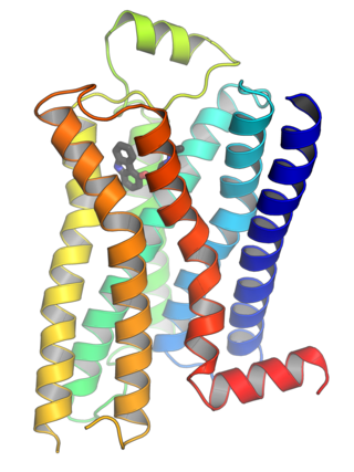
Crystal structure of G protein coupled receptor kinase 1 (GRK1) bound to ATP. [1]
Identifiers
EC no. 2.7.11.16
Databases
IntEnz IntEnz view
BRENDA BRENDA entry
ExPASy NiceZyme view
KEGG KEGG entry
MetaCyc metabolic pathway
PRIAM profile
PDB structures RCSB PDB PDBe PDBsum
Gene Ontology AmiGO / QuickGO
Search
PMC articles
PubMed articles
NCBI proteins

G protein-coupled receptor kinases (GPCRKs, GRKs) are a family of protein kinases within the AGC (protein kinase A, protein kinase G, protein kinase C) group of kinases. Like all AGC kinases, GRKs use ATP to add phosphate to Serine and Threonine residues in specific locations of target proteins. In particular, GRKs phosphorylate intracellular domains of G protein-coupled receptors (GPCRs). GRKs function in tandem with arrestin proteins to regulate the sensitivity of GPCRs for stimulating downstream heterotrimeric G protein and G protein-independent signaling pathways. [2] [3]

Contents

Types of GRKs

NameNotes Gene OMIM
G protein-coupled receptor kinase 1 Rhodopsin kinase GRK1 180381
G protein-coupled receptor kinase 2 β-Adrenergic receptor kinase 1 (βARK1) ADRBK1 109635
G protein-coupled receptor kinase 3 β-Adrenergic receptor kinase 2 (βARK2) ADRBK2 109636
G protein-coupled receptor kinase 4 Polymorphism associated with hypertension [4] GRK4 137026
G protein-coupled receptor kinase 5 Polymorphism associated with cardioprotection [5] GRK5 600870
G protein-coupled receptor kinase 6 Knockout mice are supersensitive to dopaminergic drugs [6] GRK6 600869
G protein-coupled receptor kinase 7 Cone opsin kinase GRK7 606987

GRK activity and regulation

GRKs reside normally in an inactive state, but their kinase activity is stimulated by binding to a ligand-activated GPCR (rather than by regulatory phosphorylation as is common in other AGC kinases). Because there are only seven GRKs (only 4 of which are widely expressed throughout the body) but over 800 human GPCRs, GRKs appear to have limited phosphorylation site selectivity and are regulated primarily by the GPCR active state. [3]

G protein-coupled receptor kinases phosphorylate activated G protein-coupled receptors, which promotes the binding of an arrestin protein to the receptor. Phosphorylated serine and threonine residues in GPCRs act as binding sites for and activators of arrestin proteins. Arrestin binding to phosphorylated, active receptor prevents receptor stimulation of heterotrimeric G protein transducer proteins, blocking their cellular signaling and resulting in receptor desensitization. Arrestin binding also directs receptors to specific cellular internalization pathways, removing the receptors from the cell surface and also preventing additional activation. Arrestin binding to phosphorylated, active receptor also enables receptor signaling through arrestin partner proteins. Thus the GRK/arrestin system serves as a complex signaling switch for G protein-coupled receptors. [3]

GRKs can be regulated by signaling events in cells, both in direct feedback mechanisms where receptor signals alter GRK activity over time, and due to signals emanating from distinct pathways from a particular GPCR/GRK system of interest. For example, GRK1 is regulated by the calcium sensor protein recoverin: calcium-bound recoverin binds directly to GRK1 to inhibit its ability to phosphorylate and desensitize rhodopsin, the visual GPCR in the retina, in light-activated retinal rod cells since light activation raises intracellular calcium in these cells, whereas in dark-adapted eyes, calcium levels are low in rod cells and GRK1 is not inhibited by recoverin. [7] The non-visual GRKs are inhibited instead by the calcium-binding protein calmodulin. [2] GRK2 and GRK3 share a carboxyl terminal pleckstrin homology (PH) domain that binds to G protein beta/gamma subunits, and GPCR activation of heterotrimeric G proteins releases this free beta/gamma complex that binds to GRK2/3 to recruit these kinases to the cell membrane precisely at the location of the activated receptor, augmenting GRK activity to regulate the activated receptor. [2] [3] GRK2 activity can be modulated by its phosphorylation by protein kinase A or protein kinase C, and by post-translational modification of cysteines by S-nitrosylation. [8] [9]

GRK Structures

X-ray crystal structures have been obtained for several GRKs (GRK1, GRK2, GRK4, GRK5 and GRK6), alone or bound to ligands. [10] Overall, GRKs share sequence homology and domain organization in which the central protein kinase catalytic domain is preceded by a domain with homology to the active domain of Regulator of G protein Signaling proteins, RGS proteins (the RGS-homology – RH – domain) and is followed by a variable carboxyl terminal tail regulatory region. [3] In the folded proteins, the kinase domain forms a typical bi-lobe kinase structure with a central ATP-binding active site. [3] The RH domain is composed of alpha-helical region formed from the amino terminal sequence plus a short stretch of sequence following the kinase domain that provides 2 additional helices, and makes extensive contacts with one side of the kinase domain. [10] Modeling and mutagenesis suggests that the RH domain senses GPCR activation to open the kinase active site. [11]

GRK physiological functions

GRK1 is involved with rhodopsin phosphorylation and deactivation in vision, together with arrestin-1, also known as S-antigen. Defects in GRK1 result in Oguchi stationary night blindness. GRK7 similarly regulates cone opsin phosphorylation and deactivation in color vision, together with cone arrestin, also known as arrestin-4 or X-arrestin. [3]

GRK2 was first identified as an enzyme that phosphorylated the beta-2 adrenergic receptor, and was originally called the beta adrenergic receptor kinase (βARK, or ββARK1). GRK2 is overexpressed in heart failure, and GRK2 inhibition could be used to treat heart failure in the future. [12]

Polymorphisms in the GRK4 gene have been linked to both genetic and acquired hypertension, acting in part through kidney dopamine receptors. [4] GRK4 is the most highly expressed GRK at the mRNA level, in maturing spermatids, but mice lacking GRK4 remain fertile so its role in these cells remains unknown. [13]

In humans, a GRK5 sequence polymorphism at residue 41 (leucine rather than glutamine) that is most common in individuals with African ancestry leads to elevated GRK5-mediated desensitization of airway beta2-adrenergic receptors, a drug target in asthma. [14] In zebrafish and in humans, loss of GRK5 function has been associated with heart defects due to heterotaxy, a series of developmental defects arising from improper left-right laterality during organogenesis. [15]

In the mouse, GRK6 regulation of D2 dopamine receptors in the striatum region of the brain alters sensitivity to psychostimulant drugs that act through dopamine, and GRK6 has been implicated in Parkinson's disease and in the dyskinesia side effects of anti-parkinson therapy with the drug L-DOPA. [16] [17]

Non-GPCR functions of GRKs

GRKs also phosphorylate non-GPCR substrates. GRK2 and GRK5 can phosphorylate some tyrosine kinase receptors, including the receptor for platelet-derived growth factor (PDGF) and insulin-like growth factor (IGF). [18] [19]

GRKs also regulate cellular responses independent of their kinase activity. In particular, G protein-coupled receptor kinase 2 is known to interact with a diverse repertoire of non-GPCR partner proteins, but other GRKs also have non-GPCR partners. [20] The RGS-homology (RH) domain of GRK2 and GRK3 binds to heterotrimeric G protein subunits of the Gq family, but despite these RH domains being unable to act as GTPase-activating proteins like traditional RGS proteins to turn off G protein signaling, this binding reduces Gq signaling by sequestering active G proteins away from their effector proteins such as phospholipase C-beta. [21]

See also

Related Research Articles

<span class="mw-page-title-main">G protein-coupled receptor</span> Class of cell surface receptors coupled to G-protein-associated intracellular signaling

G protein-coupled receptors (GPCRs), also known as seven-(pass)-transmembrane domain receptors, 7TM receptors, heptahelical receptors, serpentine receptors, and G protein-linked receptors (GPLR), form a large group of evolutionarily related proteins that are cell surface receptors that detect molecules outside the cell and activate cellular responses. They are coupled with G proteins. They pass through the cell membrane seven times in the form of six loops of amino acid residues, which is why they are sometimes referred to as seven-transmembrane receptors. Ligands can bind either to the extracellular N-terminus and loops or to the binding site within transmembrane helices. They are all activated by agonists, although a spontaneous auto-activation of an empty receptor has also been observed.

<span class="mw-page-title-main">Rod cell</span> Photoreceptor cells that can function in lower light better than cone cells

Rod cells are photoreceptor cells in the retina of the eye that can function in lower light better than the other type of visual photoreceptor, cone cells. Rods are usually found concentrated at the outer edges of the retina and are used in peripheral vision. On average, there are approximately 92 million rod cells in the human retina. Rod cells are more sensitive than cone cells and are almost entirely responsible for night vision. However, rods have little role in color vision, which is the main reason why colors are much less apparent in dim light.

Visual phototransduction is the sensory transduction process of the visual system by which light is detected by photoreceptor cells in the vertebrate retina. A photon is absorbed by a retinal chromophore, which initiates a signal cascade through several intermediate cells, then through the retinal ganglion cells (RGCs) comprising the optic nerve.

<span class="mw-page-title-main">Pleckstrin homology domain</span> Protein domain

Pleckstrin homology domain or (PHIP) is a protein domain of approximately 120 amino acids that occurs in a wide range of proteins involved in intracellular signaling or as constituents of the cytoskeleton.

<span class="mw-page-title-main">Beta-1 adrenergic receptor</span> Protein-coding gene in the species Homo sapiens

The beta-1 adrenergic receptor, also known as ADRB1, can refer to either the protein-encoding gene or one of the four adrenergic receptors. It is a G-protein coupled receptor associated with the Gs heterotrimeric G-protein that is expressed predominantly in cardiac tissue. In addition to cardiac tissue, beta-1 adrenergic receptors are also expressed in the cerebral cortex.

<span class="mw-page-title-main">Arrestin</span> Family of proteins

Arrestins are a small family of proteins important for regulating signal transduction at G protein-coupled receptors. Arrestins were first discovered as a part of a conserved two-step mechanism for regulating the activity of G protein-coupled receptors (GPCRs) in the visual rhodopsin system by Hermann Kühn, Scott Hall, and Ursula Wilden and in the β-adrenergic system by Martin J. Lohse and co-workers.

<span class="mw-page-title-main">G protein-coupled receptor kinase 2</span> Enzyme

G-protein-coupled receptor kinase 2 (GRK2) is an enzyme that in humans is encoded by the ADRBK1 gene. GRK2 was initially called Beta-adrenergic receptor kinase, and is a member of the G protein-coupled receptor kinase subfamily of the Ser/Thr protein kinases that is most highly similar to GRK3(βARK2).

Rhodopsin kinase is a serine/threonine-specific protein kinase involved in phototransduction. This enzyme catalyses the following chemical reaction:

<span class="mw-page-title-main">Arrestin beta 2</span> Protein-coding gene in the species Homo sapiens

Beta-arrestin-2, also known as arrestin beta-2, is an intracellular protein that in humans is encoded by the ARRB2 gene.

<span class="mw-page-title-main">Regulator of G protein signaling</span>

Regulators of G protein signaling (RGS) are protein structural domains or the proteins that contain these domains, that function to activate the GTPase activity of heterotrimeric G-protein α-subunits.

<span class="mw-page-title-main">Arrestin beta 1</span> Human protein and coding gene

Arrestin, beta 1, also known as ARRB1, is a protein which in humans is encoded by the ARRB1 gene.

<span class="mw-page-title-main">GRK6</span> Protein-coding gene in the species Homo sapiens

This gene encodes a member of the G protein-coupled receptor kinase subfamily of the Ser/Thr protein kinase family, and is most highly similar to GRK4 and GRK5. The protein phosphorylates the activated forms of G protein-coupled receptors to regulate their signaling.

<span class="mw-page-title-main">GRK5</span> Protein-coding gene in the species Homo sapiens

G protein-coupled receptor kinase 5 is a member of the G protein-coupled receptor kinase subfamily of the Ser/Thr protein kinases, and is most highly similar to GRK4 and GRK6. The protein phosphorylates the activated forms of G protein-coupled receptors to regulate their signaling.

<span class="mw-page-title-main">SAG (gene)</span>

S-arrestin is a protein that in humans is encoded by the SAG gene.

<span class="mw-page-title-main">GRK4</span> Protein-coding gene in the species Homo sapiens

G protein-coupled receptor kinase 4 (GRK4) is an enzyme that is encoded by the GRK4 gene in humans.

<span class="mw-page-title-main">Homologous desensitization</span> When a receptor decreases its response to an agonist at high concentration

Homologous desensitization occurs when a receptor decreases its response to an agonist at high concentration. It is a process through which, after prolonged agonist exposure, the receptor is uncoupled from its signaling cascade and thus the cellular effect of receptor activation is attenuated.

<span class="mw-page-title-main">G beta-gamma complex</span>

The G beta-gamma complex (Gβγ) is a tightly bound dimeric protein complex, composed of one Gβ and one Gγ subunit, and is a component of heterotrimeric G proteins. Heterotrimeric G proteins, also called guanosine nucleotide-binding proteins, consist of three subunits, called alpha, beta, and gamma subunits, or Gα, Gβ, and Gγ. When a G protein-coupled receptor (GPCR) is activated, Gα dissociates from Gβγ, allowing both subunits to perform their respective downstream signaling effects. One of the major functions of Gβγ is the inhibition of the Gα subunit.

G-protein-coupled receptor kinase 7 is a serine/threonine-specific protein kinase involved in phototransduction. This enzyme catalyses the phosphorylation of cone (color) photopsins in retinal cones during high acuity color vision primarily in the fovea.

Heterologous desensitization is the term for the unresponsiveness of cells to one or more agonists to which they are normally responsive. Typically, desensitization is a receptor-based phenomenon in which one receptor type, when bound to its ligand, becomes unable to further influence the signalling pathways by which it regulates cells and, in the case of cell surface membrane receptors, may thereafter be internalized. The desensitized receptor is degraded or freed of its activating ligand and re-cycled to a state where it is again able to respond to cognate ligands by activating its signalling pathways.

<span class="mw-page-title-main">G protein-coupled receptor kinase 3</span> Protein-coding gene in the species Homo sapiens

G-protein-coupled receptor kinase 3 (GRK3) is an enzyme that in humans is encoded by the ADRBK2 gene. GRK3 was initially called Beta-adrenergic receptor kinase 2 (βARK-2), and is a member of the G protein-coupled receptor kinase subfamily of the Ser/Thr protein kinases that is most highly similar to GRK2.
References

  1. PDB: 3C4W ; Singh P, Wang B, Maeda T, Palczewski K, Tesmer JJ (May 2008). "Structures of rhodopsin kinase in different ligand states reveal key elements involved in G protein-coupled receptor kinase activation". J. Biol. Chem. 283 (20): 14053–62. doi: 10.1074/jbc.M708974200 . PMC   2376226 . PMID   18339619.
  2. 1 2 3 Ribas C, Penela P, Murga C, Salcedo A, García-Hoz C, Jurado-Pueyo M, Aymerich I, Mayor F Jr (2007). "The G protein-coupled receptor kinase (GRK) interactome: role of GRKs in GPCR regulation and signaling". Biochim Biophys Acta. 1768 (4): 913–922. doi: 10.1016/j.bbamem.2006.09.019 . PMID   17084806.
  3. 1 2 3 4 5 6 7 Gurevich VV, Gurevich EV (2019). "GPCR Signaling Regulation: The Role of GRKs and Arrestins". Front Pharmacol. 10: 125. doi: 10.3389/fphar.2019.00125 . PMC   6389790 . PMID   30837883.
  4. 1 2 Yang J, Villar VA, Jones JE, Jose PA, Zeng C (2015). "G protein-coupled receptor kinase 4: role in hypertension". Hypertension. 65 (6): 1148–1155. doi:10.1161/HYPERTENSIONAHA.115.05189. PMC   6350509 . PMID   25870190.
  5. Dorn GW 2nd, Liggett SB (2008). "Pharmacogenomics of beta-adrenergic receptors and their accessory signaling proteins in heart failure". Clin Transl Sci. 1 (3): 255–262. doi:10.1111/j.1752-8062.2008.00059.x. PMC   5350665 . PMID   20443857.
  6. Gainetdinov RR, Bohn LM, Sotnikova TD, et al. (April 2003). "Dopaminergic supersensitivity in G protein-coupled receptor kinase 6-deficient mice". Neuron . 38 (2): 291–303. doi: 10.1016/S0896-6273(03)00192-2 . PMID   12718862.
  7. Chen CK, Inglese J, Lefkowitz RJ, Hurley JB (July 1995). "Ca(2+)-dependent interaction of recoverin with rhodopsin kinase". The Journal of Biological Chemistry. 270 (30): 18060–6. doi: 10.1074/jbc.270.30.18060 . PMID   7629115.
  8. Willets JM, Challiss RA, Nahorski SR (2003). "Non-visual GRKs: are we seeing the whole picture?" (PDF). Trends Pharmacol Sci. 24 (12): 626–633. doi:10.1016/j.tips.2003.10.003. PMID   14654303.
  9. Whalen EJ, Foster MW, Matsumoto A, Ozawa K, Violin JD, Que LG, Nelson CD, Benhar M, Keys JR, Rockman HA, Koch WJ, Daaka Y, Lefkowitz RJ, Stamler JS (2007). "Regulation of beta-adrenergic receptor signaling by S-nitrosylation of G-protein-coupled receptor kinase 2". Cell. 129 (3): 511–522. doi: 10.1016/j.cell.2007.02.046 . PMID   17482545.
  10. 1 2 Homan KT, Tesmer JJ (2015). "Molecular basis for small molecule inhibition of G protein-coupled receptor kinases". ACS Chem Biol. 10 (1): 246–256. doi:10.1021/cb5003976. PMC   4301174 . PMID   24984143.
  11. He Y, Gao X, Goswami D, Hou L, Pal K, Yin Y, Zhao G, Ernst OP, Griffin P, Melcher K, Xu HE (2017). "Molecular assembly of rhodopsin with G protein-coupled receptor kinases". Cell Res. 27 (6): 728–747. doi:10.1038/cr.2017.72. PMC   5518878 . PMID   28524165.
  12. Tesmer, J. J.; Koch, W. J.; Sklar, L. A.; Cheung, J. Y.; Gao, E.; Song, J.; Chuprun, J. K.; Huang, Z. M.; Hinkle, P. M. (2012). "Paroxetine is a direct inhibitor of g protein-coupled receptor kinase 2 and increases myocardial contractility". ACS Chemical Biology. 7 (11): 1830–1839. doi:10.1021/cb3003013. ISSN   1554-8929. PMC   3500392 . PMID   22882301.
  13. Premont RT, Macrae AD, Stoffel RH, et al. (1996). "Characterization of the G protein-coupled receptor kinase GRK4. Identification of four splice variants". J. Biol. Chem. 271 (11): 6403–10. doi: 10.1074/jbc.271.11.6403 . PMID   8626439.
  14. Wang WC, Mihlbachler KA, Bleecker ER, Weiss ST, Liggett SB (2008). "A polymorphism of G-protein coupled receptor kinase5 alters agonist-promoted desensitization of beta2-adrenergic receptors". Pharmacogenet Genomics. 18 (8): 729–732. doi:10.1097/FPC.0b013e32830967e9. PMC   2699179 . PMID   18622265.
  15. Lessel D, Muhammad T, Casar Tena T, Moepps B, Burkhalter MD, Hitz MP, Toka O, Rentzsch A, Schubert S, Schalinski A, Bauer UM, Kubisch C, Ware SM, Philipp M (2016). "The analysis of heterotaxy patients reveals new loss-of-function variants of GRK5". Sci Rep. 6: 33231. Bibcode:2016NatSR...633231L. doi:10.1038/srep33231. PMC   5020398 . PMID   27618959.
  16. Gainetdinov RR, Bohn LM, Sotnikova TD, Cyr M, Laakso A, Macrae AD, Torres GE, Kim KM, Lefkowitz RJ, Caron MG, Premont RT (2003). "Dopaminergic supersensitivity in G protein-coupled receptor kinase 6-deficient mice". Neuron. 38 (2): 291–303. doi: 10.1016/S0896-6273(03)00192-2 . PMID   12718862.
  17. Ahmed MR, Berthet A, Bychkov E, Porras G, Li Q, Bioulac BH, Carl YT, Bloch B, Kook S, Aubert I, Dovero S, Doudnikoff E, Gurevich VV, Gurevich EV, Bezard E (2010). "Lentiviral overexpression of GRK6 alleviates L-dopa-induced dyskinesia in experimental Parkinson's disease". Sci Transl Med. 2 (28): 28ra28. doi:10.1126/scitranslmed.3000664. PMC   2933751 . PMID   20410529.
  18. Wu JH, Goswami R, Cai X, Exum ST, Huang X, Zhang L, Brian L, Premont RT, Peppel K, Freedman NJ (2006). "Regulation of the platelet-derived growth factor receptor-beta by G protein-coupled receptor kinase-5 in vascular smooth muscle cells involves the phosphatase Shp2". J Biol Chem. 281 (49): 37758–37772. doi: 10.1074/jbc.M605756200 . PMID   17018529.
  19. Zheng H, Worrall C, Shen H, Issad T, Seregard S, Girnita A, Girnita L (2012). "Selective recruitment of G protein-coupled receptor kinases (GRKs) controls signaling of the insulin-like growth factor 1 receptor". Proc Natl Acad Sci USA. 109 (18): 7055–7060. Bibcode:2012PNAS..109.7055Z. doi: 10.1073/pnas.1118359109 . PMC   3345003 . PMID   22509025.
  20. Evron T, Daigle TL, Caron MG (March 2012). "GRK2: multiple roles beyond G protein-coupled receptor desensitization". Trends Pharmacol. Sci. 33 (3): 154–64. doi:10.1016/j.tips.2011.12.003. PMC   3294176 . PMID   22277298..
  21. Tesmer VM, Kawano T, Shankaranarayanan A, Kozasa T, Tesmer JJ (2005). "Snapshot of activated G proteins at the membrane: the Galphaq-GRK2-Gbetagamma complex". Science. 310 (5754): 1686–1690. Bibcode:2005Sci...310.1686T. doi:10.1126/science.1118890. PMID   16339447. S2CID   11996453.

Further reading