Myosin light-chain kinase

Last updated
Myosin Light-Chain kinase, smooth muscle
Identifiers
Symbol MYLK
NCBI gene 4638
HGNC 7590
OMIM 600922
RefSeq NM_053025
UniProt Q15746
Other data
EC number 2.7.11.18
Locus Chr. 3 qcen-q21
Search for
Structures Swiss-model
Domains InterPro
myosin light-chain kinase 2, skeletal muscle
SkMLCK.png
Crystal structure of the S. dubia centrin / human skeletal muscle myosin light-chain complex. [1]
Identifiers
Symbol MYLK2
NCBI gene 85366
HGNC 16243
OMIM 606566
RefSeq NM_033118
UniProt Q9H1R3
Other data
Locus Chr. 20 q13.31
Search for
Structures Swiss-model
Domains InterPro
myosin light-chain kinase 3, cardiac
Identifiers
Symbol MYLK3
NCBI gene 91807
HGNC 29826
OMIM 612147
RefSeq NM_182493
UniProt Q32MK0
Other data
Locus Chr. 16 q11.2
Search for
Structures Swiss-model
Domains InterPro
Human Myosin Light-Chain Kinase
2X4F.pdb.png
The Crystal Structure of the Human Myosin Light Chain Kinase Loc340156. [2]
Identifiers
Symbol MYLK4
NCBI gene 340156
HGNC 27972
RefSeq NM_001012418
UniProt Q86YV6
Search for
Structures Swiss-model
Domains InterPro

Myosin light-chain kinase also known as MYLK or MLCK is a serine/threonine-specific protein kinase that phosphorylates a specific myosin light chain, namely, the regulatory light chain of myosin II. [3]

Contents

General structural features

While there are numerous differing domains depending on the cell type, there are several characteristic domains common amongst all MYLK isoforms. MYLK’s contain a catalytic core domain with an ATP binding domain. On either sides of the catalytic core sit calcium ion/calmodulin binding sites. Binding of calcium ion to this domain increases the affinity of MYLK binding to myosin light chain. This myosin binding domain is located at the C-Terminus end of the kinase. On the other side of the kinase at the N-Terminus end, sits the actin-binding domain, which allows MYLK to form interactions with actin filaments, keeping it in place. [4] [5]

Isoforms

Four different MYLK isoforms exist: [6]

Function

These enzymes are important in the mechanism of contraction in muscle. Once there is an influx of calcium cations (Ca2+) into the muscle, either from the sarcoplasmic reticulum or from the extracellular space, contraction of smooth muscle fibres may begin. First, the calcium will bind to calmodulin. [7] After the influx of calcium ions and the binding to calmodulin, pp60 SRC (a protein kinase) causes a conformational change in MYLK, activating it and resulting in an increase in phosphorylation of myosin light chain at serine residue 19. The phosphorylation of MLC will enable the myosin crossbridge to bind to the actin filament and allow contraction to begin (through the crossbridge cycle). Since smooth muscle does not contain a troponin complex, as striated muscle does, this mechanism is the main pathway for regulating smooth muscle contraction. Reducing intracellular calcium concentration inactivates MLCK but does not stop smooth muscle contraction since the myosin light chain has been physically modified through phosphorylation(and not via ATPase activity). To stop smooth muscle contraction this change needs to be reversed. Dephosphorylation of the myosin light chain (and subsequent termination of muscle contraction) occurs through activity of a second enzyme known as myosin light-chain phosphatase (MLCP). [8]

Upstream Regulators

Protein kinase C and ROC Kinase are involved in regulating Calcium ion intake; these Calcium ions, in turn stimulate a MYLK, forcing a contraction. [9] Rho kinase also modulates the activity of MYLK by downregulating the activity of MYLK's counterpart protein: Myosin Light Chain Phosphatase (MYLP). [10] In addition to downregulation of MYLK, ROCK indirectly strengthens actin/myosin contraction through inhibiting Cofilin, a protein which depolymerizes actin stress fibers. [11] Similar to ROCK, Protein Kinase C regulates MYLK via the CPI-17 protein, which downregulates MYLP. [12]

Structural Diagram and Regulation of MYLK Myosin Light Chain Kinase Regulation + Structural Motifs.png
Structural Diagram and Regulation of MYLK

Mutations and resulting diseases

Some pulmonary disorders have been found to arise due to an inability of MYLK to function properly in lung cells. Over-activity in MYLK creates an imbalance in mechanical forces between adjacent endothelial and lung tissue cells. An imbalance may result in acute respiratory distress syndrome, in which fluid is able to pass into the alveoli. [13] Within the cells, MYLK provides an inward pulling force, phosphorylating myosin light chain causing a contraction of the myosin/actin stress fiber complex. Conversely, cell-cell adhesion via tight and adherens junctions, along with anchoring to extra cellular matrix (ECM) via integrins and focal adhesion proteins results in an outward pulling force. Myosin light chain pulls the actin stress fiber attached to the cadherin, resisting the force of the adjacent cell's cadherin. However, when the inward pulling force of the actin stress fiber becomes greater than the outward pulling force of the cell adhesion molecules due to an overactive MYLK, tissues can become slightly pulled apart and leaky, leading to passage of fluid into the lungs. [14]

Another source of smooth muscle disorders like ischemia–reperfusion, hypertension, and coronary artery disease arise when mutations to protein kinase C (PKC) result in excessive inhibition of MYLP, which counteracts the activity of MYLK by dephosphorylating myosin light chain. Because myosin light chain has no inherent phosphate cleaving property over active PKC prevents the dephosphorylation of myosin light protein leaving it in the activated conformation, causing an increase in smooth muscle contraction. [12]

See also

Related Research Articles

<span class="mw-page-title-main">Calmodulin</span> Messenger protein

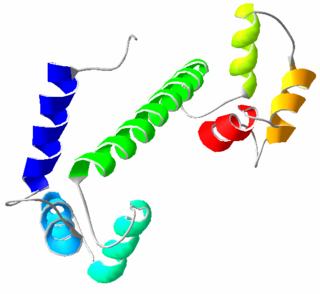
Calmodulin (CaM) (an abbreviation for calcium-modulated protein) is a multifunctional intermediate calcium-binding messenger protein expressed in all eukaryotic cells. It is an intracellular target of the secondary messenger Ca2+, and the binding of Ca2+ is required for the activation of calmodulin. Once bound to Ca2+, calmodulin acts as part of a calcium signal transduction pathway by modifying its interactions with various target proteins such as kinases or phosphatases.

<span class="mw-page-title-main">Smooth muscle</span> Involuntary non-striated muscle

Smooth muscle is an involuntary non-striated muscle, so-called because it has no sarcomeres and therefore no striations. It is divided into two subgroups, single-unit and multiunit smooth muscle. Within single-unit muscle, the whole bundle or sheet of smooth muscle cells contracts as a syncytium.

<span class="mw-page-title-main">Myofibril</span> Contractile element of muscle

A myofibril is a basic rod-like organelle of a muscle cell. Skeletal muscles are composed of long, tubular cells known as muscle fibers, and these cells contain many chains of myofibrils. Each myofibril has a diameter of 1–2 micrometres. They are created during embryonic development in a process known as myogenesis.

<span class="mw-page-title-main">Sarcomere</span> Repeating unit of a myofibril in a muscle cell

A sarcomere is the smallest functional unit of striated muscle tissue. It is the repeating unit between two Z-lines. Skeletal muscles are composed of tubular muscle cells which are formed during embryonic myogenesis. Muscle fibers contain numerous tubular myofibrils. Myofibrils are composed of repeating sections of sarcomeres, which appear under the microscope as alternating dark and light bands. Sarcomeres are composed of long, fibrous proteins as filaments that slide past each other when a muscle contracts or relaxes. The costamere is a different component that connects the sarcomere to the sarcolemma.

<span class="mw-page-title-main">Myosin</span> Superfamily of motor proteins

Myosins are a superfamily of motor proteins best known for their roles in muscle contraction and in a wide range of other motility processes in eukaryotes. They are ATP-dependent and responsible for actin-based motility.

<span class="mw-page-title-main">Muscle contraction</span> Activation of tension-generating sites in muscle

Muscle contraction is the activation of tension-generating sites within muscle cells. In physiology, muscle contraction does not necessarily mean muscle shortening because muscle tension can be produced without changes in muscle length, such as when holding something heavy in the same position. The termination of muscle contraction is followed by muscle relaxation, which is a return of the muscle fibers to their low tension-generating state.

Voltage-gated calcium channels (VGCCs), also known as voltage-dependent calcium channels (VDCCs), are a group of voltage-gated ion channels found in the membrane of excitable cells (e.g., muscle, glial cells, neurons, etc.) with a permeability to the calcium ion Ca2+. These channels are slightly permeable to sodium ions, so they are also called Ca2+–Na+ channels, but their permeability to calcium is about 1000-fold greater than to sodium under normal physiological conditions.

CAMK, also written as CaMK or CCaMK, is an abbreviation for the Ca2+/calmodulin-dependent protein kinase class of enzymes. CAMKs are activated by increases in the concentration of intracellular calcium ions (Ca2+) and calmodulin. When activated, the enzymes transfer phosphates from ATP to defined serine or threonine residues in other proteins, so they are serine/threonine-specific protein kinases. Activated CAMK is involved in the phosphorylation of transcription factors and therefore, in the regulation of expression of responding genes. CAMK also works to regulate the cell life cycle (i.e. programmed cell death), rearrangement of the cell's cytoskeletal network, and mechanisms involved in the learning and memory of an organism.

<span class="mw-page-title-main">Myofilament</span> The two protein filaments of myofibrils in muscle cells

Myofilaments are the three protein filaments of myofibrils in muscle cells. The main proteins involved are myosin, actin, and titin. Myosin and actin are the contractile proteins and titin is an elastic protein. The myofilaments act together in muscle contraction, and in order of size are a thick one of mostly myosin, a thin one of mostly actin, and a very thin one of mostly titin.

<span class="mw-page-title-main">Telokin</span> Protein domain

Telokin is an abundant protein found in smooth-muscle. It is identical to the C-terminus of myosin light-chain kinase. Telokin may play a role in the stabilization of unphosphorylated smooth-muscle myosin filaments. Because of its origin as the C-terminal end of smooth muscle myosin light chain kinase, it is called "telokin".

Calmodulin-binding proteins are, as their name implies, proteins which bind calmodulin. Calmodulin can bind to a variety of proteins through a two-step binding mechanism, namely "conformational and mutually induced fit", where typically two domains of calmodulin wrap around an emerging helical calmodulin binding domain from the target protein.

<span class="mw-page-title-main">Caldesmon</span> Mammalian protein found in Homo sapiens

Caldesmon is a protein that in humans is encoded by the CALD1 gene.

<span class="mw-page-title-main">Myosin-light-chain phosphatase</span>

Myosin light-chain phosphatase, also called myosin phosphatase (EC 3.1.3.53; systematic name [myosin-light-chain]-phosphate phosphohydrolase), is an enzyme (specifically a serine/threonine-specific protein phosphatase) that dephosphorylates the regulatory light chain of myosin II:

<span class="mw-page-title-main">Myosin light chain</span> Small polypeptide subunit of myosin

A myosin light chain is a light chain of myosin. Myosin light chains were discovered by Chinese biochemist Cao Tianqin when he was a graduate student at the University of Cambridge in England.

<span class="mw-page-title-main">CAMK2G</span> Protein-coding gene in the species Homo sapiens

Calcium/calmodulin-dependent protein kinase type II gamma chain is an enzyme that in humans is encoded by the CAMK2G gene.

<span class="mw-page-title-main">MYLK</span> Gene of the immunoglobulin superfamily

Myosin light chain kinase, smooth muscle also known as kinase-related protein (KRP) or telokin is an enzyme that in humans is encoded by the MYLK gene.

<span class="mw-page-title-main">MYLK3</span> Protein-coding gene in the species Homo sapiens

Myosin light chain kinase 3 also known as MYLK3, is an enzyme which in humans is encoded by the MYLK3 gene.

<span class="mw-page-title-main">MYLK2</span> Protein-coding gene in the species Homo sapiens

Myosin light chain kinase 2 also known as MYLK2 is an enzyme which in humans is encoded by the MYLK2 gene.

<span class="mw-page-title-main">Rho-associated protein kinase</span>

Rho-associated protein kinase (ROCK) is a kinase belonging to the AGC family of serine-threonine specific protein kinases. It is involved mainly in regulating the shape and movement of cells by acting on the cytoskeleton.

<span class="mw-page-title-main">Calponin 1</span> Protein-coding gene in the species Homo sapiens

Calponin 1 is a basic smooth muscle protein that in humans is encoded by the CNN1 gene.

References

  1. Radu, L.; Assairi, L.; Blouquit, Y.; Durand, D.; Miron, S.; Charbonnier, J.B.; Craescu, C.T. (2011). "RCSB Protein Data Bank - Structure Summary for 3KF9 - Crystal structure of the SdCen/skMLCK complex". Worldwide Protein Data Bank. doi:10.2210/pdb3kf9/pdb.
  2. Muniz, J.R.C.; Mahajan, P.; Rellos, P.; Fedorov, O.; Shrestha, B.; Wang, J.; Elkins, J.M.; Daga, N.; Cocking, R.; Chaikuad, A.; Krojer, T.; Ugochukwu, E.; Yue, W.; von Delft, F.; Arrowsmith, C.H.; Edwards, A.M.; Weigelt, J.; Bountra, C.; Gileadi, O.; Knapp, S. (2010). "RCSB Protein Data Bank - Structure Summary for 2X4F - The Crystal Structure of the Human Myosin Light Chain Kinase Loc340156". Worldwide Protein Data Bank. doi:10.2210/pdb2x4f/pdb.
  3. Gao Y, Ye LH, Kishi H, Okagaki T, Samizo K, Nakamura A, Kohama K (June 2001). "Myosin light chain kinase as a multifunctional regulatory protein of smooth muscle contraction". IUBMB Life. 51 (6): 337–44. doi:10.1080/152165401753366087. PMID   11758800. S2CID   46180993.
  4. Khapchaev AY, Shirinsky VP (December 2016). "Myosin Light Chain Kinase MYLK1: Anatomy, Interactions, Functions, and Regulation". Biochemistry. Biokhimiia. 81 (13): 1676–1697. doi:10.1134/S000629791613006X. PMID   28260490. S2CID   11424747.
  5. Stull JT, Lin PJ, Krueger JK, Trewhella J, Zhi G (December 1998). "Myosin Light Chain Kinase: Functional Domains and Structural Motifs". Acta Physiologica. 164 (4): 471–482. doi: 10.1111/j.1365-201X.1998.tb10699.x . PMID   9887970.
  6. Manning G, Whyte DB, Martinez R, Hunter T, Sudarsanam S (December 2002). "The protein kinase complement of the human genome". Science. 298 (5600): 1912–34. Bibcode:2002Sci...298.1912M. doi:10.1126/science.1075762. PMID   12471243. S2CID   26554314.
  7. Robinson A, Colbran R (2013). "Calcium/Calmodulin-Dependent Protein Kinases". In Lennarz W, Lane D (eds.). Encyclopedia of Biological Chemistry (2nd ed.). Elsevier inc. pp. 304–309. ISBN   978-0-12-378631-9.
  8. Feher J (2017). "Smooth Muscle". Quantitative Human Physiology (2nd ed.). Elsevier inc. pp. 351–361. ISBN   978-0-12-800883-6.
  9. Anjum I (January 2018). "Calcium sensitization mechanisms in detrusor smooth muscles". Journal of Basic and Clinical Physiology and Pharmacology. 29 (3): 227–235. doi:10.1515/jbcpp-2017-0071. PMID   29306925. S2CID   20486807.
  10. Amano M, Nakayama M, Kaibuchi K (September 2010). "Rho-kinase/ROCK: A key regulator of the cytoskeleton and cell polarity". Cytoskeleton. 67 (9): 545–54. doi:10.1002/cm.20472. PMC   3038199 . PMID   20803696.
  11. Dudek SM, Garcia JG (October 2001). "Cytoskeletal regulation of pulmonary vascular permeability". Journal of Applied Physiology. 91 (4): 1487–500. doi:10.1152/jappl.2001.91.4.1487. PMID   11568129. S2CID   7042112.
  12. 1 2 Ringvold HC, Khalil RA (2017). "Protein Kinase C as Regulator of Vascular Smooth Muscle Function and Potential Target in Vascular Disorders". Vascular Pharmacology - Smooth Muscle. Advances in Pharmacology. Vol. 78. pp. 203–301. doi:10.1016/bs.apha.2016.06.002. ISBN   978-0-12-811485-8. PMC   5319769 . PMID   28212798.
  13. Szilágyi KL, Liu C, Zhang X, Wang T, Fortman JD, Zhang W, Garcia JG (February 2017). "Epigenetic contribution of the myosin light chain kinase gene to the risk for acute respiratory distress syndrome". Translational Research. 180: 12–21. doi:10.1016/j.trsl.2016.07.020. PMC   5253100 . PMID   27543902.
  14. Cunningham KE, Turner JR (July 2012). "Myosin Light Chain Kinase: Pulling the Strings of Epithelial Tight Junction Function". Annals of the New York Academy of Sciences. 1258 (1): 34–42. Bibcode:2012NYASA1258...34C. doi:10.1111/j.1749-6632.2012.06526.x. PMC   3384706 . PMID   22731713.

Further reading

This article incorporates text from the United States National Library of Medicine, which is in the public domain.